Diseases of heart:
Lab 3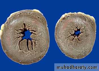
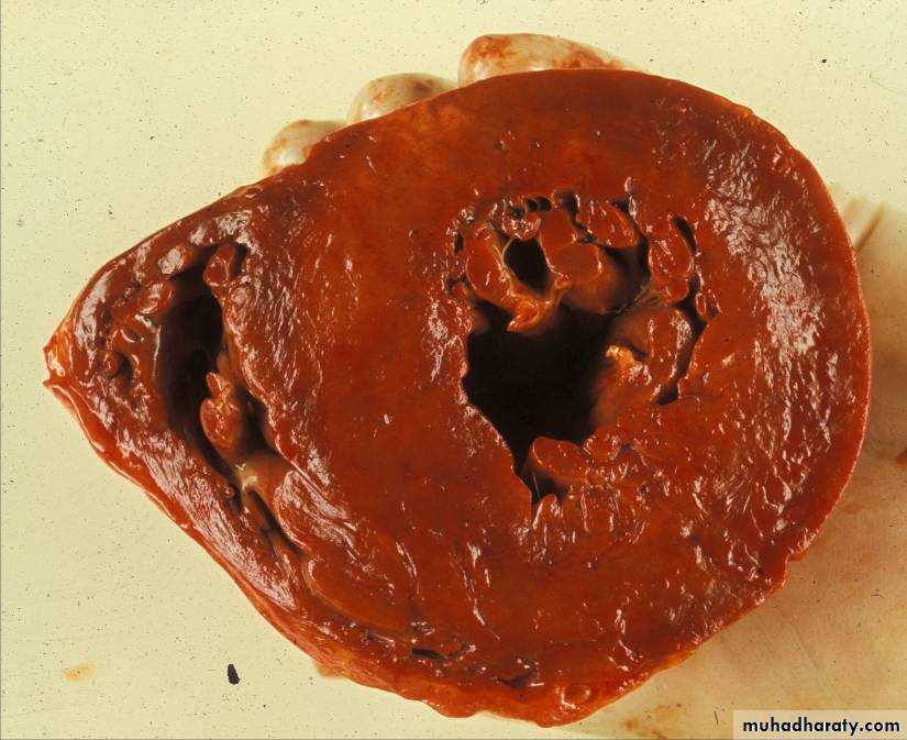
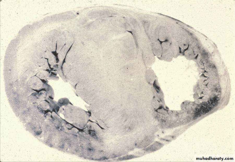
1-Heart, concentric left ventricular hypertrophy - Gross, cross sections, cut surfaces
1- Heart, concentric left ventricular H- Gross, cross sections, cut surfaces:Two cross sections of left ventricle from a patient with severe long-standing hypertension.
There is severe left ventricular hypertrophy.
The hypertrophy is designated concentric because the left ventricle and septum are of approximately equal thickness.
The walls of the ventricles are thickened.
The cavity size is decreased.
2-A- Heart, aortic valve – calcific aortic stenosis: Gross
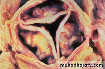
• 2- Calcific aortic stenosis:
• Large, dense, calcific nodules arise in aortic valve cusps.• The calcific masses approach, but usually do not involve, the free edges of the cusps.
• The cusps are distorted and frozen by the densely calcified nodules.
• In this particular case, the commissures of the valve are not fused. Fusion of the commissures would suggest a pre-existing inflammatory process, such as prior rheumatic heart disease or a prior bout of infective endocarditis.
2-B- Heart, left ventricular hypertrophy in calcific aortic stenosis - Gross, longitudinal section, cut surface
6 - Heart, rheumatic heart disease, rheumatic aortic stenosis - Gross
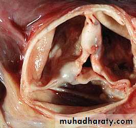
6 - Heart, rheumatic heart disease, rheumatic aortic stenosis - GrossThis valve depicts postrheumatic aortic stenosis.
The severe cuspal thickening and fusion of the commissures have distorted the three cusps of the valve so that they appear as a single fused unit, fixed in an open but stenotic configuration.
This valve would generate a complex murmur with components of both aortic stenosis (AS) and aortic insufficiency (AI).
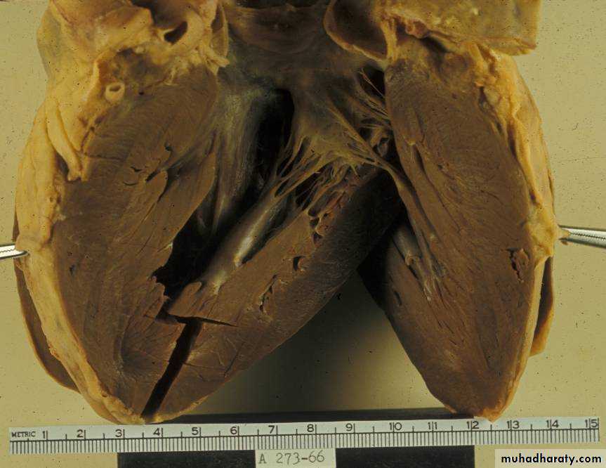
7- Heart, rheumatic mitral and aortic stenosis - Gross, longitudinal section, cut surface
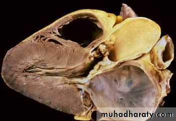
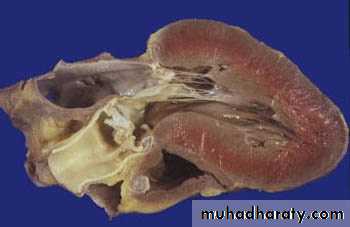
7- Heart, rheumatic mitral and aortic stenosis - Gross, longitudinal section, cut surface
Both the mitral and the aortic valves are affected.Severe, long-standing, aortic stenosis has resulted in left ventricular hypertrophy.
The severe left atrial dilation is a result of mitral stenosis.
1- Dilated right ventricle. 2- Stenotic aortic valve. 3- Dilated left atrium. 4- Stenotic mitral valve. 5- Hypertrophy of left ventricle.
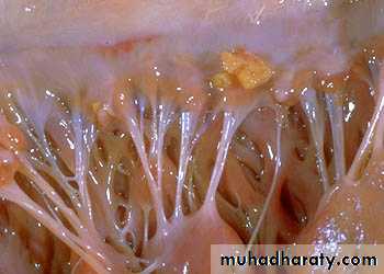
8- Heart, nonbacterial thrombotic endocarditis - Gross
8- Heart, nonbacterial thrombotic endocarditis - Gross
A small yellow-tan vegetation is present on the mitral valve.There is no apparent destruction of the underlying valve leaflet.
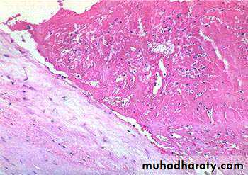
10- Heart, mitral valve, nonbacterial thrombotic endocarditis - High power
10- Heart, mitral valve, nonbacterial thrombotic endocarditis - High powerThis is the microscopic picture of the valve illustrated in the previous image.
Note the normal valve (not inflamed or damaged) with the overlying vegetation consisting of pink strands of fibrin with rare enmeshed leukocytes.
No bacteria or inflammation can be seen.
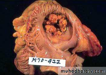
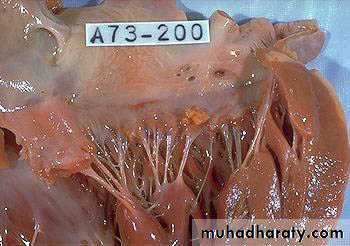
11- Heart, infective endocarditis of the tricuspid valve - Gross, from opened right atrium
11- Heart, infective endocarditis of the tricuspid valve - Gross, from opened right atrium
There are large, bulky vegetations on all the leaflets of this valve.
The valve may have been regurgitant.
The patient may have developed pulmonary symptoms from septic emboli arising from these vegetations.
12- Heart, bacterial endocarditis - Very low power
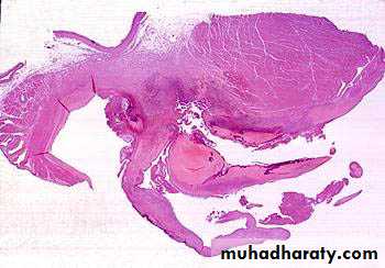
12- Heart, bacterial endocarditis - Very low power
This is a mitral valve vegetation in bacterial endocarditis.There are destructive vegetations on both sides of the valve and an abscess-like process is burrowing into the myocardium.
The bacteria are visible as a dark blue zone on the surface of the vegetations.
13- Heart, bacterial endocarditis - High power
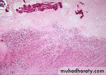
13- Heart, bacterial endocarditis - High power
The vegetation is composed of fibrin, inflammatory cells, and bacteria.The bacteria appear as a blue zone on the surface of the vegetation.
Cardiomyopathy:
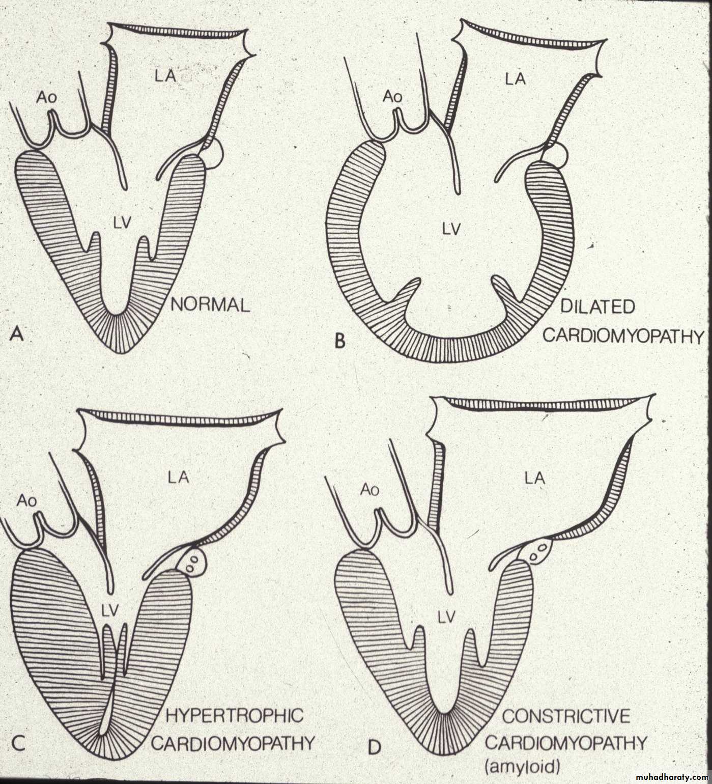
Dilated Cardiomyopathy
Gross – increased weight, dilatation, endocardial fibrosis, normal valves and coronary arteriesMicroscopic – myocyte hypertrophy, myofibrillar loss and interstitial fibrosis
Etiology – viral, genetic, toxins
Clinical significance – heart failure & death
Dilated cardiomyopathy
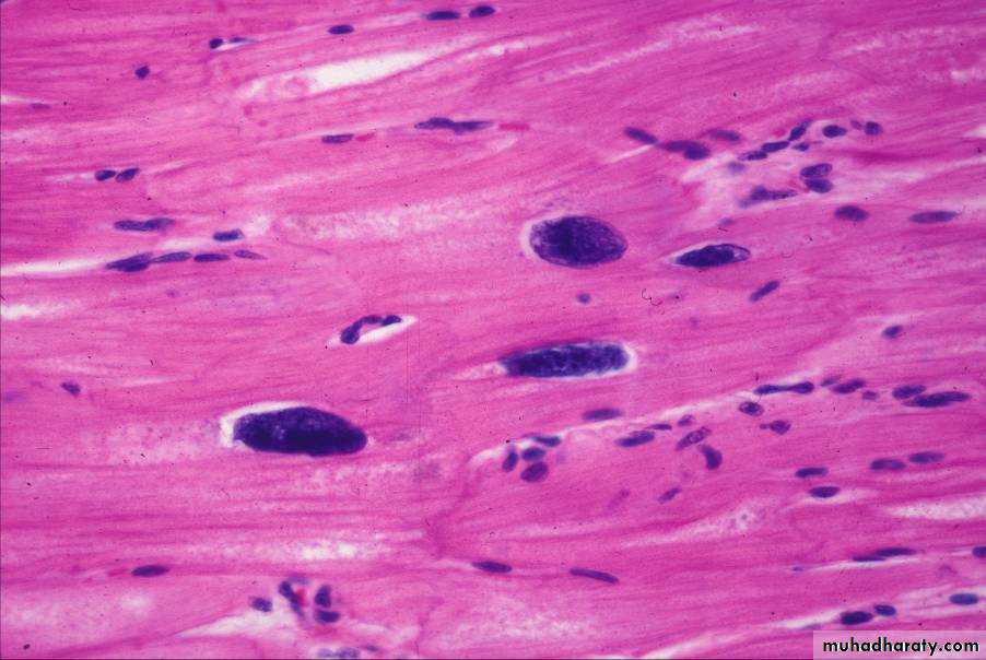
Cardiomyopathy – loss of myofibrils
Hypertrophic CardiomyopathyHypertrophy of ventricular septum.
Disarray of myofibers.
Volume reduction of ventricles.
Endocardial thickening of LV.
Mitral valve leaflet thickening.
Dilated atria.
Abnormal intramural coronaries.
Hypertrophic cardiomyopathy
Hypertrophic cardiomyopathy
Hypertrophic cardiomyopathy
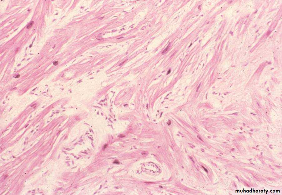
Hypertrophic cardiomyopathy – myofiber dysarray – not all fibers are pulling
the same direction. Thus the contraction is ineffective. However, the cardiacconduction system can have these same problems, which might cause the
arrhythmias and sudden death these patients tend to die of.