
THE Cardiovascular system
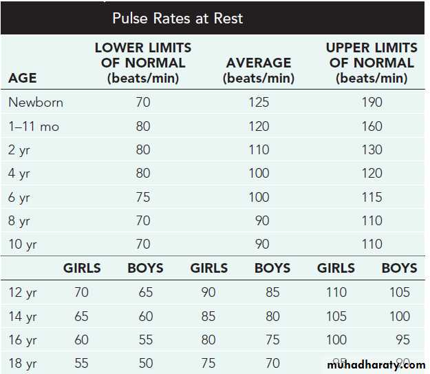
Objectives: The following lectures aim to teach the student about:Normal values of pulse rate in pediatrics
Congenital heart disease
Cyanotic: TOF,TGA,TA,EA.
Acyanotic: ASD,VSD, PDA,
Obstructive: Coarctation of Aorta.
Heart failure in infancy and childhood: Etiology, presentation, diagnosis,& treatment.
Rheumatic fever: Etiology, Diagnosis, Treatment, Prevention.
Infective endocarditis : Etiology, Diagnosis, Treatment, Prevention.
Cardiomyopathies : types with special focus on dilated CMP.
History
Children do not present with the typical features of congestive heart failure as seen in adults.Age is very important when assessing child.
Infants:
Feeding difficulties
Easily fatigued
Sweating while feeding
Rapid respirations
Older children:
Shortness of breath
Dyspnea on exertion
Physical examination:
• Tachycardia
• Rapid respiration• Tender hepatomegaly
• Pulmonary rales
REMEMBER THAT During physical examination:
− Need to refer to normal heart and respiratory rates for ages to determine tachycardia and tachypnea.− Height and weight should be assessed to determine proper growth.
− Always get upper and lower extremity blood pressures and pulses.
− Hepatosplenomegaly suggests right-sided heart failure.
− Always palpate for femoral pulses and compare with radials.
− Cyanosis and clubbing result from hypoxia.
Diagnostic tests
Chest radiograph for:° Heart size
° Lung fields
° Ribs for notching
° Position of great vessels
Electrocardiogram
Echocardiography definitive diagnosis
Other : MRI, cardiac catheterization, angiography, exercise testing
Congenital Heart Diseases
Classification of congenital heart diseases
• Group I : Left to right shunts (acyanotic): ASD, VSD, PDA• Group II: Right to lefts shunts (cyanotic): TOF, TGA,TA,EA
• Group III: Obstructive lesions: AS,PS,COA
Incidence : 8/1000 births
Etiology:Multifactorial
Some are associated with chromosomal disorders, or single gene defects.
Teratogens & maternal drugs.
Maternal metabolic disease.
Acyanotic Congenital Heart Disease
• Left-to-Right Shunt Lesions• Atrial Septal Defect (ASD)
• Ventricular Septal Defect (VSD)
• Atrioventricular Septal Defect (AV Canal)
• Patent Ductus Arteriosus (PDA)
Atrial Septal Defect
• ASD is an opening in the atrial septum permitting free communication of blood between the atria. Seen in 10% of all CHD.
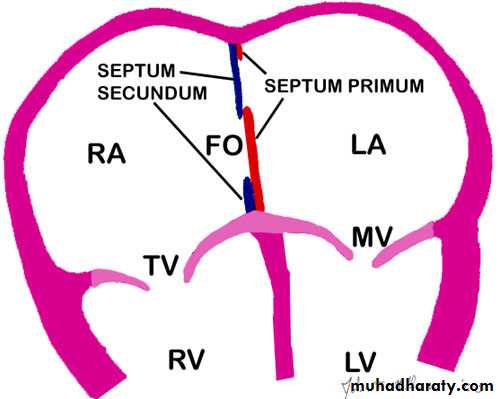
There are 3 major types:
• Secundum ASD – at the Fossa Ovalis, most common.• • Primum ASD – lower in position.
• • Sinus Venosus ASD – high in the atrial septum, associated with anomalous venous return & the least common.
• • Most are asymptomatic but may have easy fatigability or mild growth failure.
• • Cyanosis does not occur unless pulmonary HTN is present.• Examination:
• • Hyperactive precordium, RV heave, fixed widely split S2.
• • II-III/VI systolic ejection murmur at LSB.
• • Mid-diastolic murmur heard over LLSB indicates large defect.
Diagnosis
• Chest x-ray—varying heart enlargement (right ventricular and right atrial); Increased pulmonary vessel markings, edema• ECG—right-axis deviation and RVH
• Echocardiogram definitive
• Treatment:
• Surgical or Catheterization laboratory closure is generally recommended for secundum ASD with a Qp:Qs ratio >2:1.
• • Closure is performed electively between ages 2 & 5 yrs to avoid late complications.
• • Surgical correction is done earlier in children with CHF or significant Pulmonary hypertension( HTN).
• • Once pulmonary HTN with shunt reversal occurs this is considered too late (Eisnmenger syndrome).
• • Mortality is < 1%.
• N.B. Infective Endocarditis is very rare in ASD.
Ventricular Septal Defect
VSD – is an abnormal opening in the ventricular septum, which allows free communication between the Rt & Lt ventricles. Accounts for 25% of CHD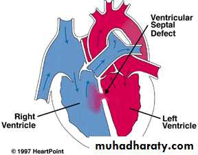
• Clinical Signs & Symptoms
• • Small - moderate VSD, 3-6mm, are usually asymptomatic and 50% will close spontaneously by age 2yrs.• • Moderate – large VSD, almost always have symptoms(dyspnea, feeding difficulties, poor growth, sweating, pulmonary infection, heart failure) and will require surgical repair
• Symptoms develop between 1 – 6 months.
• • CHF, FTT, Respiratory infections, exercise intolerance hyperactive precordium• • II-III/VI harsh pansystolic murmur heard along the LSB, more prominent with small VSD.
Imaging Studies
ECG and CX-ray findings depend on the size of the VSD.
• Small VSDs usually have normal studies.
• Larger VSDs, ECG findings of left atrial and ventricular enlargement and hypertrophy.
• A CXR may reveal cardiomegaly, enlargement of the left ventricle, an increase in thepulmonary artery silhouette, and increased pulmonary bloodflow.
• Pulmonary hypertension due to either increased flow or increased pulmonary vascular resistance may lead to right ventricular enlargement and hypertrophy.
• Treatment
• • Small VSD - no surgical intervention, nophysical restrictions, just reassurance and periodic follow-up and endocarditis prophylaxis.• • Symptomatic VSD - Medical treatment initially with afterload reducers & diuretics( ± digoxin).
• • Prophylaxis against infective endocarditis(after dental or GU procedures).
Indications for Surgical Closure:
• Large VSD with medically uncontrolled symptomatology & continued FTT.• Ages 6-12 mo with large VSD & Pulmonary HTN.
• Age > 24 mo w/ Qp:Qs ratio > 2:1.
• Supracristal VSD of any size.
Complications
Large defects lead to heart failure, failure to thriveEndocarditis
Pulmonary hypertension
Patent Ductus Arteriosus
Persistence of the normal fetal vessel that joins the PA to the Aorta.
• Normally closes in the 1st wk of life.
• Accounts for 10% of all CHD, seen in 10% of other congenital heart lesions and can often play a critical role in some lesions.• Female : Male ratio of 2:1
• Often associated with coarctation & VSD.
• Can be caused by congenital Rubella.
• Clinical Signs & Symptoms
• Small PDA’s are usually asymptomatic.• Large PDA’s can result in symptoms of CHF, growth restriction, FTT.
• Collapsing arterial pulses.
• Widened pulse pressure .
• Enlarged heart, prominent apical impulse.
• Classic continuous machinary systolic murmur.
• Mid-diastolic murmur at the apex.
Imaging Studies
• ECG and chest x-ray findings are normal with small PDAs• moderate to large shunts may result in a full pulmonary artery silhouette and increased pulmonary vascularity.
• ECG findings vary from normal to evidence of LVH. If pulmonary HTN is present, there is also RVH.
• Treatment:
• Indomethacin, inhibitor of prostaglandin synthesis can be used in premature infants.
• PDA requires surgical or catheter closure.
• Closure is required for heart failure & to prevent pulmonary vascular disease.
• Usually done by ligation & division or intra vascular coil.
• Prophylaxis against infective endocarditis(after dental or GU procedures).
Obstructive Heart Lesions
• Pulmonary Stenosis• Aortic Stenosis
• Coarctation of the Aorta
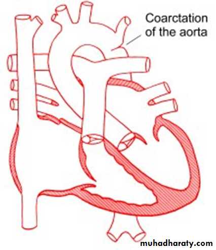
Coarctation of the Aorta
is narrowing of the aorta at varying points anywhere from the transverse arch to the iliac bifurcation. It is commonly associated with bicuspid valve.More common in Turner’s syndrome.
• 98% of coarctations are juxtaductal
• Male: Female ratio 3:1.
• Accounts for 7 % of all CHD
• The obstruction to blood flow will lead to LVH.
• Clinical Signs & Symptoms
• Classic signs of coarctation are diminution or absence of femoral pulses.• Higher BP in the upper extremities as compared to the lower extremities.
• 90% have systolic hypertension of the upper extremities.
• With severe coarctation, HF and shock.
• Differential cyanosis if ductus is still open.
• II/VI systolic ejection murmur at LSB.
• Cardiomegaly, rib notching on X-ray.
notching of ribs in
coarctation.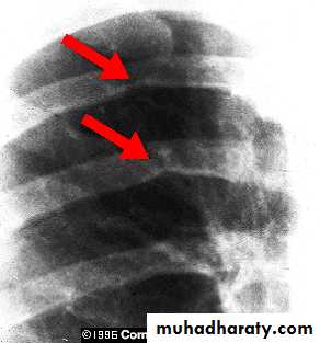
• Treatment
• With severe coarctation maintaining the ductus with prostaglandin E is essential.• Surgical intervention, to prevent LV dysfunction.
• Angioplasty is used by some centers.
• Re-coarctation can occur, balloon angioplasty is the procedure of choice.
• Prophylaxis against infective endocarditis(after dental or GU procedures).
Cyanotic congenital heart disease
Tetralogy of Fallot
It is the most common cyanotic congenital heart diseaseComponents:
• Pulmonary stenosis and infundibular stenosis (obstruction to right ventricular outflow)• VSD
• Overriding aorta (overrides the VSD)
• Right ventricular hypertrophy
Hemodynamics
Pulmonary stenosis plus hypertrophy of subpulmonic muscle (crista supraventricularis)→ Varying degrees of right ventricular outflow obstruction
→ Blood shunted right-to-left across the VSD with varying degrees of arterial desaturation and cyanosis.
Clinical Picture
• Symptomatic any time after birth• Paroxysmal attacks of dyspnea
• Anoxic spells
• Predominantly after waking up
• Child cry
• Dyspnea& cyanosis
• Loss of consciousness
• Convulsions
• Frequency varies from once a few days to many attack everyday
Examination
Varying degrees of cyanosis and a murmur, a single S2 and right ventricular impulse at the left sternal border are typical findings.
Clubbing of fingers & toes.
Cyanosis may not be apparent for the first few weeks of life until pulmonary stenosis reach a critical level that cause reduction in pulmonary blood flow.
Therefore; the only finding in the first few days may be the murmur of PS.
Hypoxic (Tet) spells
1. Restlessness and agitation2. Inconsolable cry
3. Squatting:
increases the peripheral vascular resistance, which diminishes the right-to-left shunt
increases pulmonary blood flow.
4. Predominantly after waking up
5. Dyspnea& cyanosis6. Loss of consciousness
7. Convulsions
8. Loss of the murmur
Frequency varies from once a few days to many attack everyday
Imaging studies
ECG : RAD &RVH
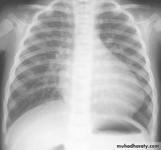
CXR : Boot-shaped heart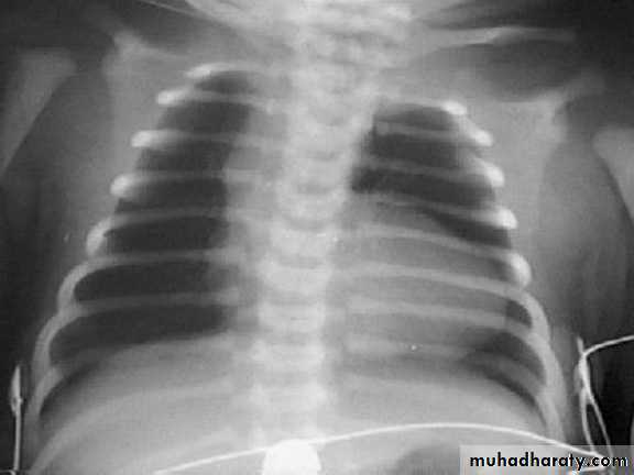
Complications:
• Each anoxic spell is potentially fatal• Polycythemia may lead to Cerebrovascular thrombosis
• Anoxic infaction of CNS
• Brain Abcess
• Infective endocarditis
Treatment
Management of anoxic spell:Knee chest position
Humified O2
Be careful not to provoke the child
Morphine 0.1 -0.2 mg/kg subcutaneously
Correct acidosis – sodium bicarb IV 1 mmol/kg slowly
Propanolol start (0.1mg/kg/IV) slowly during spells followed by (0.5 to 1.0) mg/kg/ 4-6hourly orally
Vasopressors: Methoxamine or Phenylephrine IM or IV drip
GA is the last resort
Surgery
Palliative procedure: Blalock Taussig shunt
Subclavian artery – Pulmonary artery anastomosis
Definitive operation: complete surgical repair with VSD closure and removal or patching of the pulmonary stenosis can be performed in
infancy
Transposition of Great Areries (TGA)
Aorta originating from the right ventricle, and pulmonary artery originating from the left ventricleAccounts for 5-7% of all congenital heart disease
Survival is dependent on the presence of mixing between the pulmonary and systemic circulation
Associated ASD, VSD, or PDA are essential for survival .
50% of patients have a VSD
Usually presents in the first day of life with profound cyanosis
More common in boys
Examination :
Cyanosis in an otherwise healthy looking babyLoud S2
Loud VSD murmur
CXR : Egg on side & narrow mediastinum
Acute management in newborn baby
Initial medical management includes prostaglandin E1 to maintain ductal patency. If significant hypoxia persists on prostaglandin therapy, a balloon atrial septostomy improves mixing between the two circulations.Surgical repair Aterial switch (old style).
Arterial switch (new)TRICUSPID ATRESIA:
The absence of the tricuspid valve results in a hypoplastic right ventricle. All systemic venous return must cross the atrial septum into the left atrium.A PDA or VSD is necessary for pulmonary blood flow and
survival.
Clinical Manifestations
Usually severely cyanosed.Single S2.
If a VSD is present, there may be a murmur.
ECG: LVH& LAD.
Treatment:PGE-1, and minimal O2 to maintain ductal patency
Palliative procedure: Blalock-Taussig procedure
Definitive: bidirectional cavopulmonary shunt (bidirectional Glenn) and Fontan procedure.
Ebstein anomaly
Downward displacement of abnormal tricuspid valve into right ventricle; the right ventricle gets divided into two parts: an atrialized portion, which is thin-walled, and smaller normal ventricular myocardium.
• Right atrium is huge; tricuspid valve regurgitant
• Right ventricular output is decreased because:
− Poorly functioning, small right ventricle
− Tricuspid regurgitation
− Variable right ventricular outflow obstruction—abnormal anterior tricuspid valve leaflet. Therefore, increased right atrial volume shunts blood through foramen Ovale or ASD → cyanosis.
Clinical presentation
− Severity and presentation depend upon degree of displacement of valve and degree of right ventricular outflow obstruction° May not present until adolescence or adulthood
° If severe in newborn → marked cyanosis, huge heart
− Pansystolic murmur of tricuspid insufficiency over most of anterior left chest (most characteristic finding)
− Chest x-ray—heart size varies from normal to massive (increased right atrium); if severe, decreased pulmonary blood flow.
− ECG—tall and broad P waves, RBBB, and a normal or prolonged PR interval
• Treatment:
− PGE1− Systemic-to-pulmonary shunt
− Then staged surgery