
1
Fifth stage
Pediatric
Lec-1
.د
مازن
1/1/2014
Pediatric oncology
At the end of lectures, the students should be able to:
1-Know the epidemiology, predisposing factors and the common presentation of childhood
malignancies.
2-Diagnose ALL, know the C/P, treatment protocols and to identify relapses.
3-Diagnose and know the outlines of treatment of NHL and HD.
General Principles of Cancer in Children
Epidemiology
Cancer is the most common cause of disease-related death in childhood and has an annual
incidence of 1 in 600 for those younger than 15 years.
Types
Common types of cancer in children :Leukemia and tumors of the central nervous system
represent the majority of childhood neoplasms, followed by a wide array of solid tumors.
Uncommon types of cancer in children: Typical adult-type epithelial-derived tumors such as
carcinomas of the lung, colon, and breast are extremely rare during childhood.
Predisposing factors
Most childhood malignancies are of unknown cause and occur in otherwise healthy
children.
Certain children, however, are at an increased risk for cancer because of their
constitutional makeup or exposure to cancer-causing agents.
1-Genetic syndromes.
1-Trisomy 21 is associated with a 15-fold increased risk of developing leukemia.
2-Individuals with overgrowth syndromes (e.g., Beckwith-Wiedemann) are at increased risk
for the development of Wilms tumor.
3-Children with Fanconi anemia often have higher chance to develope leukemia.

2
2-Immunodeficiencies:
Wiskott-Aldrich syndrome is an X-linked disorder characterized by progressive T-cell
dysfunction, eczema, thrombocytopenia, and a propensity to development of lymphomas.
Common variable immunodeficiency predisposes affected individuals to stomach cancer
and lymphomas.
X-linked lymphoproliferative syndrome in males results in severe infections with the
Epstein-Barr virus; if the acute infection does not end fatally, induction of lymphoma
occurs.
3-Infections. There are primarily two viruses that infect cells of the immune system that
have been associated with childhood malignancy.
1-Epstein-Barr virus has been associated with the development of endemic Burkitt
lymphoma. This malignancy involves a specific chromosomal change involving translocation
of a portion of the long arm of chromosome onto the area of another chromosome (14, 2,
or 22.)
2-Human immunodeficiency virus (HIV) infection may lead to viral destruction of helper T
cells and acquired immune deficiency syndrome (AIDS), which is characterized by an
increased susceptibility to opportunistic infections and malignancies .
Pediatric patients who have AIDS are susceptible to lymphoid malignancies such as Burkitt
lymphoma, and few children have developed Kaposi sarcoma.
4-Environmental factors
Although many environmental carcinogens and toxic exposures are associated with the
development of cancer in adults, childhood cancers caused by environmental factors and
toxic exposures are rare.
One known risk factor for childhood cancer is prior treatment of malignancy in a child with
chemotherapeutic agents, ionizing radiation, or both.
The Leukemias
Classification:
Leukemias are classified as lymphoblastic leukemias, which are proliferations of cells of
lymphoid lineage, and myeloblastic leukemias, which are proliferations of cells of
granulocyte, monocyte, erythrocyte, or platelet lineage .
Both are divided to acute and chronic.

3
Acute leukemias constitute 97% of all childhood leukemias. If untreated, they are rapidly
fatal within weeks to a few months of the diagnosis; however, with treatment, they often
are curable.
-Acute lymphoblastic leukemia (ALL) is the most common pediatric neoplasm and accounts
for 80% of all childhood acute leukemia cases.
-Acute myeloblastic leukemia (AML) is also called acute myelogenous leukemia and
accounts for the remaining 20% of acute leukemia cases in children.
Chronic leukemias represent 3% of childhood leukemias; even without treatment, patients
can survive for many months to years.
All chronic leukemias in children are of nonlymphoid lineage. The leukemic cells are more
mature and functional than the blasts of acute leukemias.
Acute lymphoblastic leukemia
ALL is a malignant disease in which abnormal lymphoid blasts accumulate in the bone
marrow and replace the normal hematopoietic elements.
They are also released into peripheral blood, through which they spread throughout the
body and infiltrate all organ systems.
ALL is the most common type of childhood leukemia.
It is more common in white children than in black children and more common in males
than in females (1.2–1.3 times).
ALL is associated with a peak incidence in the 3- 5 year-old age group .
Etiology
The malignant lymphoblasts of each patient who has ALL are all thought to be generated
from a single abnormal precursor cell, and as such represent a clone.
They demonstrate a unique pattern of gene rearrangement that is identical in all the cells
of the clone, and which distinguishes them from the clones of other ALL patients.
Classification of ALL
Morphologic classification of ALL is based on the following features:
1-Appearance of the leukemic lymphoblasts. According to the French-American-British
(FAB) classification, leukemic lymphoblasts are subdivided into three categories.
-L1 lymphoblasts are small, with scant cytoplasm and absent or inconspicuous nucleoli .
-L2 lymphoblasts are larger, with more abundant cytoplasm and one or more prominent
nucleoli.

4
-L3 lymphoblasts are large, with deeply basophilic and vacuolated cytoplasm and prominent
nucleoli.
2-Enzymatic evaluation. Terminal deoxynucleotidyl transferase (TdT) is a unique DNA
polymerase that is found in almost all lymphoblasts but only rarely in AML blasts.
3-Histochemical evaluation shows:
-Absence of surface markers typical of AML blasts
-In many cases, accumulations of glycogen on periodic acid-Schiff stain ( PAS).
Immunologic classification regards ALL as a heterogeneous group of malignancies
characterized by accumulation of immature lymphoid cells arrested at various stages of
development. On the basis of immunophenotype, ALL is divided into the following
subtypes:
1-B-cell precursor ALL accounts for 84% of all cases.
2-Mature B-cell ALL accounts for 1% of cases. This is the most mature form of ALL of B-cell
lineage and is closely related to Burkitt lymphoma.
3-T-cell ALL accounts for the remaining 15% of ALL cases.
T-cell ALL has a characteristic clinical presentation that includes:
1. Occurrence in older children and teenagers
2. Predilection for males
3. High white blood cell count, often more than 100,000/mm3
4. Presence of anterior mediastinal mass
5. Early dissemination to meninges and testes
Clinical features
1-Bone marrow failure. Replacement of the normal hematopoietic elements by the
leukemic cell population .
-Pallor, irritability, or fatigue secondary to anemia
-Petechiae, ecchymoses in the skin, or epistaxis secondary to thrombocytopenia.
-Fever associated with infection due to a paucity of functional white blood cells, especially
granulocytes
2-Reticuloendothelial system infiltration
Lymphadenopathy is common, especially in ALL, and may be so massive as to resemble that
in the lymphomas.

5
Hepatosplenomegaly may also be present. Both the liver and the spleen can be minimally
to massively enlarged.
3-Bone pain .
4-Involvement of sanctuary sites. Sanctuary sites are rarely involved at the time of diagnosis
but may be involved with the recurrence of disease. These sites are the:
CNS, where involvement manifests as diffuse meningeal infiltration with signs of increased
intracranial pressure .
Testes, one or both of which may be involved, with infiltration producing enlargement that
is out of proportion to the child's sexual development
Investigation
1- CBC , blood film and ESR.
2- Bone marrow examination for morphology ,immunohistochemical and special stain
study.
3- CSF examination.
4- CXR, Abdominal ultrasound.
5- Liver Function Test, Renal function test, LDH, serum electrolytes and uric acid.
Therapy
A-Antileukemic chemotherapy is administered in distinct phases with distinct objectives.
B-Supportive care. Treatment of any complications in the child who has newly diagnosed
leukemia is essential and lifesaving.
1-Transfusion therapy : blood and platelets transfusion.
2- Treatment of infection.
3-Metabolic support is necessary in patients who have large tumor burdens as represented
by a high white blood cell count or significant organ infiltration.
These patients are at risk for tumor lysis syndrome:
-Hyperuricemia
-Hyperkalemia
-Hyperphosphatemia and hypocalcemia.
-Renal failure.

6
Prevention of tumor lysis syndrome:
1-Vigorous hydration to promote uric acid excretion.
2-Allopurinol, is used to block uric acid formation.
3-In cases of severe uric acid elevations, urate oxidase, an enzyme that converts urate into
the water-soluble allantoin, has been used to successfully decrease the uric acid to a safe
and very low level .
4-Alkalinization of urine to increase uric acid solubility
4-Treatment of hyperviscosity.
White blood cell counts > 100,000/mm3 in patients who have AML can cause significant
hyperviscosity, leading to an increased risk of CNS hemorrhagic stroke or pulmonary
infarction.
The white blood cell count may be lowered by exchange transfusions or leukapheresis.
5-Treatment of superior vena cava syndrome.
A mass in the anterior mediastinum may produce compressive symptoms with airway
compromise and obstruction of the superior vena cava, which results in a syndrome
consisting of facial plethora, venous distention, and increased intracranial pressure.
If the mass and compressive symptoms do not decrease with the institution of
chemotherapy, radiotherapy to the mass may be effective.
PROGNOSTIC INDICATORS
1-Demographics:
Standard risk patients are aged between 1 and 9 years with a presenting white cell count of
< 50000/dl, while high risk patients are aged 10 years or less than one year, or have a
presenting white cell count of ≥ 50000/dl. Approximately 75% of patients with B lineage
leukemia are standard risk, while 75% of patients with T lineage leukemia are in the high
risk category.
Boys have a worse prognosis than girls
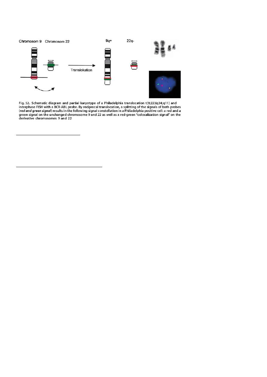
2-Chromosomes
An unfavorable prognosis is associated with specific chromosomal translocations within
the leukemic cells.
They include the Philadelphia chromosome t(9;22); or t(4;11); and t(1;19).
-Hypodiploidy, due to fewer than 46 chromosomes in the leukemic cells.
7
Favorable prognosis is associated with t(12;21), and hyperdibloidy especially if there are 53
or more chromosomes within the leukemic cells
3-Response to treatment
High-risk group includes all children irrespective of initial risk group, who have a slow early
response to treatment, (10-12% of children).
4-Minimal residual disease (MRD)
Improved techniques for detection of MRD and long term prospective study of large
number of patients has shown that clearance of MRD is an independent prognostic factor in
childhood ALL.
This has opened the way for clinical application of MRD measurement in the management
of ALL.
MRD detected prior to bone marrow transplant predicts relapse post-transplant and would
allow pretransplant intensification.
Chemotherapy
Remission induction. This initial phase lasts at least 4 weeks, during which maximal
cytoreduction is achieved.
At least three drugs—including steroids, vincristine, and asparaginase—are employed.
Anthracyclines are added for patients who have a high risk of relapse.
Consolidation or intensification consists of continued systemic therapy designed to kill
additional leukemic cells to prevent systemic relapse along with CNS-directed therapy to
prevent CNS relapse.
Intrathecal therapy is the current mainstay of CNS prophylaxis.

8
Maintenance or continuation therapy is the longest and last phase of therapy, typically
lasting at least 2 to 3 years.
The objective in this phase is to continue the remissions achieved in the previous phases
and to produce whatever additional cytoreduction is necessary to cure the leukemia.
Relapses
Relapses still occur in approximately 20% to 25% of patients.
At least one half of relapses occur while initial chemotherapy is still being administered.
Relapse can occur in the:
-Bone marrow, which is the most common site of recurrence
-CNS, which was formerly the most common site of recurrence before CNS prophylaxis
-Testes, which are becoming the most common site of extramedullary relapse
Childhood lymphoma
Lymphoma is the third most common pediatric tumor .
It’s a malignant tumor of lymph nodes.
-Non Hodgkin lymphoma (NHL)
-Hodgkin disease (HD)
NHL
-Male: Female = 3:1
-The peak incidence is 7-11 years.
-All cases of NHL are highly malignant ,diffuse and aggressive with little differentiation.
-NHL may associate with immunodeficiency, autoimmune dis., and EBV inf.
Histological types of NHL
-Lymphoblastic NHL: most are T cell.
-Non lymphoblastic:
1-small non cleaved( Burkitt, non Burkitt): B cell.

9
2-large cell : B cell.
3-anaplastic type : T cell.
Clinical pictures of NHL:
1- The most common presentation is painless LN enlargement.
2-The abdomen, head and neck are the most common sites for B cell NHL.
3-The anterior mediastinum and peripheral nodes are the primary sites for T cell NHL.
4- In abdominal presentation the child may complain from abdominal pain, nausea ,
vomiting , acute abdomen due to intussuception and large abdominal mass (in Burkitt
type).
5- Large mediastinal mass which may lead to air way obstruction or SVC syndrome and
pleural effusion
6-unussual presentation : thyroid and parotid glands swelling, spinal cord compression and
proptosis.
7-systemic symptoms: fever and weight loss.
Investigations:
1-Diagnosis is established by LN or tissue biopsy or ascetic or pleural fluid analysis.
2- Bone marrow aspiration and bone biopsy examination to role out BM involvement.
3- CSF examination to role out CNS involvement.
4- CBP, S .uric acid, LDH.
5-Whole body CT Scan , PET scan
Treatment
1- T cell lymphoblastic lymphoma and T cell anablastic large cell lymphoma are generally
treated with aggressive regimens similar to those used in ALL.
2- B cell NHL with localized disease require minimal amounts of chemotherapy.
3- Disseminated NHL or CNS or BM involvement require aggressive high dose
chemotherapy.
4- Radiation therapy is rarely used in children with NHL.

10
Hodgkin Disease
Most commonly seen in adolescent.
HD if affect younger children it will carry poor prognosis.
Sub type include:
1-lymphocyte predominant 10-20%.
2-nodular sclerosis 40-60%.
3-mixed cellularity 20-40%.
4-lymphocyte depletion <5%.
Clinical pictures:
1- painless firm Lymphadenopathy usually in the cervical or supraclavicular areas.
2- mediastinal LN may lead to SVC syndrome or respiratory distress.
3- B symptoms: high fever ,weight loss and drenching night sweating.
Staging: Ann Arbor classification
Stage I: single lymphatic (I) or extra lymphatic (IE) organ.
Stage II: two or more (II) or (IIE) organ on the same site of diaphragm.
Stage III: affect organs or LN on both sites of diaphragm with or with out spleen
involvement.
Stage IV: disseminated involvement.
Diagnosis:
1-high ESR : related to the prognosis.
2-tissue biopsy from involved LN or organs to identify RS cells.
3-CT scan of the neck, chest, abdomen and pelvis .
4-gallium scan.
5-BM aspiration and biopsy.
Treatment Combination of chemotherapy and low – dose involved field radiotherapy.