Diseases of the pleuraPleural tumorsChest tubeThoracoscopy
Diseases of the pleura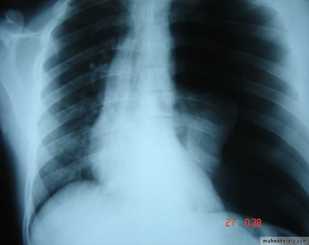
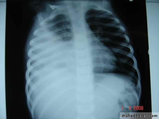
• 1-Spontaneous pneumothorax
Is the accumulation of air inside the pleural cavity , occurring without any known etiology .More in males ,more on the right side .It can be bilateral
• Causes 1- Ruptured pulmonary bleb.2-Ruptured of a cystic defect in the pleura.3-Teared visceral pleura 4-No cause can be demonstrated in (15-20%).Complications:-1-pleural effusion2-empyema 3-tension pneumothorax which leads to mediastinal shift &circulatory collapse.4-Respiratory failure in elderly patient with COAD .
• Treatment :-
• 1-Bed rest ,O2 administration &observation in limited pneumothorax.
• 2-Aspiration
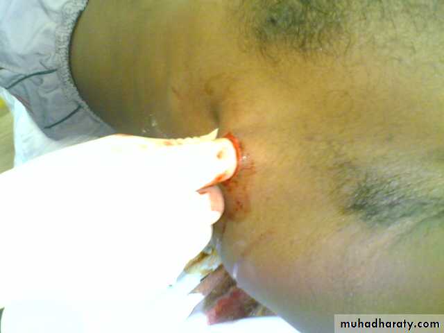
• 3-Chest tube (thoracostomy tube or ICD intercostal drain in a safety triangle which is bounded by pectoralis muscle anteriorly &lattismus muscle posteriorly and the superior border of the nipple.in the fifth intercostal space just anterior to the mid axillary line to avoid the long thoracic nerve .
• 4-bronchoscopy is indicated if the lung fail to expand
• 5-Chemical pleurodesis.by injecting sclerosing agent as Tetra cycline
• 6-Surgery pleurectomy by thoracotomy or thoracoscopically if the lung fail to re expand
• 2-Spontaneous haemothorax
• Is the presence of blood inside the pleural cavity
• Causes:-
• 1-pulmonary causes ----------TB , AV malformation
• 2-pleural causes -----------torn vascular adhesion
• 3-pulmonary malignancy ….primary or metastatic
• 4-blood dyscrasia ……………..hemophilia
• 5-abdominal pathology ……….. haemo peritoneum
• 6-thoracic causes ………ruptured great vessels
• Clinical featuresdyspnea , chest pain ,syncopesigns of hypovolaemic shock blood inside the pleural cavity may leads to deposition of fibrin on the pleural surface leading to fibrosis (trapped lung syndrome) .
• Treatment
• 1-Resuscitation
• 2-Tube thoracostomy
• 3-May needs thoracotomy if excessive bleeding
• initial bleeding more than 1.5 liter
• Or continuous bleeding more than 200 ml/hour for more than 4 hours
• 3-Chylo –thorax
• Is the presence of lymph in the pleural space• Causes
• A-Congenital atresia of the thoracic duct , birth trauma
• B-Traumatic
• C-Neoplastic malignancy
• D-Infection TB
• Diagnosis milky pleural effusion that does not clot and contains fat , fat soluble vitamins & antibodies
• Treatment
• 1-Conservative consists of insertion of tube thoracostomy to drain the effusion , correction of the fluid and electrolytes with nutritional supplement.
• 2-Surgery consists of ligation of the thoracic duct if the effusion continues for more than two weeks .
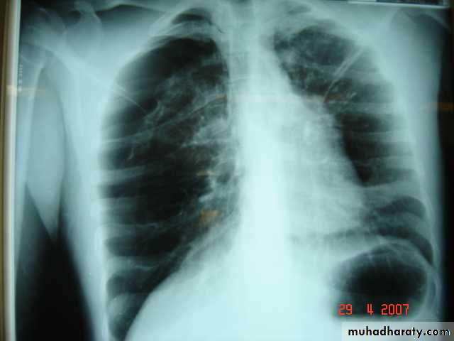
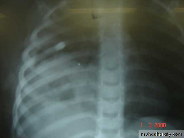
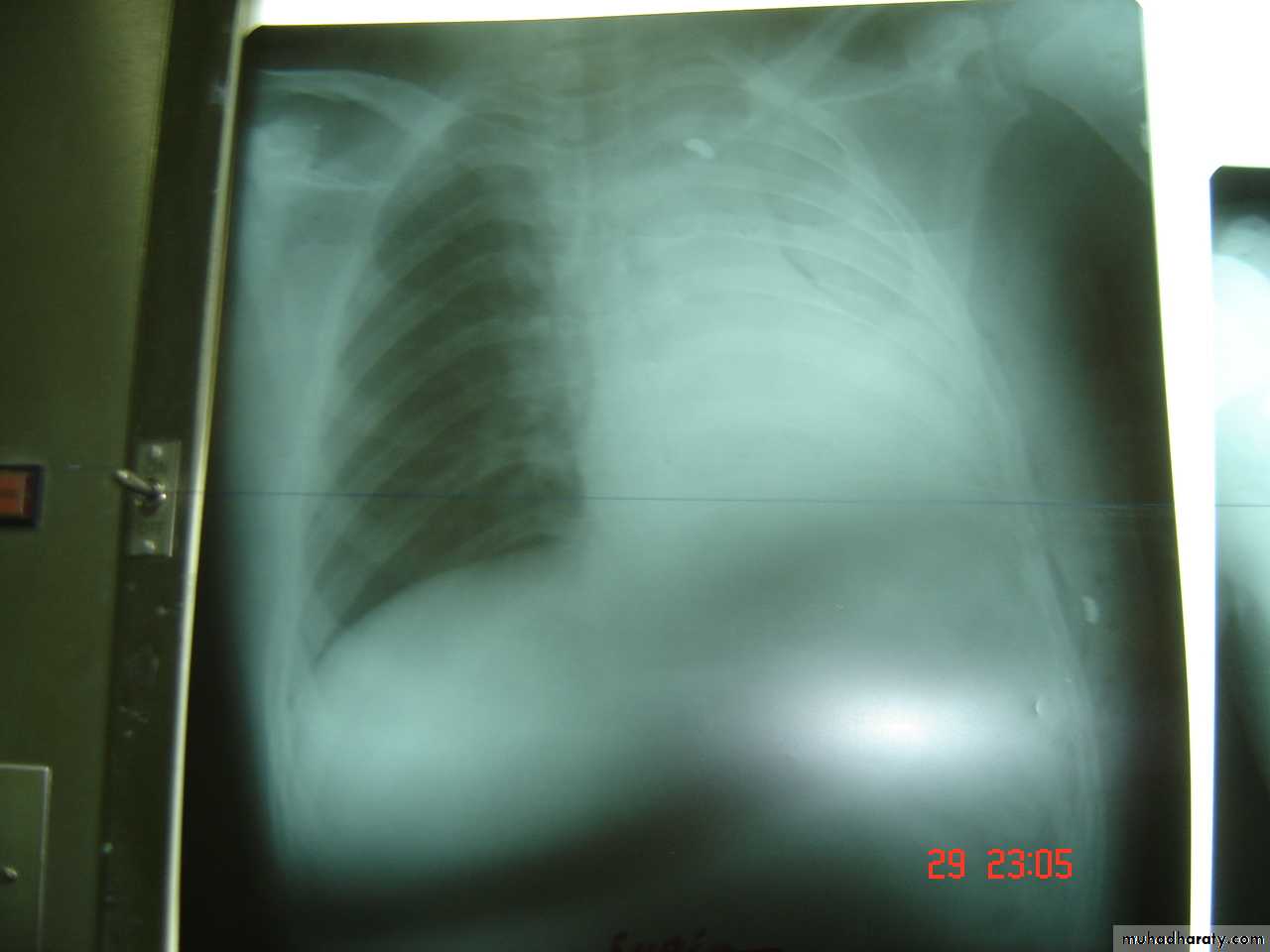
• 4-Pleural effusion
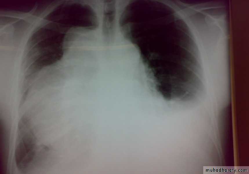
• Is the accumulation of fluid in the pleural space excessive transudation or exudation of the interstitial fluid from the pleural surface. It is signfrom ify pleural or systemic disease .
• Its effect depends on its size (mild , moderate or massive ) & the state of the underlying lung .It is classified as transudate when the protein content is less than 3g/100ml, or exudates when protein content is more than 3 gm /100ml.Clinically patients will present with dyspnea & pleuritic chest pain
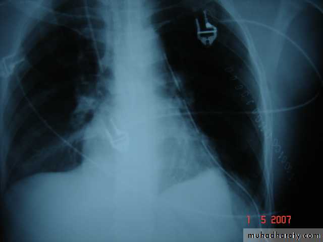
• Radio logically (concave meniscus sign)
• Transudate as in CHF
• Exudate as in malignancy
• Treatment :-1-aspiration (thoracentesis) 2-tube thoracostomy
• 3-chemical pleurodesis 4-pleuectomy to remove the pleura to stop the effusion.
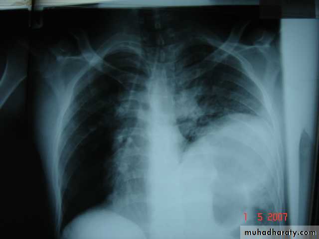
• 5-Empyaema
• Is the accumulation of pus in the pleural space , it passes into three stages
• 1-Acute phase with the clinical manifestation of fever & toxicity .• 2-Transitional phase with the increased turbidity of the fluid & decrease the size of the lung .
• 3-Chronic phase with the pleural thickening ,decrease amount of the fluid & the development of the trapped lung syndrome .
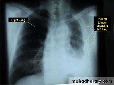
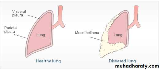
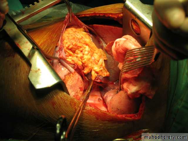
Pleural Tumors
Classified as primary and secondary tumors .Primary Pleural tumors are Mesotheiloma which may be
1-Localized benign 2- Diffuse Malignant
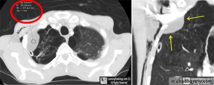
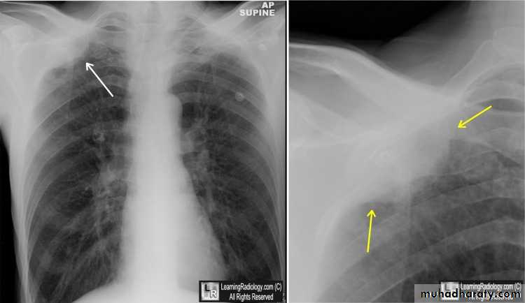
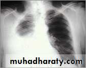
Malignant Mesothelioma causes chest pain , bloody pleural effusion and chest X-Ray findings of diffused pleural thickening with nodularity and limited pleural effusion .There is a possible relationship with asbestos exposure .
Metastases are uncommon .Death usually occurs within 1-2 years .It has a poor response to surgery , radiotherapy and chemotherapy .
Pleural involvement by metastatic diseases is more common than primary tumor and usually comes from lung , breast and stomach .
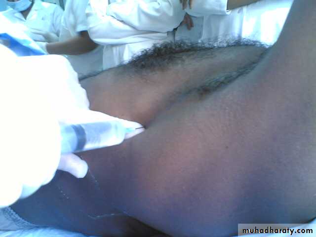
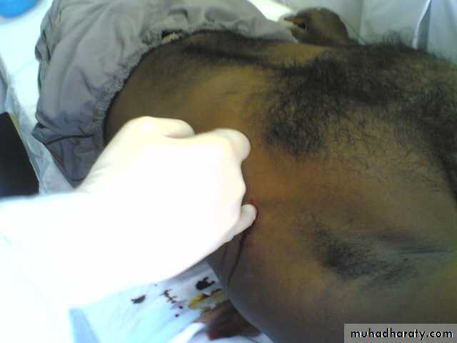
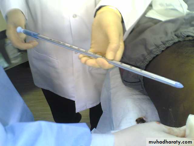
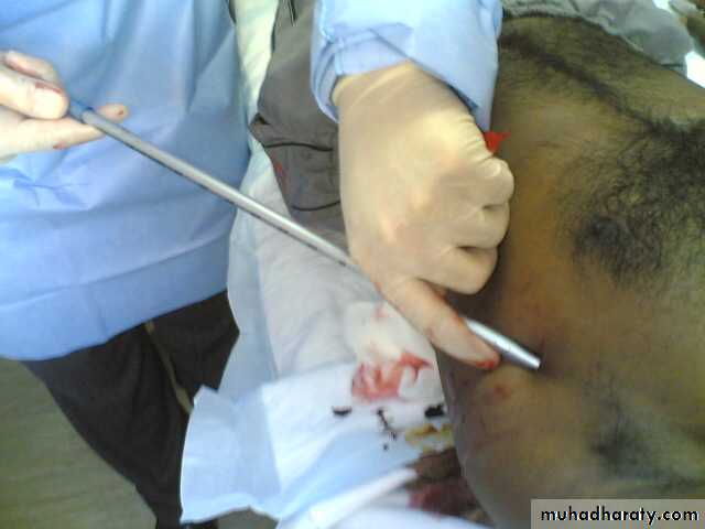
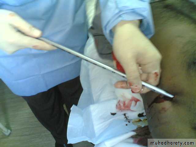
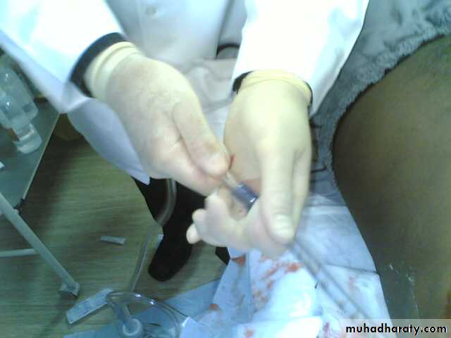
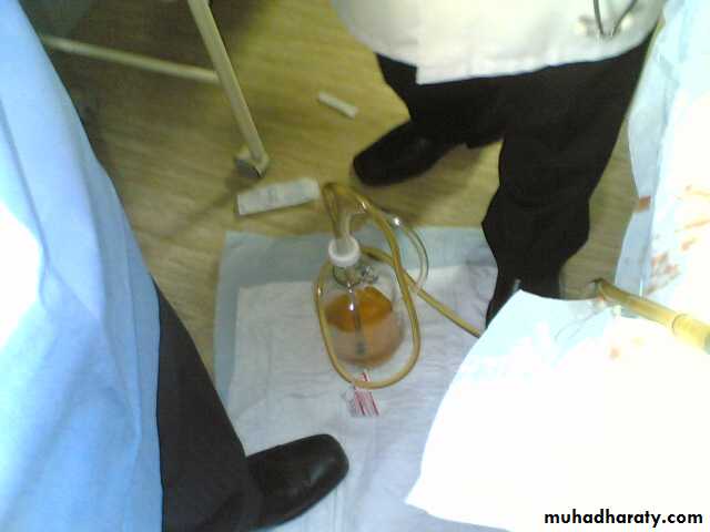
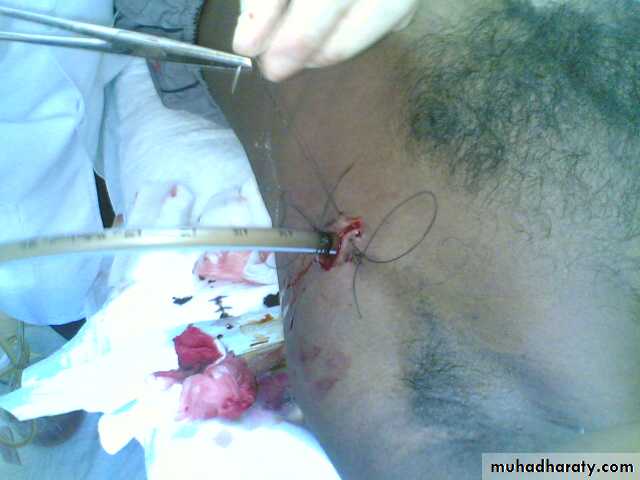
• Tube Thoracostomy Tube thoracostomy or Chest Tube or ICD(Intercostal drain) Is a flexible hollow plastic tube that is inserted through the chest wall into the pleural space and connected to a bedside drainage container
• Indications:-
• 1-Pnemothorax
• 2-Pleural Effusion This effusion may be A-Empyema
• 2-Hemothorax Traumatic or Malignant Effusion
• 3-Hydro thorax 4-Chylothorax 5-Thoracic Operation (Tube Thoracostomy without trocar ) .On the lung or Mediastinum Or The esophagus 6-Postoperative (Collection or Infected space ).7-malignant Effusion drainage and giving medication through it.
• Contra-Indications:-Refractory coagulopathy Lack of cooperation by the patientDiaphragmatic Hernia Lobar EmphysemaSurgical Emphysema without underlying pneumothorax
• Technique:-
• LA or GA
• Surgical Set
• The tube may be inserted in the Emergency Dept. , ICU ,Operating Room or General Hospital Room
• Size infantile , pediatric ,adult (8 FG ------ 40 FG)
• Roughly ---- the size of adult index finger
• Sites ----
• Safe zone
• The free end of the tube ------underwater seal below the level of the chest
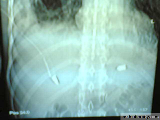
• Chest radiograph to be taken to check the location of the drain
• The tube stays in for as long as there is air or fluid
• How long is a chest tube used ?The tube remains in place until the lung is re-expand or the fluid is drained. Occasionally the patient require more than one chest tube Indications for Removal Clinical Mechanical Radiological
• Complications:-1-Minor Complications:-Severe pain during placement Subcutaneous hematoma or seroma Anxiety Shortness of breath (Dyspnea) Cough ( Rapid drainage of fluid )
• 2-Major Complications Hemorrhage ---haemothorax or haemoptysis Infection Reexpansion pulmonary edema Injury to the liver , spleen , diaphragm .Injury to the Thoracic aorta & the heart
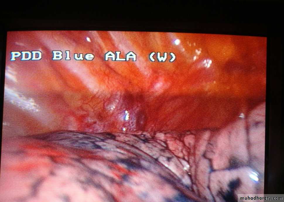
Thoracoscopy
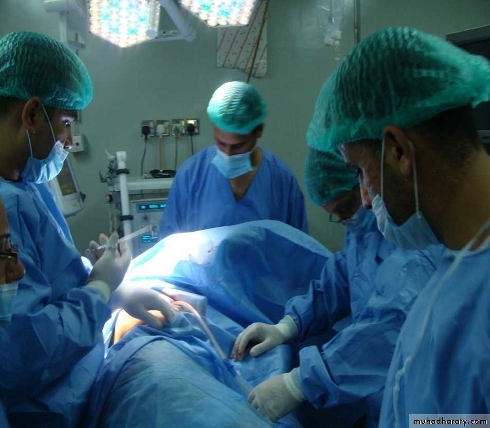
It is the examination of the pleural cavity with an endoscope .Hans Jacobaeus was the originator of the thoracoscopy in 1910 .It is done under general anesthesia with double lumen intubation ,
Indications
1-Diagnostic1-Diagnosis of pleural diseases
2-Evaluation of carcinoma of the bronchus .
3- Biopsy of a discrete pulmonary nodules .
4-Evaluation of mediastinal mass .
Therapeutics :-
1-Treatment of pleural effusion .
2-Treatment of recurrent pneumothorax .
3-Removal of intra- pleural FB .
4-Debridement of empyaema space .
5-Dorsal sympathectomy .
There is no absolute contra indications for thoracoscopy .