Introduction and technical consideration
Qais A. Altimimy, DMRD, CABMS-RAD.Lecturer, Radiology
Alkindy college of medicine, university of Baghdad
2015


Objectives:
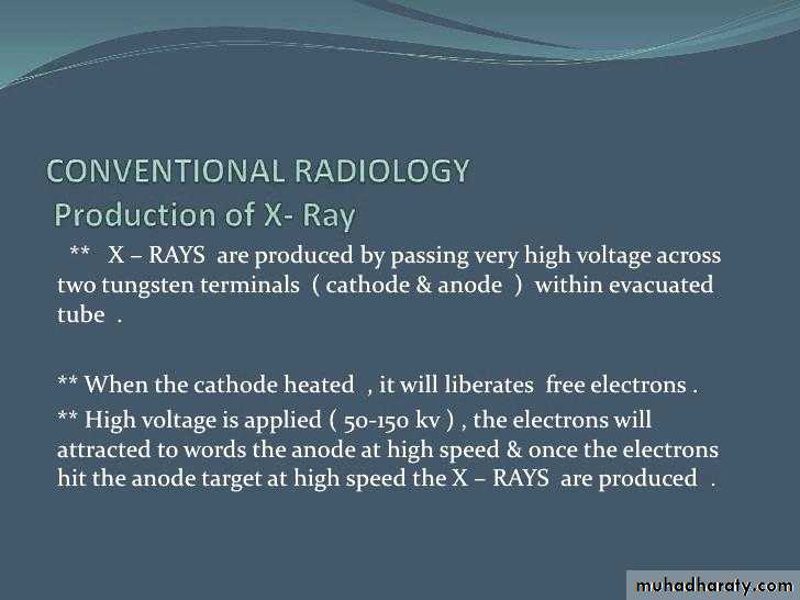
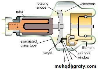
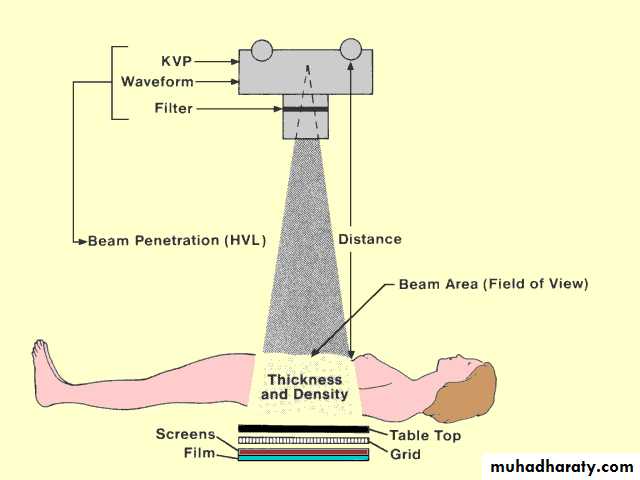
Proper use of imaging department.How X-rays are produced in Conventional Radiography.
Syllabus:
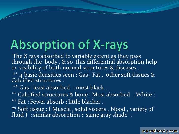
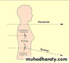
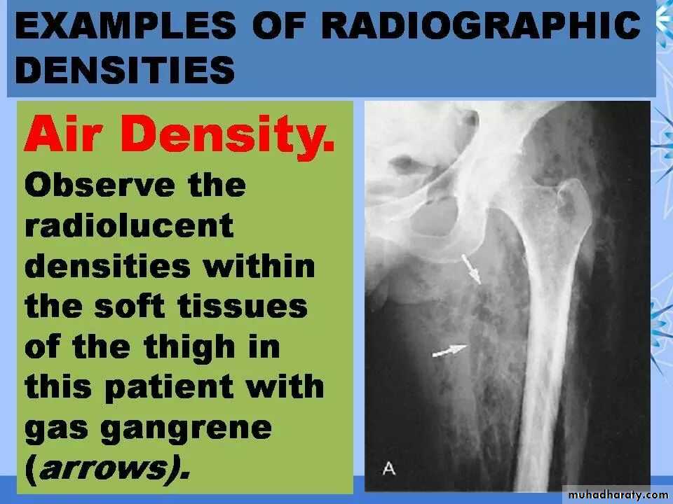
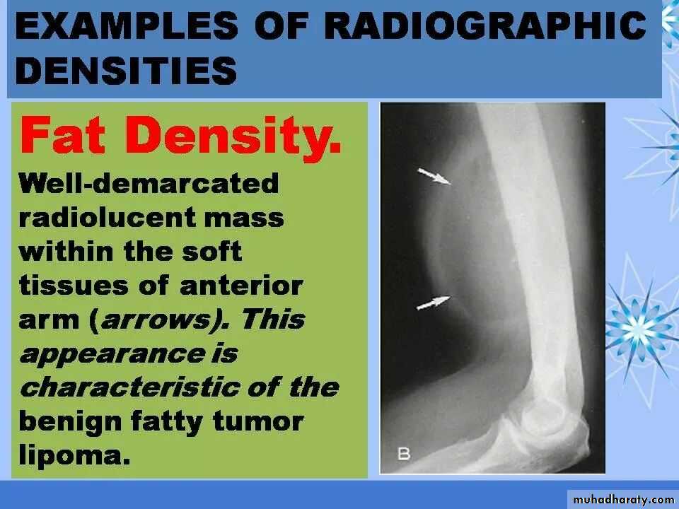
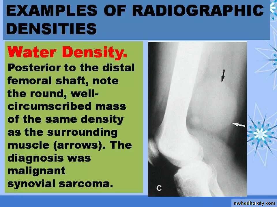
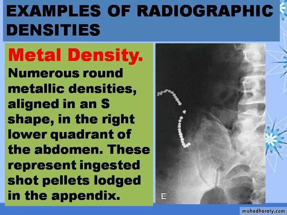
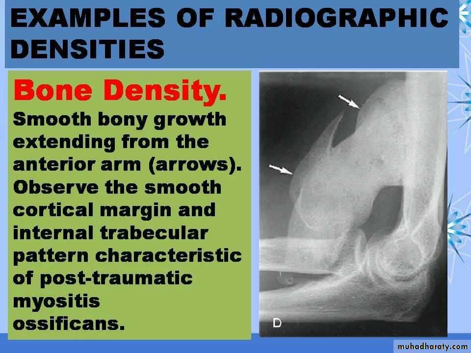
-The basic radiographic densities.
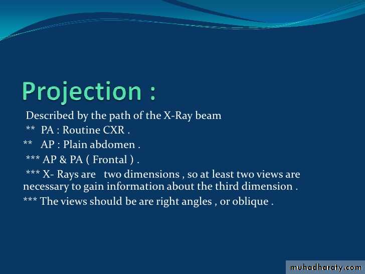
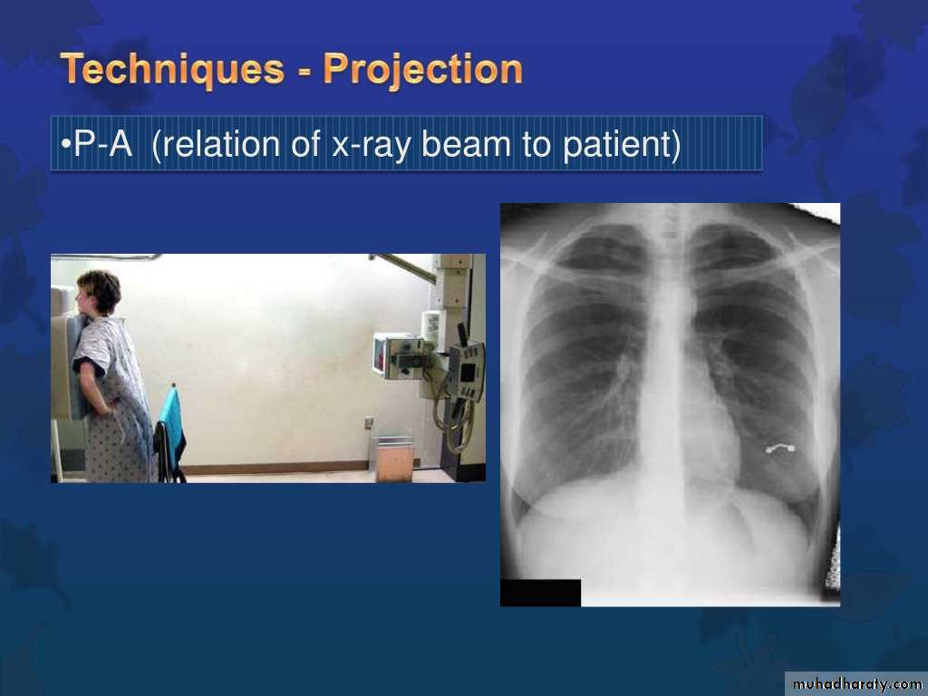
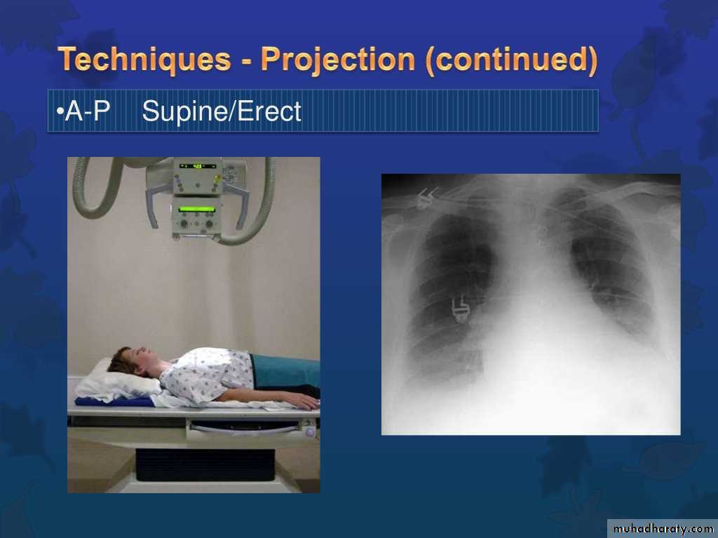
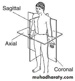
-Projections in conventional radiology:…PA. AP. Lateral, Horizontal X-ray beam, Lateral decubitus
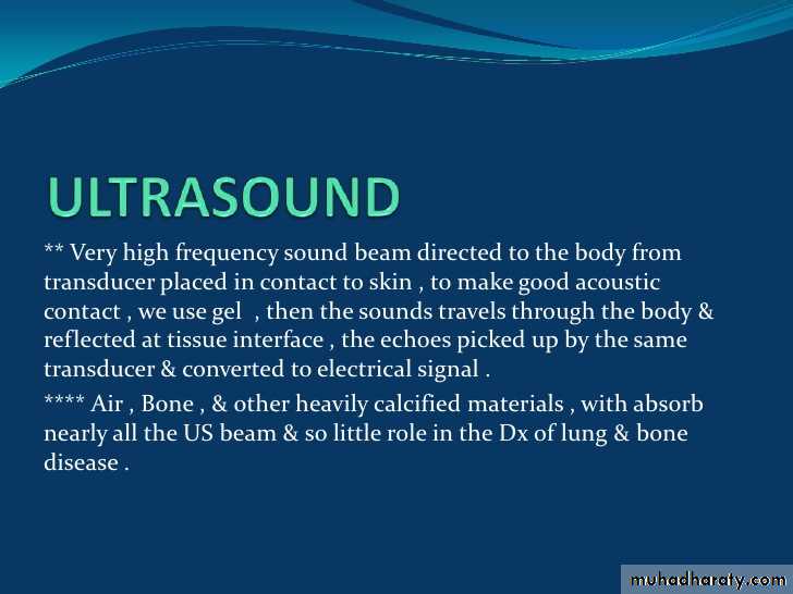
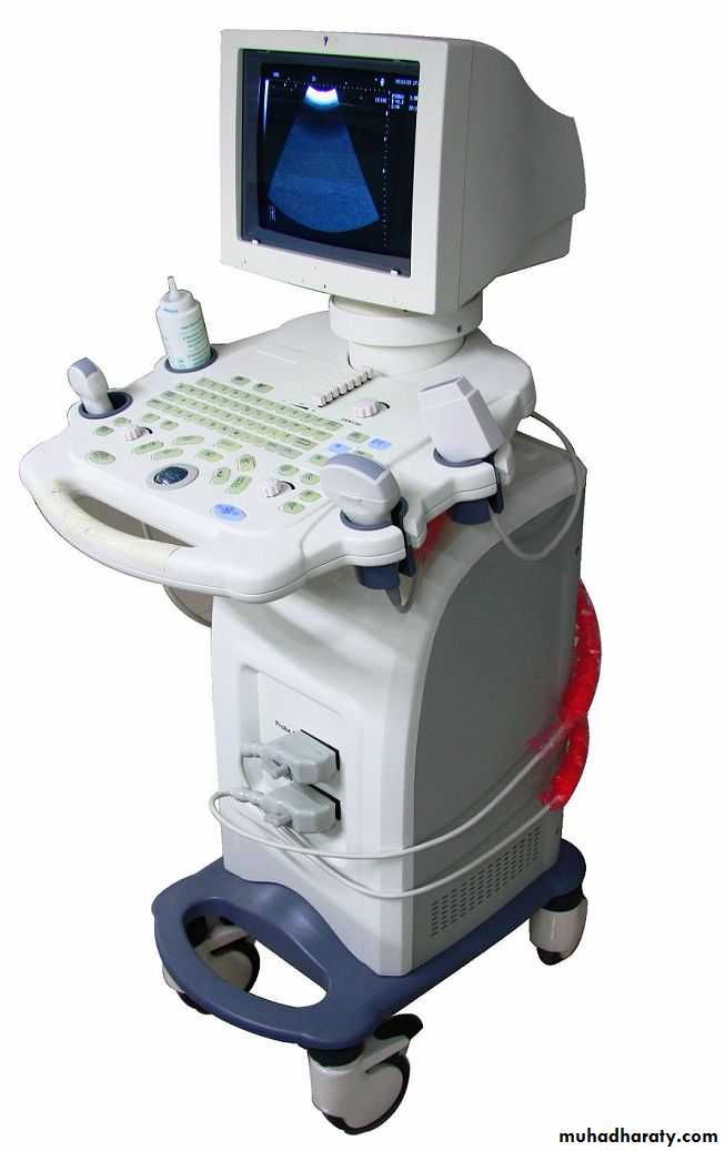
-CT Scan, Ultrasound, MRI, Radionuclide imaging & PET , Basic principles
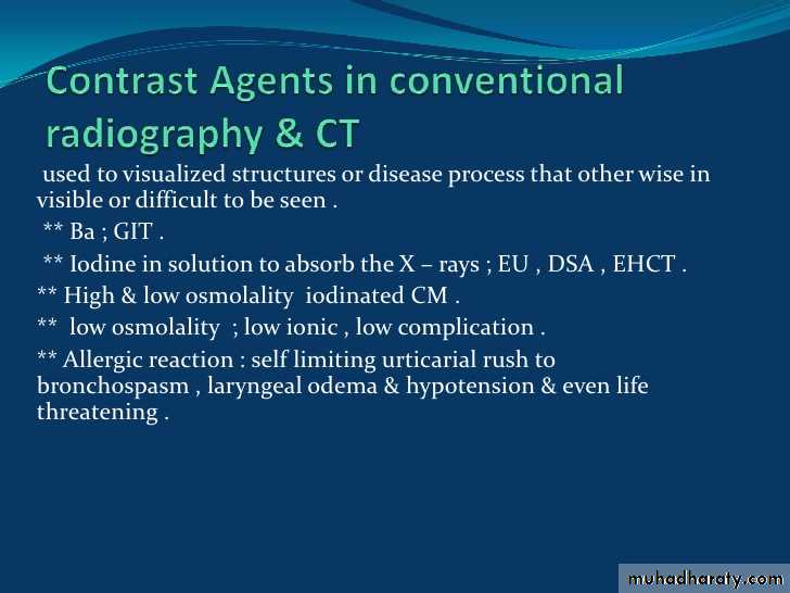
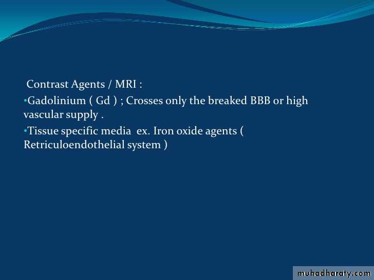
-Contrast agents in Conventional Radiography & CT.. Definition & side effects
-Radiation Hazards

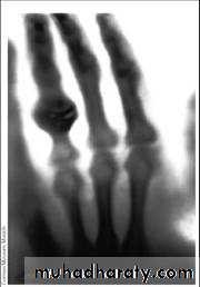
The radiograph of Bera Roentgen’s hand” taken
22 Dec. 1895• Electromagnetic spectrum :
• Electromagnetic radiation consists of energy (a particle) in small packets known as photons or quanta. They are grouped according to their wave length and frequency.•


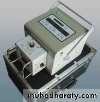
Portable
Fixed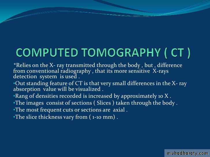


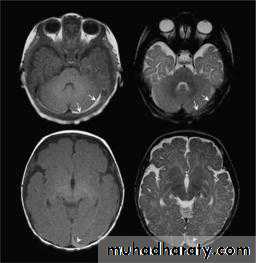
When reviewing an MR image, the easiest way to determine which pulse sequence was used, or the "weighting" of the image, is to look at the cerebrospinal fluid (CSF). If the CSF is bright (high signal), then it must be a T2-weighted imaged. If the CSF is dark, it is a T1-weighted image.
Acronym for:
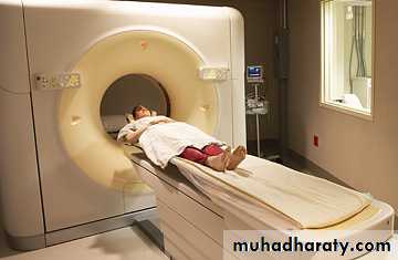
Computed tomographyMagnetic Resonance Imaging
Application:
Suited for bone injuries, Lung and Chest imaging, cancer detection
Suited for ligament and tendon injury, spinal cord injury, brain tumors
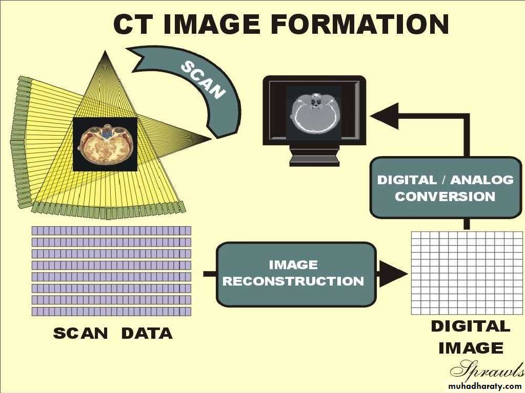
Principle:
X-ray attenuation was detected by detector & DAS system, follow by math. model (back projection model) to calculate the value of pixels then become a image.
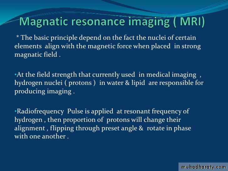
Makes use of the fact that body tissue contains lots of water (and hence protons) which gets aligned to large magnetic field to produce signal.
Ability to change the imaging plane without moving the patient:
With capability of MDCT, after helical scan with Multi-plane Reformation function, an operator can construct any plane.
MRI machines can produce images in any plane.
Cost:
cost less than MRIs.
usually more than CT
Principle used for imaging:
Uses X-rays for imaging
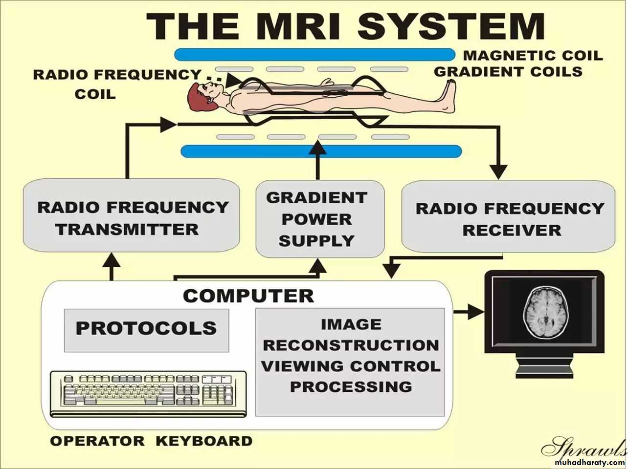
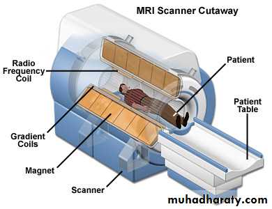
Uses large external field,RF pulse and 3 different gradient fields
Details of bony structures:
Provides good details about bony structures
Less detailed compared to CT scan
Time taken for complete scan:
Usually 2-3 minutes
Usually about 10 – 20 minutes.
Details of soft tissues:
Less tissue contrast compared to MRI
Much higher detail in the soft tissues
Effects on the body:
Despite being small, CT can pose the risk of irradiation.
No biological hazards have been reported with the use of the MRI.
Radiation exposure:
Moderate to high radiation
None
Patients with metal implants can get CT scan.
A person who is very large (e.g. over 250 kg )may not fit into the opening of a conventional CT scanner or may be over the weight limit for the moving table.
Patients with Cardiac Pacemakers, tattoos and metal implants are contraindicated due to possible injury to patient or image distortion (artifact). Patient over 160 kg may be over table's weight limit. Any ferromagnetic object may cause trauma/burn.
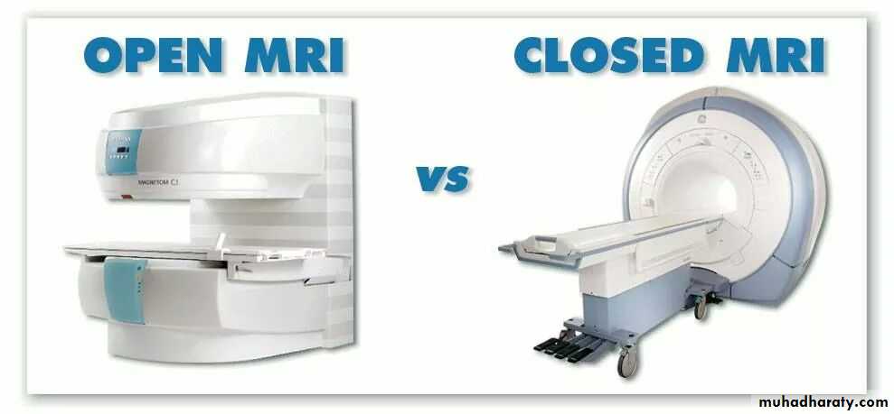
Seldom creates claustrophobia
Often creates claustrophobia in susceptible patients.
THIS PROCEDURE:
YOUR EFFECTIVE RADIATION DOSE IS:COMPARABLE TO NATURAL BACKGROUND RADIATION FOR:
ABDOMINAL REGION:
Computed Tomography (CT)-Abdomen
10 mSv3 years
Computed Tomography (CT)-Body
10 mSv
3 yearsRadiography-Lower GI Tract
4 mSv
16 monthsRadiography-Upper GI Tract
2 mSv
8 monthsBONE:
Radiography-Extremity
0.001 mSv
Less than 1 dayCHEST:
Computed Tomography (CT)-Chest
8 mSv3 years
Radiography-Chest
0.1 mSv
10 daysWOMEN'S IMAGING:
Mammography
0.7 mSv3 months