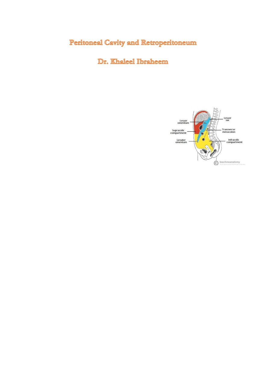
Peritoneal cavity:
potential space encompassed by the visceral and parietal layers, it divided into two main
sections, the greater sac and the lesser sac (which lies posterior to the stomach)
Ascites
Free peritoneal fluid.
With the patient supine,it collects in most dependent portions of the
peritoneal cavity: in the pelvis anterior
to the upper rectum (known as the pouch of Douglas in
females), in the space anterior to the right kidney (known
as Morrison’s pouch), and in the paracolic gutters.
Ultrasound, computed tomography (CT) and magnetic
resonance imaging (MRI) can demonstrate very small
amounts of ascites
At ultrasound , ascites is seen as anechoic regions with excellent transmission of sound, and may be used to
in order to drain the fluid
At CT, ascites is of lower density than the liver, spleen and kidney and these organs stand out clearly
compared with the adjacent fluid
Peritoneal tumours
the most frequent neoplastic disease of the peritoneum is metastases from an abdominal or pelvic tumour,
particularly carcinoma of the ovary,primary peritoneal malignancy is rare.
It presented as peritoneal nodules which are clearly demonstrated on CT and to lessor extend by ultrasound
Intraperitoneal abscesses
The common locations are subphrenic, subhepatic, paracolic and pelvic, causes include ,postoperative
,traumatic or follow perforation of viscous -duodenal leakage usually results in subhepatic or subphrenic
Abscess, left colon often results in paracolic or pelvic abscess.
CT is better than Ultrasound.
Ultrasound : They often have slightly irregular walls and may contain internal echoes due to septations or
debris.
CT: the fluid center of the abscess is identified as a homogeneous density surrounded by a definite wall of
soft tissue ,gas within abscess is very useful sign.
Retroperitoneum
Computed tomography, MRI and ultrasound all provide information about retroperitoneal structures. When
considering the retroperitoneum it is useful to appreciate the anatomy of the anterior and posterior renal
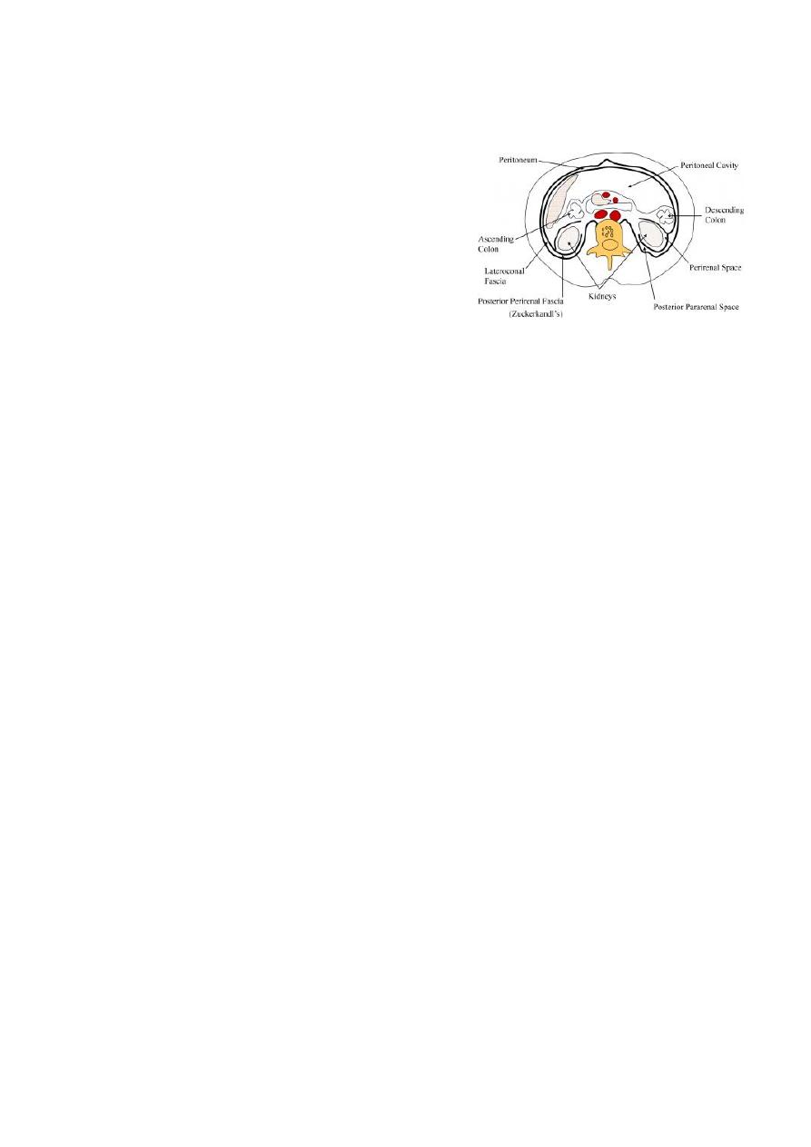
fascia which divide the retroperitoneum into three compartments: the anterior pararenal, the perinephric
and the posterior pararenal spaces.
CT: best diagnostic tool
-
Aorta and inferior vena cava (IVC) should be clearly visible
-The psoas muscles are seen as symmetrical, rounded structures outlined anteriorly by fat.
- Both adrenals are well seen in most subjects.
Ultrasound :
The aorta and inferior are easily identified ,while the adrenal glands are rarely visible
in
adults
Magnetic resonance imaging: Magnetic resonance imaging plays a small part in diagnosing
retroperitoneal disorders as it provides few advantages over CT or ultrasound.
Retroperitoneal lymphadenopathy
Nodal enlargement may occur secondary to inflammatory disease, metastatic tumour deposits and
lymphoma
It is often not possible to reliably diagnose the cause of mild nodal enlargement on cross-sectional imaging,
as the nodes have the same features and texture, regardless of the cause of enlargement, at CT, ultrasound,
and MRI
But LNs >2cm cm almost always indicates neoplasm, notably lymphoma.
Adrenal glands
The normal adrenal glands are thin, bilobed structures surrounded by fat.
Calcification of the adrenal glands may follow old intra-adrenal haemorrhage or old healed tuberculosis.
Enlargement of the adrenal glands can be recognized at CT and MRI and occasionally on ultrasound.
CT is the best routine technique for diagnosing adrenal enlargement
Tumours are either functioning (usually benign adenomas, but may be carcinomas) or nonfunctioning (also
either adenoma, carcinoma or metastasis)
Retroperitoneal tumours
The term retroperitoneal tumour covers tumours arising primarily in retroperitoneal muscles, fat or
connective tissue, the commonest being liposarcoma and fibrosarcoma.
All these tumours appear as masses on CT, ultrasound or MRI. Sometimes, the edge of the mass is well
defined,
A liposarcoma almost always contains significant amounts of recognizable fat interspersed between strands
or masses of soft-tissue density; a combination that permits a specific diagnosis to be made at CT.
Aortic aneurysm

Abdominal aortic aneurysms are readily diagnosed at ultrasound, CT and MRI, although MRI is rarely used
for this purpose .
Ultrasound is being used increasingly as a screening examination to find asymptomatic aortic aneurysms in
older men.
It is generally held that aneurysms of greater than 5.5 or 6 cm in diameter are in serious danger of rupture
Aortic aneurysms may also be recognizable on plain films of the abdomen, but only if substantial
calcification is present in the wall.
Retroperitoneal and psoas abscesses
Retroperitoneal and psoas abscesses are usually due to spread of infection from the appendix, colon, kidney,
pancreas or spine. They are often found close to the organ of origin.
Retroperitoneal abscesses have many similar features to tumours and haematomas at both CT and
ultrasound.
Usually, however, there is evidence of a fluid centre and there may be gas within the abscess. The wall of
the abscess may enhance with contrast medium, a feature that is also seen with neoplasms.
Retroperitoneal haematoma
Retroperitoneal bleeding is usually due to trauma or to bleeding from an aortic aneurysm ,It is occasionally
spontaneous in patients with bleeding disorders or in those on anti-coagulant therapy.
The diagnosis is made by CT or MRI, which show a retroperitoneal mass with the characteristics of
haematoma.