
Mohannad Al-Fallouji
, PhD (London), FRCS, FRCSI
University Staff
and
Consultant Surgeon
Baghdad’s Medical City
Teaching
Hospitals
Director:
www.ihams.org
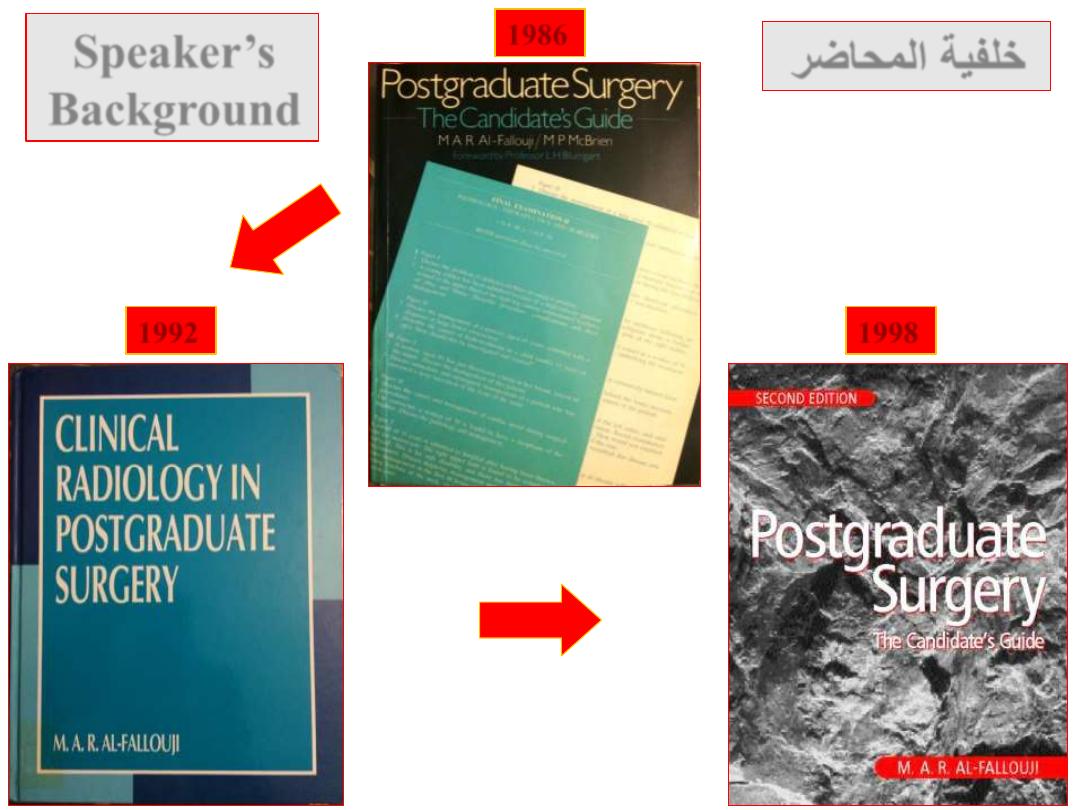
1992
1986
1998
Speaker’s
Background
خلفية المحاضر
I Love You
(in Hebrew)
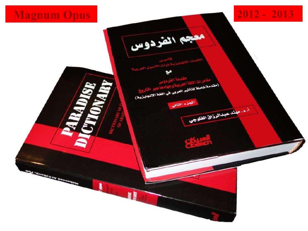
2012 - 2013
Magnum Opus
1. To reveal the surgical anatomy and the applied
physiology of the Spleen.
2. To know clinical differences between the Splenomegaly
and tumour of the left kidney.
3. To be acquainted with indications for splenectomy.
4. To be acquainted with post-splenectomy complications.
5. To sum it up in a clinical scenario of (splenic injury).
Objectives of Spleen
Surgical Anatomy:
Location
• It lies under the diaphragm in the left hypochondrium.
• It is summarised by
(
1, 3, 5, 7, 9, 11
):
- it measures
1 X 3 X 5
inches (2.5 X 7.5 X 12.5 cm).
- it weighs
7
ounces (200 gm).
- it lies beneath the
9
th
- 11
th
ribs.
Surgical Anatomy:
Relation
Spleen lies at the far left extremity of lesser sac under diaphragm.
It is related:
- to pancreatic tail,
- splenic flexure of the colon,
- left kidney.
- diaphragm.
- 6 short gastric arteries (from splenic artery) supply
the fundus of the stomach.
Surgical Physiology:
FISH(H)
Spleen is the largest lymphoid organ in the body.
F
: Filtration and removal of old, abnormal, and senescent RBCs
and recycling iron as well as removal of encapsulated
microorganisms
.مقبرة الخاليا
I
: Immunological functions (production of IgM and Opsonins)
S
: Storage Pool functions (30% of total Platelets are located
within spleen: it also receives 5% of total cardiac output
approximately 150-300 ml/min so that each RBC averages 1000
passes through spleen each day).
H
: Haematopoiesis (usually in the developing foetus, but in
adults only reactivated in myeloproliferative disorders that impair
bone marrow to produce sufficient RBCs).
H
: hormonally active?
It has recently been evoked that the spleen has an endocrine
function through the production of an immuno-potentiating peptide called
(tuftsin).
Palpate from right iliac fossa upwards towards left hypochondrium.
Splenomegaly may be mistaken for left renal tumour.
The following points will help in differentiation:
1. A renal tumour is bimanually palpable moving backwards and
forwards between one hand on the loin behind, and the other on the
anterior abdominal wall. Splenomegaly is not palpable bimanually.
2. Fingers can usually be passed between the kidney and the ribs
but not between the ribs and splenomegaly.
3. The spleen has a sharp edge with a notch. The kidney edge
always rounded and has no notch.
4. An enlarged kidney tends to bulge forwards. Peri-nephric
abscesses bulge backwards. Splenomegaly bulges towards RIF.
5. Because of overlying colonic splenic flexure percussion on
splenomegaly may be resonant.
DD of Splenomegaly from Renal tumour
Causes of Spenomegaly
Massive Splenomegaly
Spleen MUST be enlarged to 3 times its normal size before it
becomes clinically palpable.
Massive splenomegaly
is likely due to
malignant
disease
:
- Chronic myeloid leukemia,
- Myelofibrosis,
- Lymphoma (Hodgkin’s and Non-Hodgkin’s).
Hypersplenism
(see next slide)
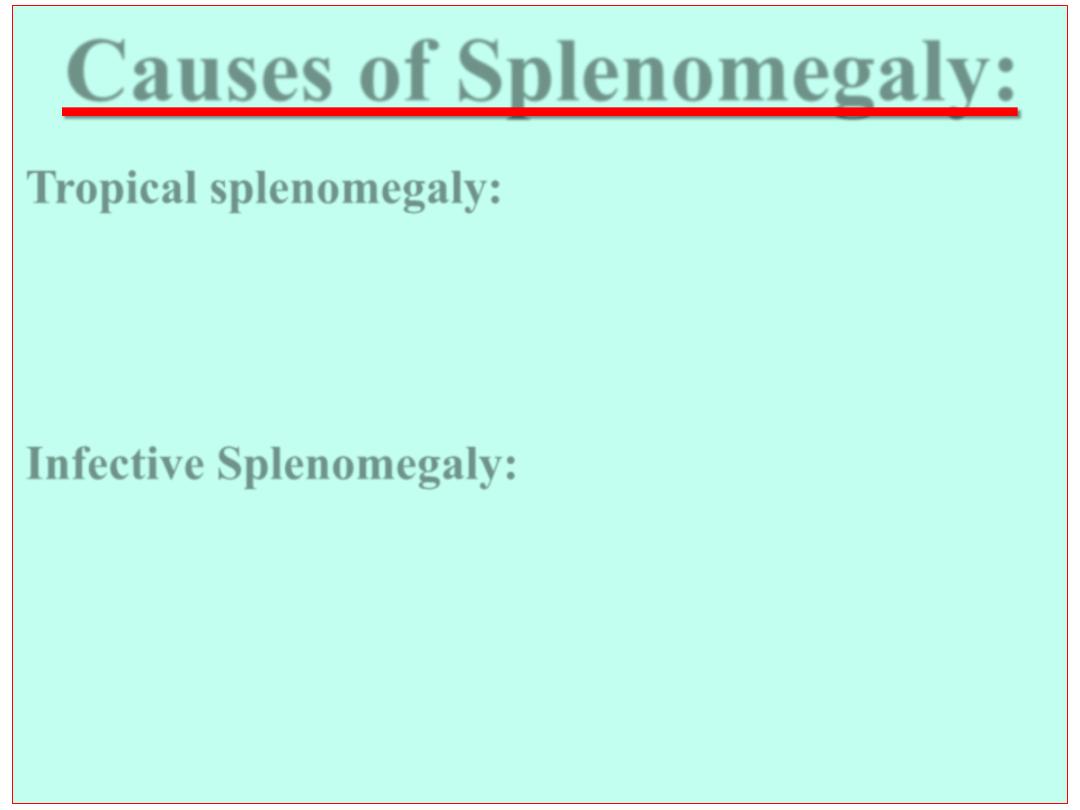
Causes of Splenomegaly:
Tropical splenomegaly
:
- Malaria
- Kala-azar
- Shistosomiasis
Infective Splenomegaly:
1. Bacterial:
Typhoid, Paratyphoid, Tuberculosis,
Splenic Abscess, Septicaemia.
2. Viral:
infectious mononucleosis, HIV, psittacosis.
3. Parasitic:
Hydatid cyst, Trypanosomiasis, Syphilis,Weil’s disease.
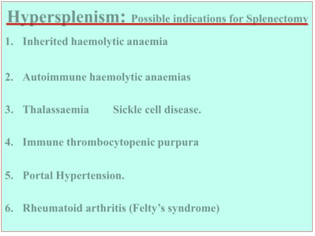
Hypersplenism
:
Possible indications for Splenectomy
1. Inherited haemolytic anaemia
(
Congenital Spherocytosis
&
Elliptocytosis
). Mechanism:
↑
RBCs fragility (Anaemia).
2. Autoimmune haemolytic anaemias
.
Mechanism: Antibodies to RBCs (Anaemia).
3. Thalassaemia
and
Sickle cell disease.
Mechanism: Abnormal haemoglobins (Anaemia).
4. Immune thrombocytopenic purpura
(primary & secondary)
Mechanism: Antibodies to platelets (Thrombocytopenia).
5. Portal Hypertension.
Mechanism:
↑
splenic venous
pressure with delayed transit of blood (Pancytopenia).
6. Rheumatoid arthritis (Felty’s syndrome)
Mechanism: Uncertain (Leucopenia or pancytopenia).
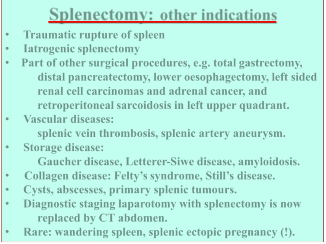
Splenectomy:
other indications
•
Traumatic rupture of spleen
•
Iatrogenic splenectomy
• Part of other surgical procedures, e.g. total gastrectomy,
distal pancreatectomy, lower oesophagectomy, left sided
renal cell carcinomas and adrenal cancer, and
retroperitoneal sarcoidosis in left upper quadrant.
•
Vascular diseases:
splenic vein thrombosis, splenic artery aneurysm.
•
Storage disease:
Gaucher disease, Letterer-Siwe disease, amyloidosis.
• Collagen disease: Felty’s syndrome, Still’s disease.
•
Cysts, abscesses, primary splenic tumours.
•
Diagnostic staging laparotomy with splenectomy is now
replaced by CT abdomen.
•
Rare: wandering spleen, splenic ectopic pregnancy (!).
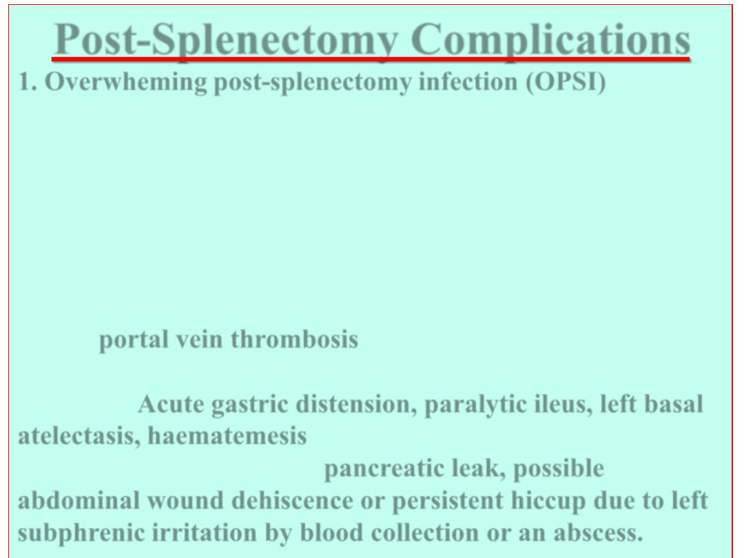
Post-Splenectomy Complications
1. Overwheming post-splenectomy infection (OPSI)
Serious late complicationdue to pneumococci, meningococci
and Haemophilus influenzae. Sepsis into fulminant infection
with fever, vomiting, dehydration and collapse. Prophylactic
immunization (pneumovac) 2 week prior to surgery,
antibiotics after emmergency surgery continued for life
(Penicillin V 250 mg b.d.).
2. Unexplained postoperative abdominal pain with fever may
herald
portal vein thrombosis
(anticoagulant and antibiotic
must be given).
3. Others:
Acute gastric distension, paralytic ileus, left basal
atelectasis, haematemesis
(due to gastric mucosal congestion
after vasa brevia ligation),
pancreatic leak, possible
abdominal wound dehiscence or persistent hiccup
due to left
subphrenic irritation by blood collection or an abscess.
Blunt trauma (slipped and fell on edge of bath-tub) 6 hr ago.
Severe upper abdominal pain
Pain in left shoulder (Kehr sign)
BP 100/60 PR: 100 Temp: 37.3 Resp Rate 18
Not in shock now - (barely haemodynamically stable)
Ecchymosis left lower chest wall
Upper abdominal tenderness
Otherwise soft abdomen
What to do next?
Chest x-ray (trachea central, chest clear but fractured rib 10)
FAST (fluid collection peri-splenic area).
What to do next?
A Clinical Scenario
of ‘Splenic Injury’ (to sum it up)
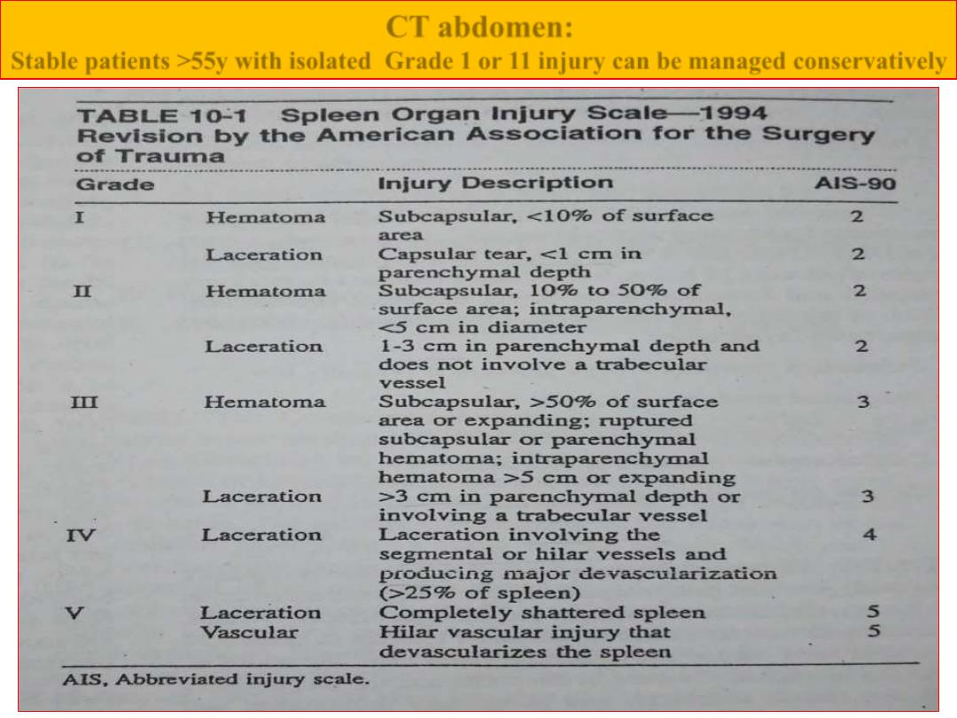
CT abdomen:
Stable patients >55y with isolated Grade 1 or 11 injury can be managed conservatively
Haemodynamically this patient became unstable.
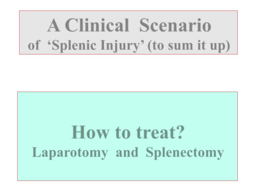
How to treat?
Laparotomy and Splenectomy
A Clinical Scenario
of ‘Splenic Injury’ (to sum it up)
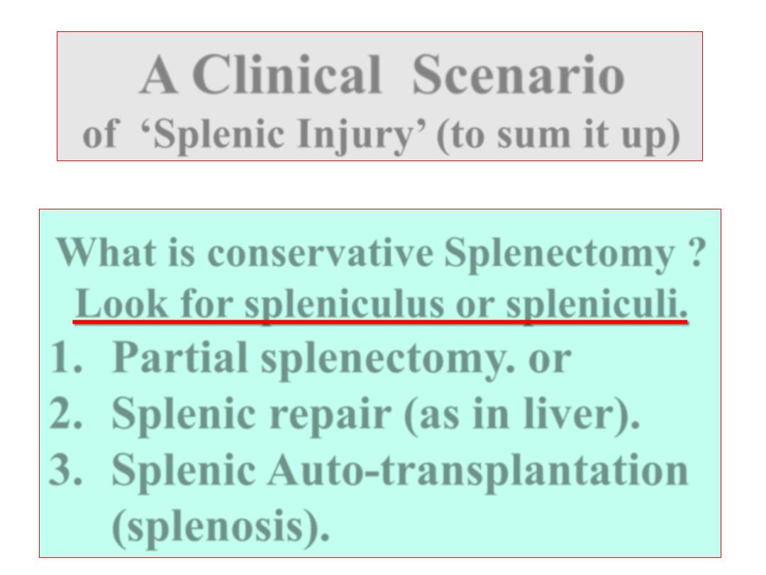
What is conservative Splenectomy ?
Look for spleniculus or spleniculi.
1. Partial splenectomy. or
2. Splenic repair (as in liver).
3. Splenic Auto-transplantation
(splenosis).
A Clinical Scenario
of ‘Splenic Injury’ (to sum it up)

Thanks for Participation