Baghdad College of Medicine / 4
th
grade
Student’s Name :
Dr. Mohammed Basil
Lec. 3
Urinary Incontinence
Thurs. 24 / 3 / 2016
DONE BY : Ali Kareem
مكتب اشور لالستنساخ
2015 – 2016

Urinary Incontinence Dr. Mohammed Basil
24-3-2016
2
©Ali Kareem 2015-2016
Urinary Incontinence
Definition
Involuntary loss of urine that is a social or hygienic problem and is objectively
demonstrable.‖ Urinary incontinence (UI) is a failure to store urine usually due to
either abnormal bladder smooth muscle or a deficient urethral sphincter. Urine
loss may also be extraurethral, secondary to anatomical abnormalities such as
ectopic ureter or vesicovaginal fistula.
Prevalence
UI has been reported to affect 12–43% of adult women and 3–11% of adult men.
Severe incontinence has a low prevalence in young women, but rapidly increases
at ages 70 through 80. Incontinence in men also increases with age, but severe
incontinence in 70- to 80-year-old men is about half that in women.
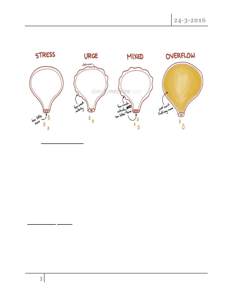
Classification
o Stress urinary incontinence (SUI) :- is involuntary urinary leakage during
effort,exertion, sneezing, or coughing, due to hypermobility of the bladder
base, pelvic floor,and/or intrinsic urethral sphincter deficiencies. In females
SUI is usually associated with multiparity. In males, SUI is most commonly
the result of prostatectomy.
o Urge urinary incontinence (UUI):- is involuntary urine leakage
accompanied or immediately preceded by a sudden, strong desire to void
(urgency).
o Mixed urinary incontinence:- is urine leakage that has characteristics of
both SUI and UUI.
o Overflow incontinence :- is leakage of urine when the bladder is
abnormally distended with large post-void residual volumes. Typically, men
Urinary Incontinence Dr. Mohammed Basil
24-3-2016
3
©Ali Kareem 2015-2016
present with chronic urinary retention and dribbling incontinence. This can
lead to hydronephrosis and renal failure in 30% of patients.
o Nocturnal enuresis:- describes any involuntary loss of urine during sleep.
The prevalence in adults is 0.5%. Approximately 750,000 children over age 7
years will regularly wet the bed. Childhood enuresis can be further
classified into primary (never been dry for longer than a 6-month period)
or secondary (re-emergence of bed wetting after a period of being dry for at least
6–12 months).
Risk factors
Predisposing factors
o Gender (female > males)
o Race (Caucasian > African American/Asian)
o Genetic predisposition
o Neurological disorders (spinal cord injury, stroke, MS, Parkinson disease)
o Anatomical disorders (vesicovaginal fistula, ectopic ureter, urethral
diverticulum)
Urinary Incontinence Dr. Mohammed Basil
24-3-2016
4
©Ali Kareem 2015-2016
o Childbirth
o Anomalies in collagen subtype
o Prostate or pelvic surgery (radical prostatectomy; radical hysterectomy;
TURP) leading to pelvic muscle and nerve injury
o Pelvic radiotherapy
Promoting factors
o Smoking (associated with chronic cough and raised intra-abdominal
pressure)
o Obesity
o UTI
o Increased fluid intake
o Medications
o Poor nutrition
o Aging
o Cognitive deficits
o Poor mobility
Pathophysiology
Bladder abnormalities
Detrusor overactivity is a urodynamic observation characterized by involuntary
bladder muscle (detrusor) contractions during the filling phase of the bladder,
which may be spontaneous or provoked, and can consequently cause urinary
incontinence. The underlying cause may be neurogenic, where there is a relevant
neurological condition, or idiopathic, where there is no defined cause .
Low bladder compliance
is characterized by a decreased volume-to-pressure relationship during a
cystometrogram and is often associated with upper tract damage. High bladder
Urinary Incontinence Dr. Mohammed Basil
24-3-2016
5
©Ali Kareem 2015-2016
pressures occur during filling because of alterations in the viscoelastic properties
of the bladder wall, or changes in bladder muscle tone (secondary to
myelodysplasia, spinal cord injury, radical hysterectomy, interstitial or radiation
cystitis).
Sphincter abnormalities
Urethral hypermobility is due to a weakness of pelvic floor support causing a
rotational descent of the bladder neck and proximal urethra during increases in
intra-abdominal pressure. If the urethra opens concomitantly, there will be urinary
leaking.
Intrinsic sphincter deficiency (ISD) describes an intrinsic malfunction of the
sphincter, regardless of its anatomical position, which is responsible for type III
SUI. Causes include inadequate urethral compression (previous urethral surgery;
aging; menopause; radical pelvic surgery) or deficient urethral support (pelvic
floor weakness; childbirth; pelvic surgery ; menopause).
Evaluation
History
Inquire about LUTS (storage or voiding symptoms), triggers for incontinence
(cough, sneezing, exercise, position, urgency), and frequency and severity of
symptoms. Establish risk factors (abdominal/pelvic surgery; neurological diseases;
obstetric and gynecological history; medications).
A validated patient-completed questionnaire may be helpful
Physical examination
Women
Urinary Incontinence Dr. Mohammed Basil
24-3-2016
6
©Ali Kareem 2015-2016
Perform a pelvic examination in the supine and standing position with a speculum
while the patient has a full bladder. Ask the patient to cough or strain, and inspect
for vaginal wall prolapse (cystocele, rectocele, enterocele),uterine or perineal
descent, and urinary leakage (stress test). Eighty percent of SUI patients will leak
with a brief squirt during cough in the supine position, while another 20% will leak
only in an inclined or standing position.
Urethral hypermobility is assessed with the Q-tip test. A lubricated cotton-tipped
applicator is introduced through the urethra to bladder neck level. Hypermobility
is defined as a resting or straining angle of >30* from horizontal.
The Bonney test is used to assess continence with manual repositioning of the
urethra and vesicle neck. Using one or two fingers to elevate the anterior vaginal
wall laterally without compressing the urethra, relief of incontinence during cough
suggests that surgical correction will be successful.

Urinary Incontinence Dr. Mohammed Basil
24-3-2016
7
©Ali Kareem 2015-2016
Both sexes
Examine the abdomen for a palpable bladder (indicating urinary retention).
A neurological examination should include assessment of anal tone and reflex,
perineal sensation, and lower limb function.
Inspect the underwear for the status of urinary collection pads; for men, a standing
or squatting ―cough test‖ gives a good indicator of the presence and severity of
stress incontinence.
Investigation
Bladder diaries
Record the frequency and volume of urine voided, incontinent episodes, pad usage,
fluid intake, and degree of urgency. Alternatively, pads can be weighed to estimate
urine loss (pad testing).
Urinalysis can exclude UTIs.
Blood tests, X-ray imaging, cystoscopy
These are indicated for persistent or severe symptoms, bladder pain, and voiding
difficulties. Cystoscopy is useful for evaluating men who have had prostatectomy—
it will show the presence of clips, stones, and strictures that may develop after
surgery that might need to be addressed concomitantly with anti-incontinence
surgery
Urodynamic investigations
Valsalva leak point pressure (VLPP) measures the abdominal pressure at which a
half-full bladder leaks during straining—normal individuals should not leak. VLPP
readings <60 cm H2O suggest ISD; VLPP readings >100 cm H2O suggest
hypermobility, while readings of 60–100 cm are indeterminant.
Detrusor leak point pressure (DLPP) measures the bladder pressure at which
leakage occurs without valsalva—DLPP >40 cm H2O puts the upper tract at risk.
Urinary Incontinence Dr. Mohammed Basil
24-3-2016
8
©Ali Kareem 2015-2016
Videourodynamics can visualize movement of the proximal urethra and bladder
neck, and establish the precise anatomical etiology of UI.
Urodynamics
Uroflowmetry testing measures the ability of the bladder to empty; a minimum
bladder volume of 150 cc is desired. A low flow rate indicates bladder outflow
obstruction or reduced bladder contractility. The volume of urine remaining in the
bladder after voiding (post-void residual) is also important(<50 mL is normal;
>200 mL is abnormal; 50–200 mL requires clinical correlation).
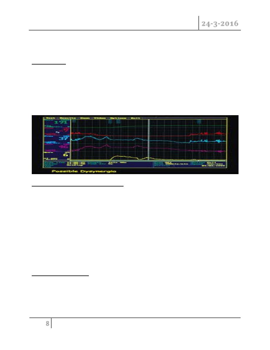
Sphincter electromyography (EMG)
EMG measures electrical activity from striated muscles of the urethra or perineal
floor and provides information on synchronization between bladder muscle
(detrusor) and external sphincter.
Treatment
Treatment for urinary incontinence depends on the type of incontinence, its
severity and the underlying cause. A combination of treatments may be needed.
Behavioral techniques
o Bladder training, to delay urination after you get the urge to go. You may
start by trying to hold off for 10 minutes every time you feel an urge to
urinate. The goal is to lengthen the time between trips to the toilet until
you're urinating only every two to four hours.
Urinary Incontinence Dr. Mohammed Basil
24-3-2016
9
©Ali Kareem 2015-2016
o Double voiding, to help you learn to empty your bladder more completely to
avoid overflow incontinence. Double voiding means urinating, then waiting
a few minutes and trying again.
o Time voiding, to urinate every two to four hours rather than waiting for the
need to go.
o Fluid and diet management, to regain control of your bladder. You may
need to cut back on or avoid alcohol, caffeine or acidic foods. Reducing
liquid consumption, losing weight or increasing physical activity also can
ease the problem.
o Pelvic floor muscle exercises : It is recommend that you do these exercises
frequently to strengthen the muscles that help control urination. Also known
as Kegel exercises, these techniques are especially effective for stress
incontinence but may also help urge incontinence.
o Medications
Anticholinergics. These medications can calm an overactive bladder
and may be helpful for urge incontinence.
Mirabegron . Used to treat urge incontinence, this medication relaxes
the bladder muscle and can increase the amount of urine your bladder
can hold. It may also increase the amount you are able to urinate at
one time, helping to empty your bladder more completely.
Alpha blockers. In men with urge or overflow incontinence, these
medications relax bladder neck muscles and muscle fibers in the
prostate
Topical estrogen.
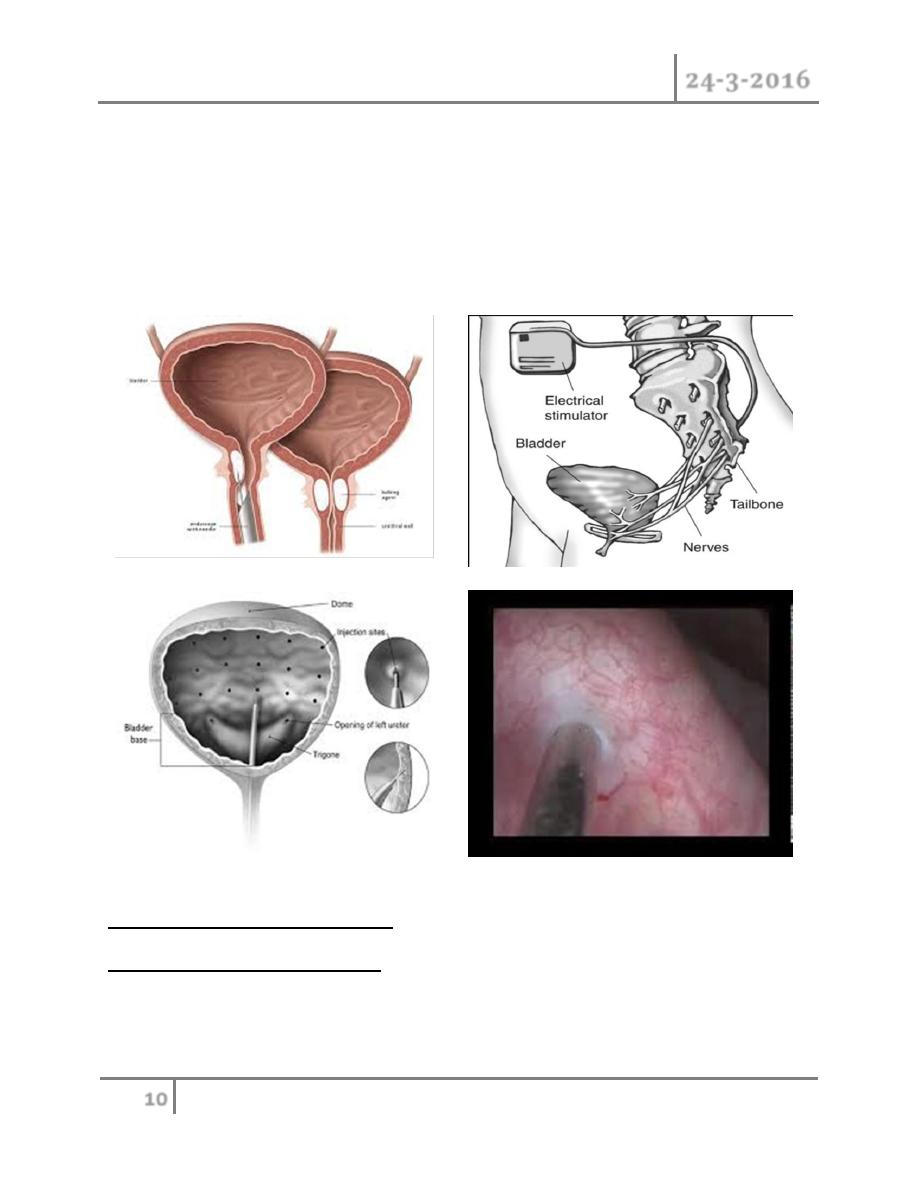
Interventional therapies
o Bulking material injections. A synthetic material is injected into tissue
surrounding the urethra.
o Botulinum toxin type A (Botox). Injections of Botox into the bladder muscle
may benefit people who have an overactive bladder. Botox is generally
prescribed to people only if other first line medications haven't been
successful.
Urinary Incontinence Dr. Mohammed Basil
24-3-2016
10
©Ali Kareem 2015-2016
o Nerve stimulators. A device resembling a pacemaker is implanted under
your skin to deliver painless electrical pulses to the nerves involved in
bladder control (sacral nerves). Stimulating the sacral nerves can control
urge incontinence if other therapies haven't worked. The device may be
implanted under the skin in your buttock and connected directly to the sacral
nerves or may deliver pulses to the sacral nerve via a nerve in the ankle.
Treatment of sphincter weakness
Incontinence: injection therapy
o The injection of bulking materials into the bladder neck and periurethral
muscles is used to increase outlet resistance.
Urinary Incontinence Dr. Mohammed Basil
24-3-2016
11
©Ali Kareem 2015-2016
o Indications
These include stress incontinence secondary to demonstrable intrinsic
sphincter deficiency (ISD), with normal bladder muscle function. Injection
therapy is used in adults and children.
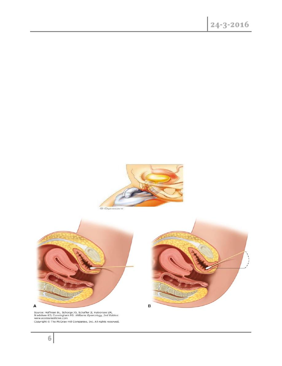
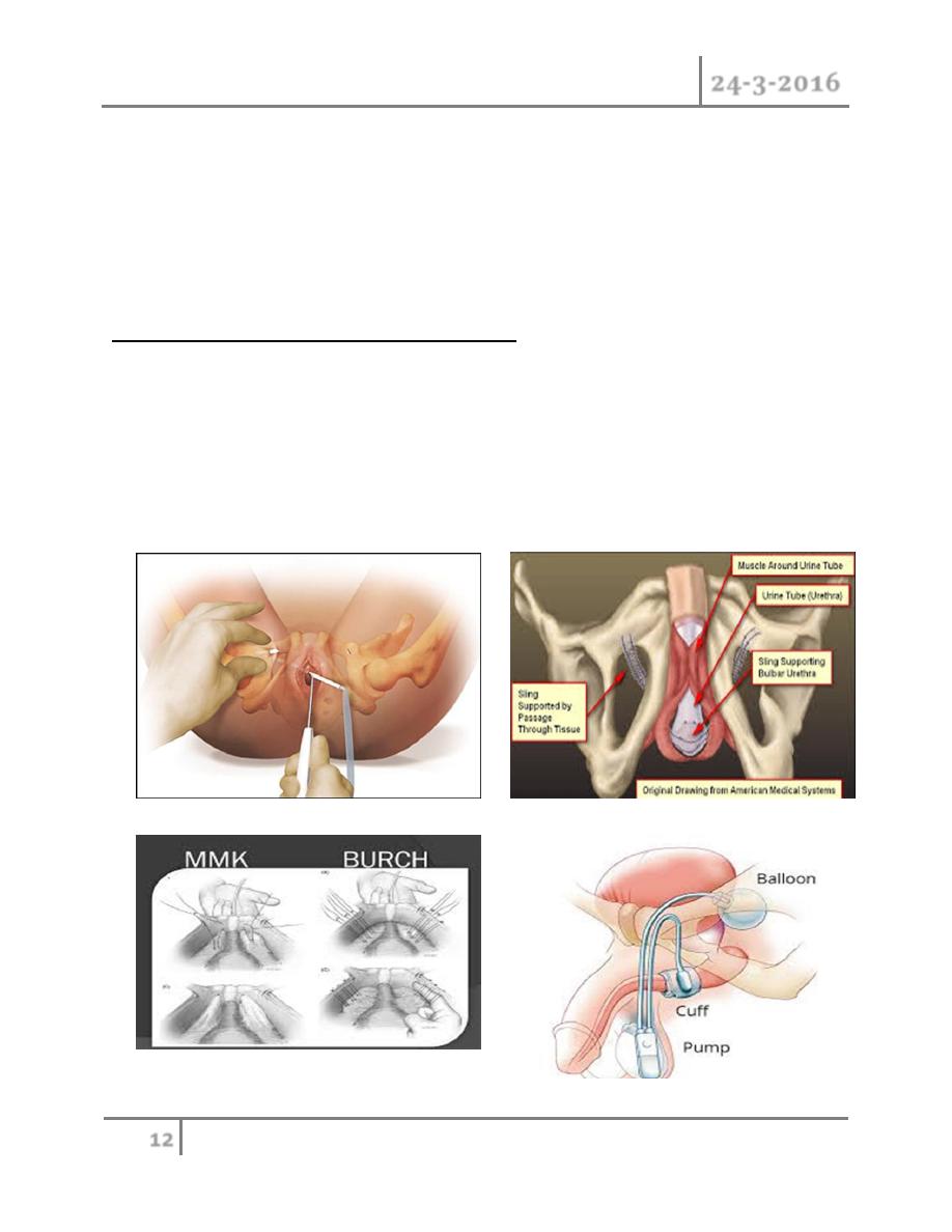
Incontinence: retropubic suspension
o Retropubic suspension procedures are used to treat female stress
incontinence caused by urethral hypermobility. The aim of surgery is to
elevate and fix the bladder neck and proximal urethra in a retropubic
position, to support the bladder neck, and to regain continence. It is
contraindicated in the presence of significant intrinsic sphincter deficiency
(ISD).
o Marshall–Marchetti–Krantz (MMK) procedure
Sutures are placed either side of the urethra around the level of the bladder
neck and then tied to the hyaline cartilage of the pubic symphysis.
o Burch colposuspension
This requires good vaginal mobility, to allow the vaginal wall to be elevated
and attached to the lateral pelvic wall where the formation of adhesions
over time secures its position. The paravaginal fascia is exposed and
approximated to the iliopectineal (Cooper) ligament of the superior pubic
rami.
o Vagino-obturator shelf/paravaginal repair
Sutures are placed by the vaginal wall and paravaginal fascia and then
passed through the obturator fascia to attach to part of the parietal pelvic
fascia below the tendinous arch (arcus tendoneus fascia). Cure rates are up
to 85%.
Incontinence: pubovaginal slings
o Indications
Sling procedures were developed mainly for female stress incontinence
associated with poor urethral function (type III or ISD) or when previous
surgical procedures have failed. The success of sling procedures, however,
has resulted in expanded applications in women with hypermobility.
Urinary Incontinence Dr. Mohammed Basil
24-3-2016
12
©Ali Kareem 2015-2016
o Types of sling
Autologous—rectus fascia, fascia lata (from the thigh), vaginal wall
slings
Nonautologous—allograft fascia lata from donated cadaveric tissue
Synthetic—monofilament ―macropore‖ polypropylene mesh (via
transobturator, transabdominal, or transvaginal needles)
Incontinence: the artificial urinary sphincter
o Indications include incontinence secondary to urethral sphincter deficiency
in patients with normal bladder capacity and compliance. In men, it is used
almost always for sphincter damage due to prostatectomy (radical
prostatectomy for prostate cancer or TURP). In women it can be used for
neuropathic sphincter weakness (e.g., spinal cord injury, spina bifida) if the
incontinence is not due to bladder overactivity.
Urinary Incontinence Dr. Mohammed Basil
24-3-2016
13
©Ali Kareem 2015-2016
Causes of transient incontinence
1) Drug side effects
2) Delirium or hypoxia
3) Impaired mobility
4) Urinary tract infection
5) Atrophic vaginitis
6) psychological problems
7) Excessive fluid intake
END OF THIS LECTURE …