
NEOPLASIA Prof. Manal A Habib
• Neoplasm = a new growth/ interchangeable with Tumor
• Oncology (Gr. oncos = tumor): study of neoplasms
• Cancer: all malignant tumors
• Neoplasm: an abnormal tissue proliferation exceeds that of adjacent
normal tissue
- Proliferation continues even after removing inciting stimulus
(Tumor autonomous)
NOMENCLATURE
1. Proliferating neoplastic cells
2. Stroma: CT + BVs: vital +Cross-talk with neoplastic cells influence growth
Variable proportions of two components determines consistency
The tumors consist predominantly of neoplastic cells are soft and
fleshy in consistency.
Conversely, abundant fibrotic stroma, referred to as desmoplasia is a hard
(scirrhous) or even a stony hard An example of the latter is carcinoma of the
breast
NOMENCLATURE OF BENIGN TUMORS
• By attaching suffix -oma to cell of origin
• Tumors of mesenchymal cells follow this rule
- fibroma
- chondroma
- osteoma
• Benign epithelial tumors more complex
- variously classified according to
Cells of origin &
Microscopic &/or
macroscopic appearance
• Adenoma
1. Forming glandular structures e.g. Renal cell adenoma
Thyroid follicular adenoma
2. Tumors derived from glands but not necessarily reproducing glandular
structures e.g. Thyroid follicular adenoma
• Papilloma: producing micro- or macro- visible finger-like (warty)
projections from epithelial surfaces, e.g. laryngeal papilloma

• Cystadenoma: adenoma forming large cystic space (es) e.g.
ovarian cystadenoma
• Papillary cystadenoma as above + papillary projections e.g.
ovarian papillary cystadenoma
• Polyp: benign/producing macro. visible projection above mucosa e.g.
Adenomatous polyp of stomach & colon
• Malignant polyps are designated polypoid cancers
NOMENCLATURE OF MALIGNANT TUMORS
•
Follows essentially same scheme used for benign neoplasms
+ certain additions
•
Sarcomas (Greek sar = fleshy):
- malignant tumors arising from or differentiating towarsd
mesenchymal cells
- generally scant CT stroma
fleshy consistency
•
Nomenclature of sarcomas relies on either
a. cell of origin
b. differentiation
- fibrosarcoma (fibroblasts)
- liposarcoma (lipocytes)
- leiomyosarcoma (SMCs)
- rhabdomyosarcoma (striated muscle cells)
• Carcinoma: malignant neoplasm of epithelial cell origin derived from
any one of three germ layers
- arising in epidermal epithelial cells (ectoderm)
- derived from renal tubules (mesoderm)
- originating from epithelial cells line GIT (endoderm)
• Further qualified according to morphology e.g.
- with a glandular growth pattern: adenoca
- with squamous cells arising in any epithelium: squamous cell ca.

• Specify organ of origin e.g.
- Renal cell Ca.
- Bronchogenic squamous cell Ca.
Undifferentiated malignant tumor c/o undifferentiated cells (no
enough
criteria to point to site of origin or differentiation
Mixed tumor: due to divergent differentiations of a single line of
neoplastic cells into other tissues e.g.
e.g. mixed tumor of salivary glands
These tumors have benign epithelial/myoepithelial cells as their basic
component. These cells show, among others, glands & tubules that scatter
within a myxoid stroma, hence by definition the tumor is adenoma. However,
they may also show, in addition, squamous nests and sometimes islands of
cartilage and even bone. All these elements are believed to arise from the
native epithelial/myoepithelial cells, that is why these neoplasms because of
their diverse morphology, are also termed pleomorphic adenomas
Teratomas
- c/o of variety of neoplastic cell types representative of > one germ
layer/usually all the three
- Arise from totipotent cells (located in gonads)
- Gonads principal site
- Totipotent cells differentiate along various germ lines producing
different tissues
skin (ectodermal) muscle/fat (mesodermal) gut epith (endodermal)
tooth/brain tissues/bronchial structures etc,
• A common example cystic teratoma (dermoid cyst)
- seen in ovary
- commonly cystic
- skin & adenexae predominant component
Inappropriate designations/do not follow above principles
ex

Melanoma (melanocarcinoma)
Seminoma (type of testicular carcinoma)
Hepatoma (hepatocellular carcinoma)
Two entities ending with –oma but not true neoplasms: anomalous
development
1. Choristoma (ectopia; heterotopia) (Gr. choristos: separated):
- presence of a normal tissue in an unexpected location e.g.
pancreatic tissue in wall of esophagus/stomach/ SI.
- may form masse mimicking neoplasm grossly
2. Hamartoma: aberrant differentiation produce mass
1. c/o disorganized but mature tissues
2. Tissues related to site of origin
- Lung hamartoma: islands of cartilage/blood vessels/bronchial mucosa
- One element can predominate purely cartilaginous/angiomatous
IMPORTANCE OF NOMENECLATURE
Specific names have specific clinical implications
- Seminoma
- tends to remain localized to testis for some time
- Spreads to para-aortic LNs
- Extremely radiosensitive/radiotherapy
- Embryonal ca
- more aggressive
- disseminates rapidly
- invades locally beyond testis
- spreads extensively throughout body
- not radiosensitive
Malignant tumors differ from benign ones by natural history (expected
behavior)

I. Malignant transformation
II. Growth rate
III. Local invasion
IV. Distant metastases
DIFFERENTIATION AND ANAPLASIA
Differentiation: extent to which neoplastic cells resemble comparable
normal cells
- Degree of differentiation represented by a spectrum
-
very well differentiated
- well differentiated
- moderately differentiated
- poorly differentiated
- undifferentiated (Anaplastic)
Very well differentiated tumor
• Neoplastic cells almost identical to native normal cells.
• A feature of benign tumors
Examples
1. SMCs in leiomyoma
2. Adipocytes in lipoma
Malignant tumors divided into
1. Well-differentiated tumors:
- C/O cells resembling comparable mature normal cells
- Certain WD follicular ca. thyroid follicles simulating normal
- Some SCC contain cells simulating normal squamous cells
• Morphologic Dx of malignancy in WD tumors can be difficult.
2. Poorly differentiated tumors

- C/O cells hardly resemble (& only focally) normal cells of origin
3. Moderately differentiated tumors
- features in-between WD/PD tumors
4. undifferentiated tumors (anaplastic tumors)
- Anaplasia: total lack of differentiation
- Undifferentiated tumors c/o primitive-appearing/unspecialized
cells/can not be assigned to any of normal cells
Morphologic features of anaplasia (Anaplastic/undifferentiated tumors
)
1. Pleomorphism: variations in size/shape of cells (and nuclei)
- large cells adjacent to small cells
2. Abnormal nuclear morphology
a. hyperchromasia
b. ↑N/C (1/1 Vs 1/4-1/6)
c. variations in shape/abnormal chromatin clumping & distrib.
d. Large nucleoli
3. Mitoses: ↑/atypical
4. Loss of polarity; disturbed orientation of cells
5. Other changes.
a. Tumor giant cells b. Large areas of ischemic necrosis
DYSPLASIA
disordered growth.”
encountered principally in epithelialT membranes ppally in epithelial
(e.g. the squamous epithelium of the cervix, skin, and
bronchial mucosa) characterized by
1. Pleomorphism
2. Loss of polarity
3. Nuclear Changes

a. hyperchromasia
b. ↑N/C
c. ↑Mitotic figures:
- Almost invariably normal
- Frequently appear in abnormal locations
• Mild/moderate/severe…….CIS……..invasion
• Dysplastic changes often found adjacent to foci of invasive ca
II. GROWTH RATE OF THE TRANSFORMED CELLS
The growth rate of neoplasms (i.e. how rapidly they increase in size)
influences not only their clinical outcome but also their response to
therapy.
Any neoplasm is now considered clonal i.e. originating from one (or at
most few) initially transformed cells (I-TC).
For the tumor to be clinically detectable (at least 1 g in wt), the I-TC and
its progeny (collectively referred to as tumor cell population) must
undergo at least 30 population doublings.
Further 10 population doublings; however, are required to produce a
mass with a maximal size compatible with survival (a weight of 1 Kg).
These calculations mean that by the time a solid tumor is clinically
detected (at least 1 g in wt); it has already completed a major portion
(75%) of its life cycle.
The larger the cancer, the more difficult it becomes to treat and control.
Accordingly, diagnostic investigations are needed to detect early cancers
& this is the prime goal of screening programs e.g. that of the cervix (Pap
smear) & breast (mammography).
GR influence :Clinical outcome & Responses to therapy
• Neoplasm originates from ITC (Clonal)…….
Divisions X 30……. Detectable (1
gm)……………. more Divisions X 10……….1 Kg
The larger the T, the more difficult to treat/control

SCREENING PROGRAMS DETECT EARLY CANCERS (pap smear for CX
cancer & mammography for breast cancer)
The cell-cycle controls are disturbed in most neoplasms and this leads to
an increase in the number of cells that enter into the replicative pool.
The size of the replicative pool relative to the total size of the tumor is
referred to as the growth fraction because this fraction is the prime
determinant of tumor expansion.
Thus, a tumor with a large growth fraction grows more rapidly than that
with a small one.
Commonly, the growth of tumors is not due to a shortening of cell-cycle
time but because more cells enter into the replicative pool of the cell cycle.
Studies suggest that during the early phase of tumor growth, the vast
majority of transformed cells are in the replicative pool.
GR determined by
1. Doubling time of tumor cells (length of cell cycle)
2. Fraction of cells in replicative pool (dividing cells) (growth fraction)
3. Rate of cell loss
• Cell-cycle controls deranged in neoplasia…… ↑↑ cells enter replicative pool
- Growth of most tumors due to > cells entering replicative pool
• During early stages: most transformed cells are in replicative pool
• As growth continues: > cells leave proliferative pool
1. Shedding
2. Lack of Blood (O
2
+ nutrients)
3. Apoptosis
4. Differentiation
5. Reversion to G0
2. Ultimately the rate at which a neoplasm grows is determined by an excess
of cell production over cell loss.

3. Some leukemias, lymphomas and small cell undifferentiated carcinomas,
have a high growth fraction, and their clinical course are, therefore,
rapid.
4. By comparison, many common tumors such as cancers of the colon and
breast have low growth fractions, and cell production exceeds cell loss
only marginally; that is why they tend to grow relatively slowly.
Ultimate GR determinant: excess of cell production over cell loss
• By time tumor detectable; most cells left replicative pool (GF 20%)
-Tumors with large GF………Rapid course
- Tumors with small GF
**
……. Slow course
Practical lessons deduced from studies of tumor cell kinetics
. Fast-growing tumors: have a high cell turnover
- High rates of proliferation & apoptosis
- Rate of proliferation > rate of apoptosis …….. ↑↑tumor growth
2. Growth fraction a determinant of susceptibility to chemotherapy
*
3. GR correlates inversely with level of differentiation
- most malignant Ts grow > rapidly than benign Ts
4. Factors affecting GR include
a. Hormonal stimulation b. Blood supply c. unknown influences
**
5. Cancers show wide variations of GR: dedifferentiation phenomenon
CANCER STEM CELLS
• Cancer stem cell (T-IC)
mitoses daughter cells (clonal)
• Stem cells initiate & sustain tumor
• Cancer stem cells (T-IC)
- identified in breast cancers & AML
- constitute 1- 2% of cell population
- initial targets of neoplastic transformation

- have low rate of replication
- probably responsible for recurrence after treatment
LOCAL INVASION
Benign tumors differ from malignant ones by the following
1. Grow as cohesive expansile masses
2. Remain localized to site of origin (no invasion or metastasis)
Malignant tumors characterized by
1. Progressive infiltration (invasion)/destruction of surrounding tissue
…………. Makes surgical resection difficult ………necessary to remove
margin of apparently normal tissues adjacent to cancer (margin of safety)
METASTASIS
• Metastases: “tumor implants discontinuous with primary
tumor”
- The only definite criterion of malignancy
- The major exceptions are
1. Most malignant gliomas of CNS (derived from glial cells)
2. Basal cell carcinoma of the skin. (Rodent ulcer)
Cancers more likely to metastasize are
1. The more aggressive & more rapidly growing
3. The larger the size
• Metastatic spread strongly reduces the possibility of cure
The invasiveness of cancers permits penetration into
1. Blood vessels
2. Lymphatic vessels
3. Body cavities
Pathways of Spread
• Dissemination of cancers may occur through one of three pathways:
1. Direct seeding of body cavities or surfaces
2. Lymphatic spread
3. Hematogenous spread

Seeding of Body Cavities and Surfaces
• Occurs when cancer penetrates into a natural "open field."
Peritoneal cavity/pleural/ pericardial/ subarachnoid/joint space
• Peritoneal cavity
- Seeding is characteristic of ovarian carcinomas
- Mucus-secreting carcinomas of appendix ………pseudomyxoma peri
• pleural cavity involved by lung carcinoma, Br. Ca etc. …………Pouring of
exudates (malignant pleural effusion)
Lymphatic Spread
• Commonest pathway for initial spread of ca (some sarc.)
• LN involvement follows natural routes of lymphatic drainage
- Br. Ca usually in UOQ
axillary LNs.
- Br. Ca inner quadrants………LNs along internal mammary As
- Later………infra-clavicular & supra-clavicular nodes
- Ca lung ……..tracheo-bronchial ……..mediastinal nodes
Axillary LNs involvement in Br. Ca very important for
1. Assessing future course
2. Selecting suitable therapy.
- Assessment of LN involvement by
a. Full axillary LN dissection ………morbidity
b. Sentinel node
- also used for spread of melanomas, colon cancers, and others
- SLN "first node in a regional lymphatic basin that receives
lymph flow from primary tumor."
• Drainage of tumor cell antigens (no cells) ……… Reactive lymph node
hyperplasia.
• Enlargement of the regional nodes may be due to caused by either
1. The spread of cancer cells (metastasis) or

2. Reactive hyperplasia to tumor antigens
So NODAL ENLARGEMENT NEAR A CANCER DOES NOT
NECESSARILY MEAN DISSEMINATION
Hematogenous Spread
Typical of sarcomas/ but seen with ca
• With venous invasion, spread follow venous flow draining site
• Liver & lungs most frequently involved by metastases
• Cancers close to vertebral colum ……..Paravertebral plexus of veins
……..Vertebral (bone) metastases (ca thyroid/prostate)
• Certain cancers show remarkable tendency to invade veins
Renal cell ca ……. branches of the renal vein……… Renal vein
……..IVC (snakelike fashion) ……….Rt. side of the heart
Hepatocellular carcinomas……..portal and hepatic veins ……..main venous
channels
• Histologic evidence of penetration of small vessels at site of primary
neoplasm is ominous feature
CHARACTERISTICS OF BENIGN AND MALIGNANT TUMORS
BENIGN TUMORS
MALIGNANT TUMORS
Mode of growth
Expansion,
Remain localized
Infiltrates locally and
Metastasizes.
Rate of growth
Slower
Faster
Histological features
[]
Similar to tissue of origin.
Nuclei are normal.
Cells uniform in size and
shape.
Many differ from tissue of
origin.
Enlarged pleomorphic
nuclei, hyerchromasia,
Prominent nucleoli,
Increased mitotic activity,
abnormal mitosis.
Cellular pleomorphism in
size and shape.
Clinical effects
Local pressure effects.
Hormone secretion.
Cured by adequate excision.
Local pressure and tissue
destructive effects.
Inappropriate hormone
secretion.
Not cured by local excision
because of metastasis.
Paraneoplastic syndromes.

PRECANCEROUS CONDITIONS
• Conditions having ↑ risk of association with cancer
• Divided into two groups
A. Non-neoplastic conditions; e.g.
1. Chronic atrophic gastritis
2. Actinic keratosis of skin
3. Chronic ulcerative colitis
4. Leukoplakia of oral cavity, vulva, and penis
5. Chronic viral B & C hepatitis
B. Benign neoplasms; e.g.
1. Villous adenoma of colon
*
2. Familial adenomatous polyposis (FAP) of colon
EFFECTS OF TUMORS ON THE HOST
• Neoplasms (Benign & malignant) may cause problems through
1. Physical progression
2. Functional activity: hormone synthesis
3. Bleeding & infections: ulceration through adjacent surfaces
4. Acute symptoms: rupture/infarction
• Metastases may produce same complications
• Cancers may also produce
5. Cachexia
6. Paraneoplastic syndromes
Local and Hormonal Effects
• GIT tumors (Obstruction, Intussusception
)
• Tumors with critical locations e.g. Pituitary adenoma:
enlargement/expansion ……destruction remaining gland
……….panhypopituitarism
• Tumors of endocrine glands may be functional

- A benign β-cell adenoma of pancreatic islets…… ↑↑insulin
……….fatal hypoglycemia
- Nonendocrine tumors may produce hormone-like substances
……….Paraneoplastic syndromes
• Destructive growth of cancers/expansile pressure of tumor on
skin/mucosa of bronchi/ GIT/bladder
Ulcerations/secondary infections
Bleeding: hemoptysis/melena/hematuria
Cancer Cachexia
• Loss of weight + weakness + anorexia + anemia
• ? Origin
- ↓intake of food + tumor parasitism + Action of TNF
**
+ ↑BMR
*
Cancer cachexia……equal loss of fat and muscle (protein)
*
PARANEOPLASTIC SYNDROMES
Manifestations not related to the physical presence of cancer (or its
metastases)
• Excluded are genuine endocrine cell tumors
• Occur in 10% of patients with malignancy
• Important to recognize because
1. Frequent (10%)
2. may be the earliest manifestations of occult cancer
3. may be serious/sometimes fatal
4. may simulate clinically metastatic disease thus confuse management
The more common syndromes are
I. Endocrinopathies
1. Cushing syndrome
2. Inappropriate ADH secretion
3. Hypercalcemia

4. Hypoglycemia
5. Polycythemia
II. Nerve and Muscle Syndromes: Myasthenia/neuropathies
III. Dermatologic Disorders
1. Acanthosis nigricans
2. Dermatomyositis
IV. Osseous, articular, and soft tissue Changes: Hypertrophic
osteoarthropathy
V. Vascular and Hematologic Changes
1. Venous thrombosis
2. Nonbacterial thrombotic endocarditis
VI. Others
1. Nephrotic synd
2. Amyloidosis
GRADING AND STAGING OF CANCERS
To assess prognosis & effectiveness of various forms of treatment
Separation of cancers into groups
each with members of high degree of similarity.
Systems of grading & staging Reflecting seriousness of various cancers & Expresses
degree of differentiation + Other micro. Features, no. of mitoses, Necrosis
Expresses extent of cancer spread
Grades 1 to 4
i.e. G1 for well-differentiated; G2 for moderatelydifferentiated,
G3 for poorly-differentiated and G4 for undifferentiated cancers.
Studies have shown that grading of cancers is of less clinical value than
staging
Staging is of great importance in selection of best therapy & has
proved to be of greater
clinical value than grading.
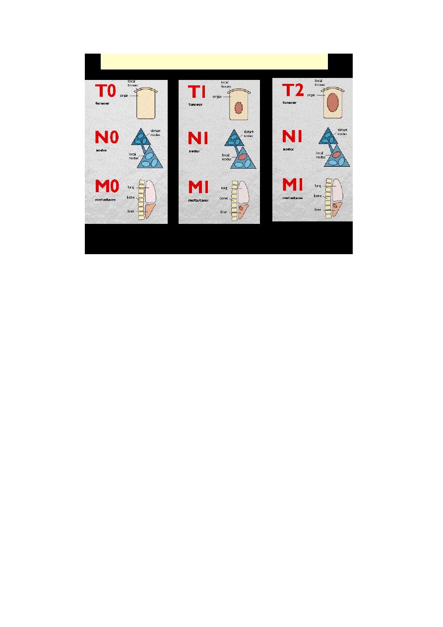
• Staging of cancers is based on
1. Size of primary tumor

2. Extent of spread to regional LNs
3. Presence/absence of blood-borne metastases
• Two major staging systems in use
1. TNM (ICC
)
2. AJC on Cancer Staging.
• The TNM varies for each specific form of cancer
General principles
- T1 to T4 with ↑size
- TIS (in situ)
- N0: no nodal involvement
- N1 to N3: involvement of an ↑number and range of nodes
- M0: no distant metastases,
- M1 or M2: presence of blood-borne metastases.
• The AJC: divides all cancers into stages 0 to IV, depending on
- Size
- Nodal spread
- Distant metastases.
TNM staging for ca breast
LABORATORY DIAGNOSIS OF CANCER
• Becomes more complex & specialized
• Several approaches to correct Dx
- Sometimes > than one approach employed
A. Histologic and Cytologic Methods
• Separating benign from malignant not usually difficult
- Difficulty is Dx of borderline tumors
- Clinical data and surgical findings very useful
• Specimen delivered to lab must be
1. Adequate
2. Representative
3. Properly preserved
• Several sampling approaches are available
1. Incisional or excisional biopsy specimen for

a. conventional histopathological diagnosis
b. frozen section diagnosis
2. Needle Biopsies
a. Fine needle aspiration material (cytology)
b. Needle-core biopsy material (histopathology)
3. Endoscopic biopsy material
4. Laparoscopic, or thoracoscopic biopsies
5. Cytologic smears from the tumor in question
• 1. Incisional biopsy
means that only a portion of the lesion is sampled, and therefore the
procedure is strictly of a diagnostic nature.
In excisional biopsies, the entire lesion is removed, usually with a rim of
normal tissue, and therefore the procedure serves both a diagnostic and a
therapeutic function.
When excision of the whole lesion is not possible, incisional biopsy is
performed, however, selection of an appropriate site for a biopsy of a
large mass by the surgeon requires awareness that the margins of the
lesion may not be representative and its center may be largely necrotic.
Requesting an intra-operative "quick-frozen section" diagnosis is
sometimes desirable, for example, in determining the nature of a mass
lesion or in evaluating the margins of an excised cancer to ascertain that
the entire neoplasm has been removed.
This method permits histologic evaluation within minutes, while the
patient is still under anesthesia. The results in such cases will modify the
course of the surgical operation.
2.Needle biopsies
Fine-needle aspiration of tumors
During fine-needle aspiration, a long, thin needle is inserted into the
suspicious area.
A syringe is used to draw out fluid and cells for analysis.
The material is then spread on a slide, stained and then examined for the
evaluation of the mass.

Core needle biopsy
A wide-bore needle with a cutting tip is used to draw a thin core of tissue
(the size of a match stick) out of a suspicious area.
The tissue obtained is processed to obtain histological sections for
evaluation.
Image-guided biopsy combines an imaging procedure, such as X-ray,
computerized tomography (CT) or ultrasound, with a needle biopsy.
Image-guided biopsy allows access to suspicious areas that cannot be felt
through the skin, such as a suspicious lesion of the liver or prostate.
Through the use of images, it possible to be sure that the biopsy needle
reaches the correct spot.
Histochemistry
The basis of surgical pathology is the examination of the specimens
following fixation in formalin, processing in graded alcohols and xylene,
embedding in paraffin, cutting of sections with a microtome, and staining
with hematoxylin-eosin (H&E).
This technique gives a lot of information quickly with a little cost.
That is why it is the standard method in all histopathology labs.
In the H&E technique, hematoxylin staining of nuclei is followed by
counterstaining of cytoplasms and various extracellular materials by
eosin.
.
Immunohistochemistry (IHC)
This is the application of immunologic principles & techniques to the
study of cells & tissues.
The availability of specific monoclonal antibodies has greatly facilitated
the identification of cell products or cell surface markers.
In several situations, the differentiation between neoplasms may be very
difficult for e.g. with the anaplastic large cell malignancies; even the most
experienced pathologists cannot tell whether the submitted tumor is a
squamous cell carcinoma, adenocarcinoma, lymphoma or a sarcoma.
Differentiation between these is of prognostic & therapeutic implications.
It is through the application of a panel of specific monoclonal antibodies,
which disclose the presence or absence of certain products or cell markers
in these cells, so that the more specific, final diagnosis can be reached.

Examples of the utility of immunohistochemistry in the diagnosis or
management of malignant neoplasms include
1. Categorization of undifferentiated malignant tumors
2. Categorization of leukemias and lymphomas
3. Determination the site of origin of metastatic tumors
4. Detection of molecules that have prognostic or therapeutic significance