1.
Entamoeba hartmanni.
2.
Entamoeba. coli.
3.
Entamoeba gingivalis.
4.
Iodamoeba butschlii.
5.
Endolimax nana.
6.Dientamoeba fragilis
(amoebic- flagellates).
These amoebae are found only in the
intestines(except
E.gingivalis).
they do not harm the body. They enter the
human body when a person swallows food or
water that has been exposed to contaminated
stool.
These amoebae can remain in a persons intestine
for weeks, months or years. Studies have shown
that these amoebae do not make people sick.
Even people who have a weakened immune
system are not affected by these amoebas.
Non-pathogenic Amoebae
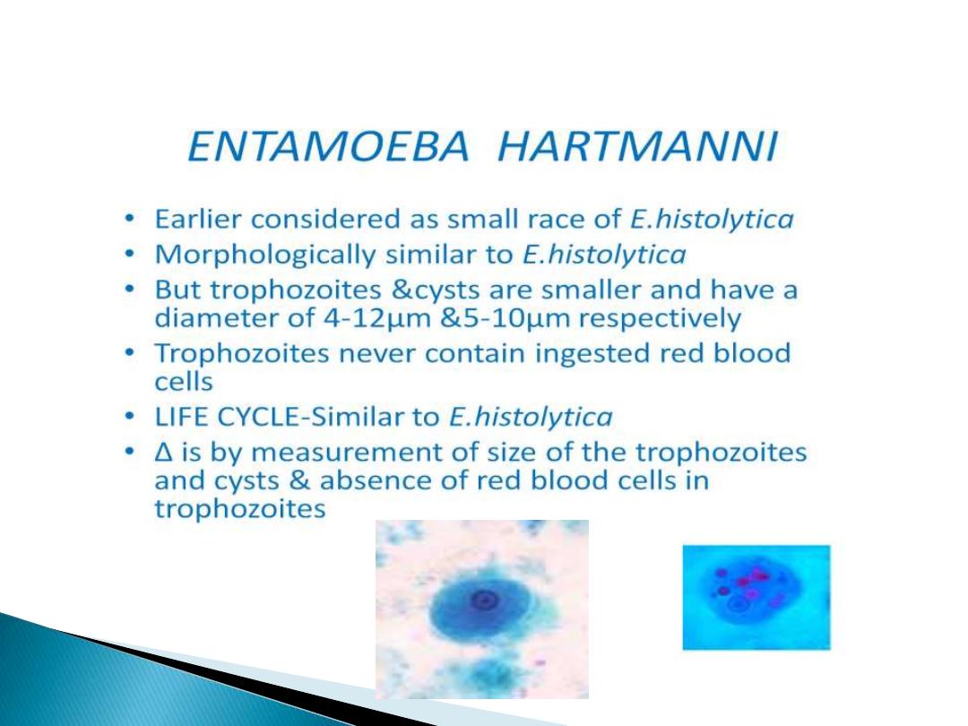
Entamoeba hartmanni
troph.
1.
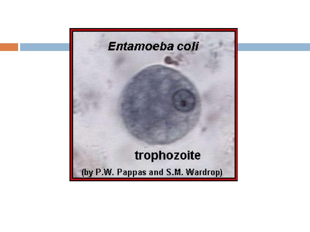
2.Entamoeba coli :
It is non pathogenic amoebae, world widely
distributed, usually the most common amoebic
parasite of man. Although it is a harmless, commensal
found in the
lumen of cecum
and
lower levels of
large intestine
, its presence in the stool of man
indicates that the patient has ingested fecally
contaminated food.
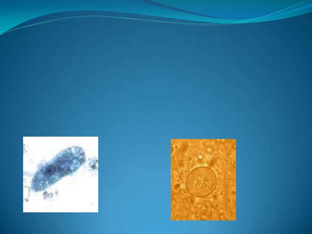
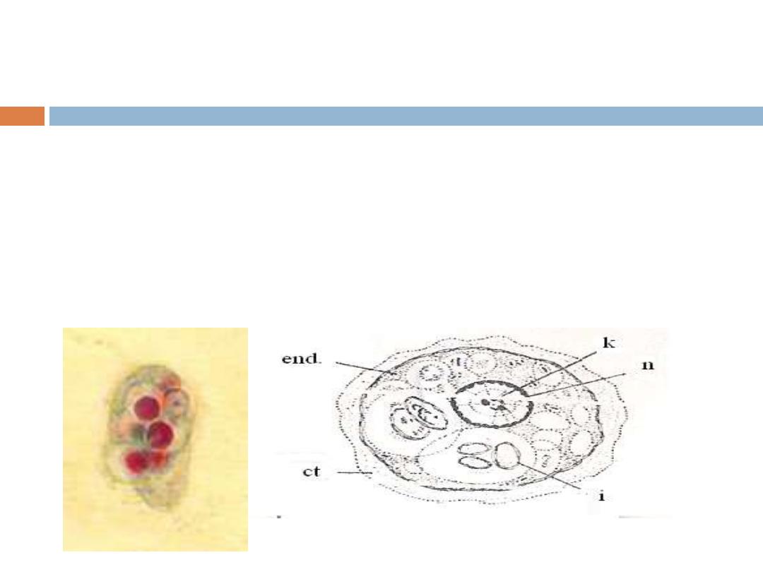
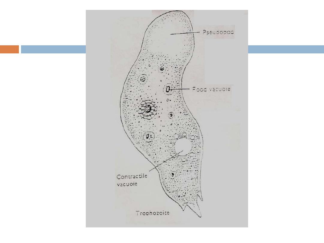
Trophozoite of E.coli
cyst of E.coli

E.coli resembles E.histolytica .These two
species are frequently misdiagnosed leading
either to unnecessary treatment for
nonpathogenic parasite or to omission of
appropriate therapy for a pathogen.
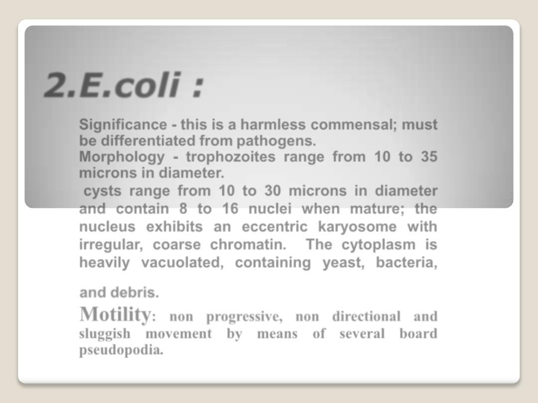
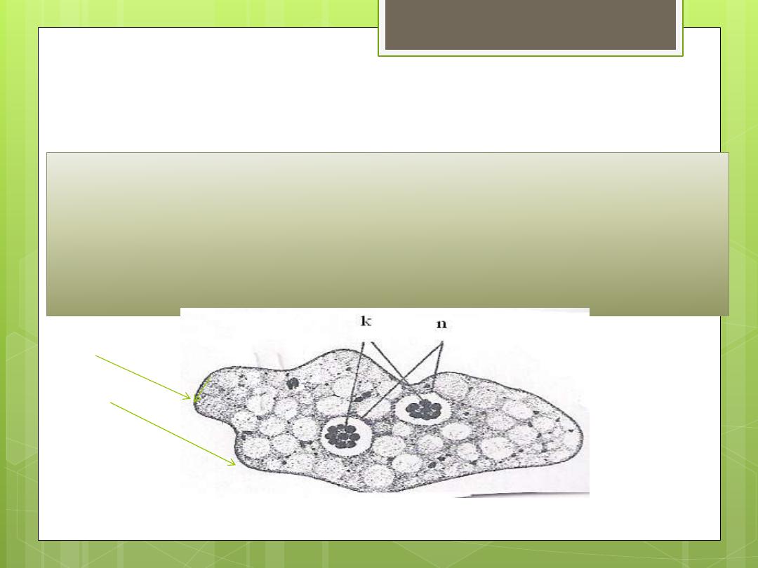
2.E.coli :
Significance - this is a harmless commensal; must
be differentiated from pathogens.
Morphology -
trophozoites
range from 10 to 35
microns in diameter.
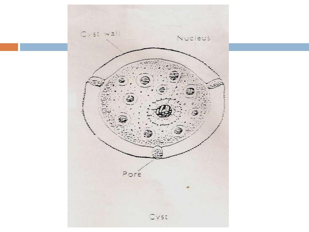
cysts
range from
10
to
30
microns in diameter
and contain
8
to
16
nuclei when mature; the
nucleus exhibits an
eccentric karyosome
with
irregular, coarse chromatin. The cytoplasm is
heavily vacuolated, containing yeast, bacteria,
and debris.
Motility
: non progressive, non directional and
sluggish movement by means of several board
pseudopodia.
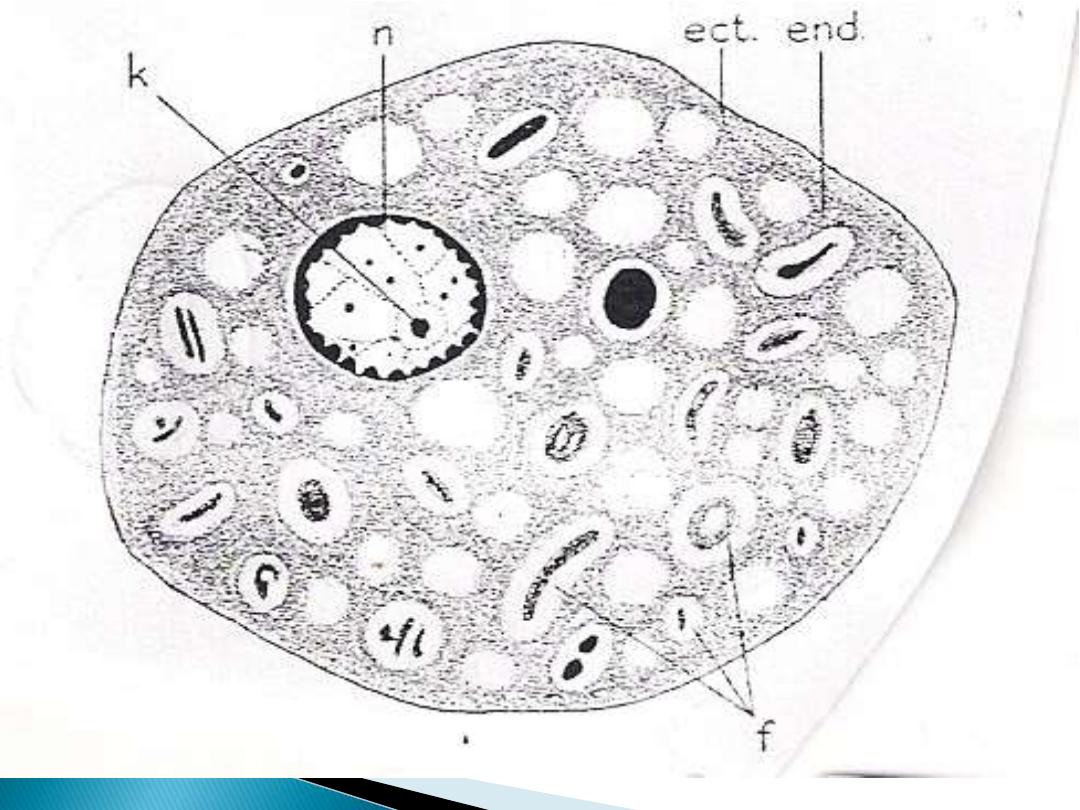
Trophozoite of
E.coli
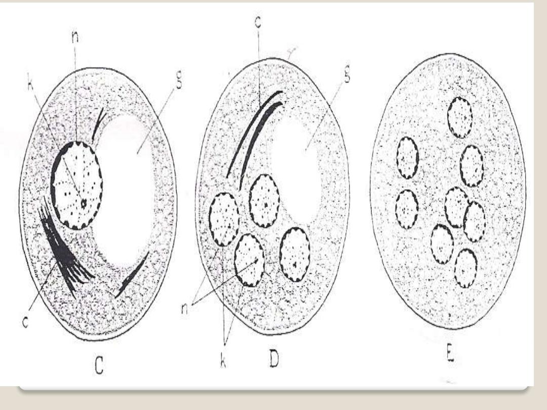
The stages of development of E.coli cyst

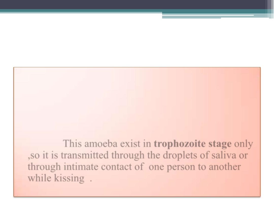
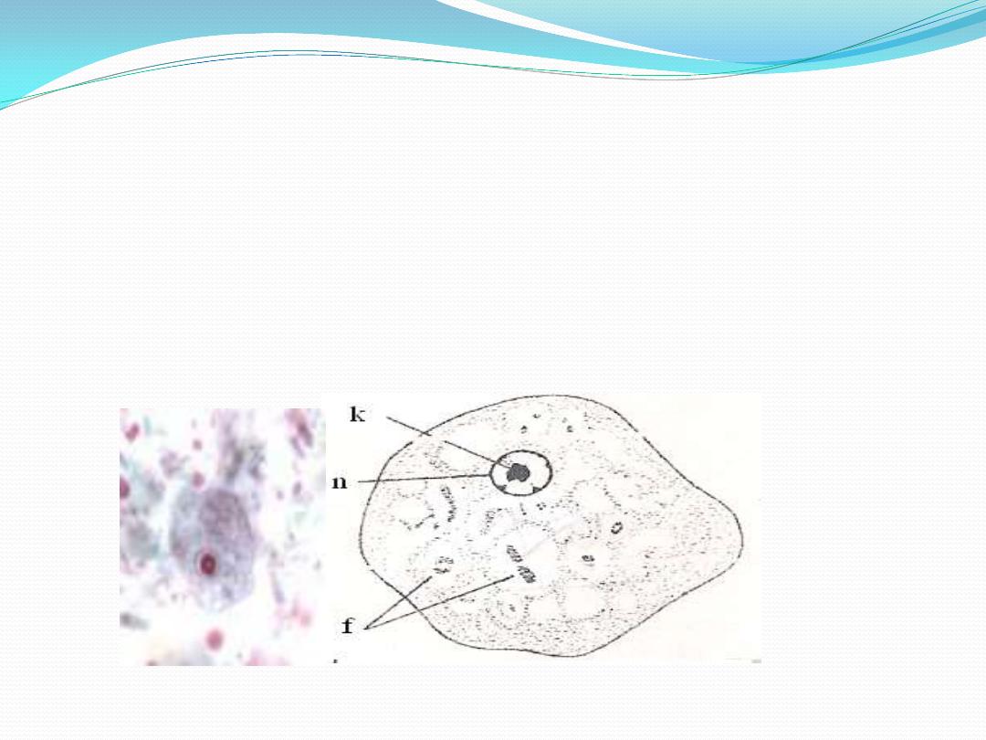
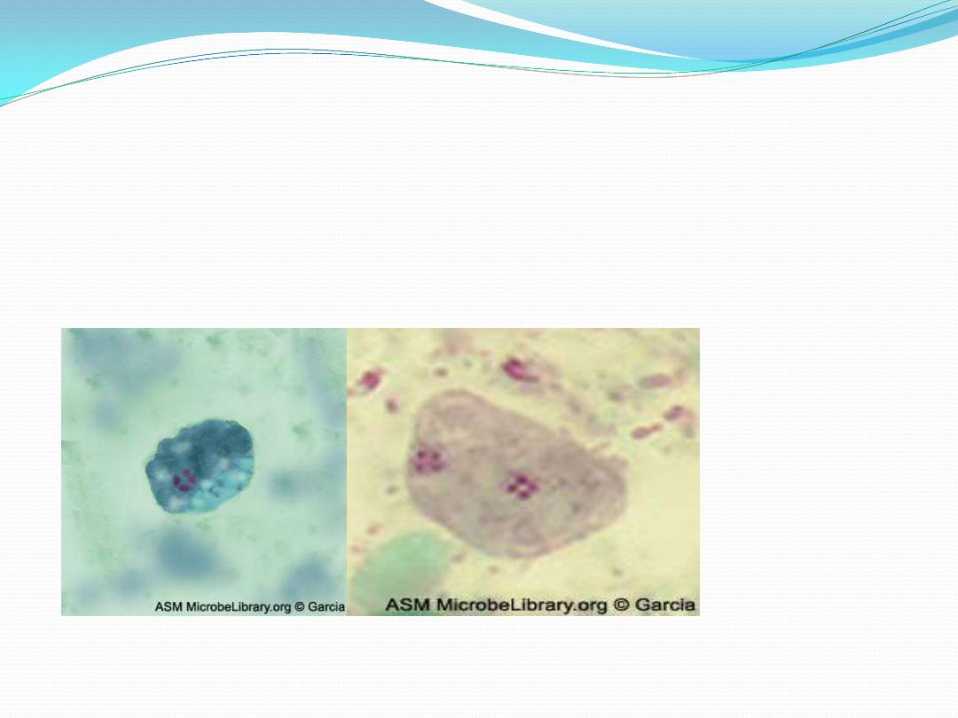
3.Entamoeba gingivalis
•
It is cosmopolitan amoebae ,the general incidence is
estimated in at least 50%of persons with diseased
gums and tonsils .It is
non pathogenic
and isolated
from healthy mouth but mainly it is isolated from
1.diseased mouth with oral abscesses ,2.dental
carries and 3.other inflammatory conditions in the
mouth. This amoeba exist in
trophozoite stage
only
,so it is transmitted through the droplets of saliva or
through intimate contact of one person to another
while kissing .
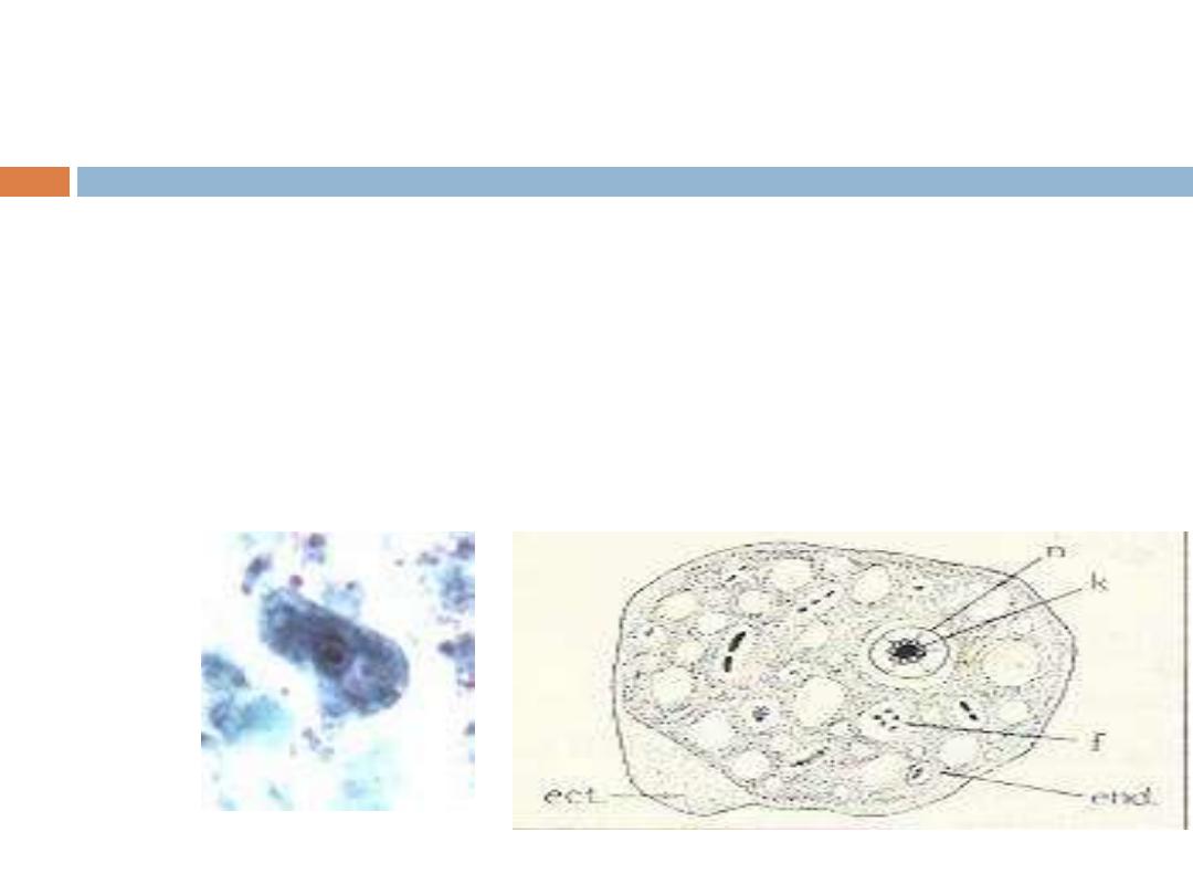
3.Entamoeba gingivalis
This amoebae is actively motile by multiple pseudopodia .
Ectoplasm is clear, well differentiated from granular
endoplasm. Endoplasm contain many food vacuoles, some of
them with diagnostic rounded dark
– staining bodies represent
the nuclei of ingested white blood cells from leucocytes are
usually recognized in stained specimens and serve to identify
the amoebae as E.gingivalis which is the only species
ingests these cells and epithelia cells. The nucleus is
histolytica type.
Trophozoite of E.gingivalis
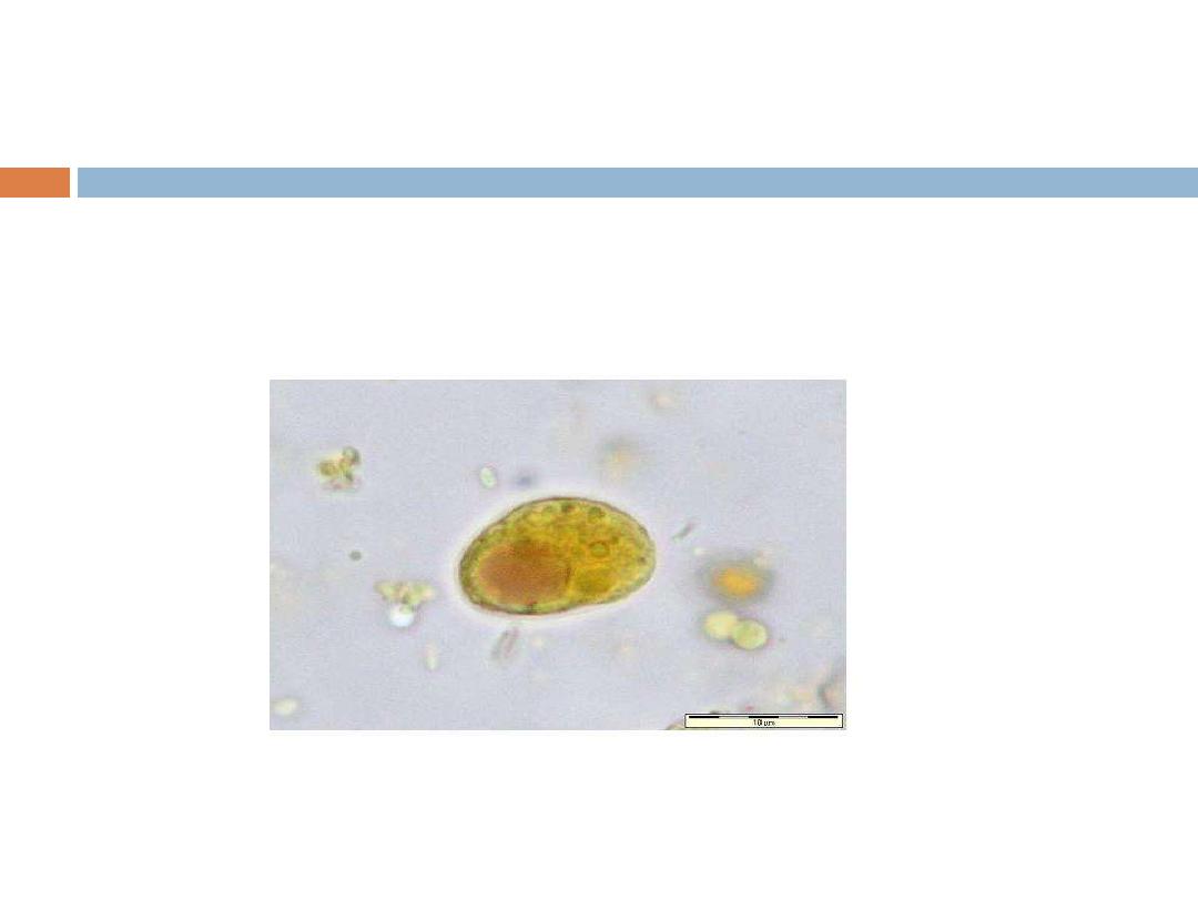
4. Iodamoeba butschlii
It is cosmopolitan amoeba, but it is less common than E. coli, it is non
pathogenic commensal living in the lumen of large intestine. This amoeba
has trophozoite and cyst stages. Trophozoite: small, sluggish, ectoplasm
no well differentiated from endoplasm. The nucleus spherical, characterized
by having central large karyosome surrounded by achromatic granules.
Iodamoeba butschlii troph.
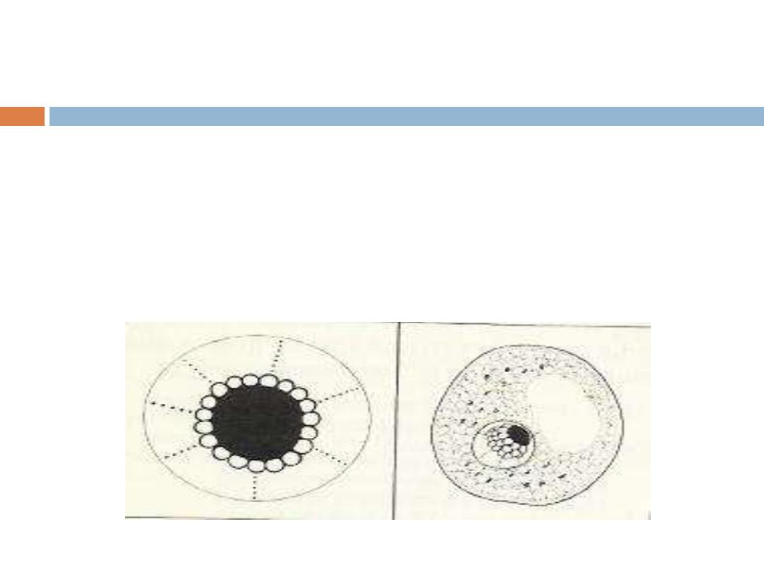
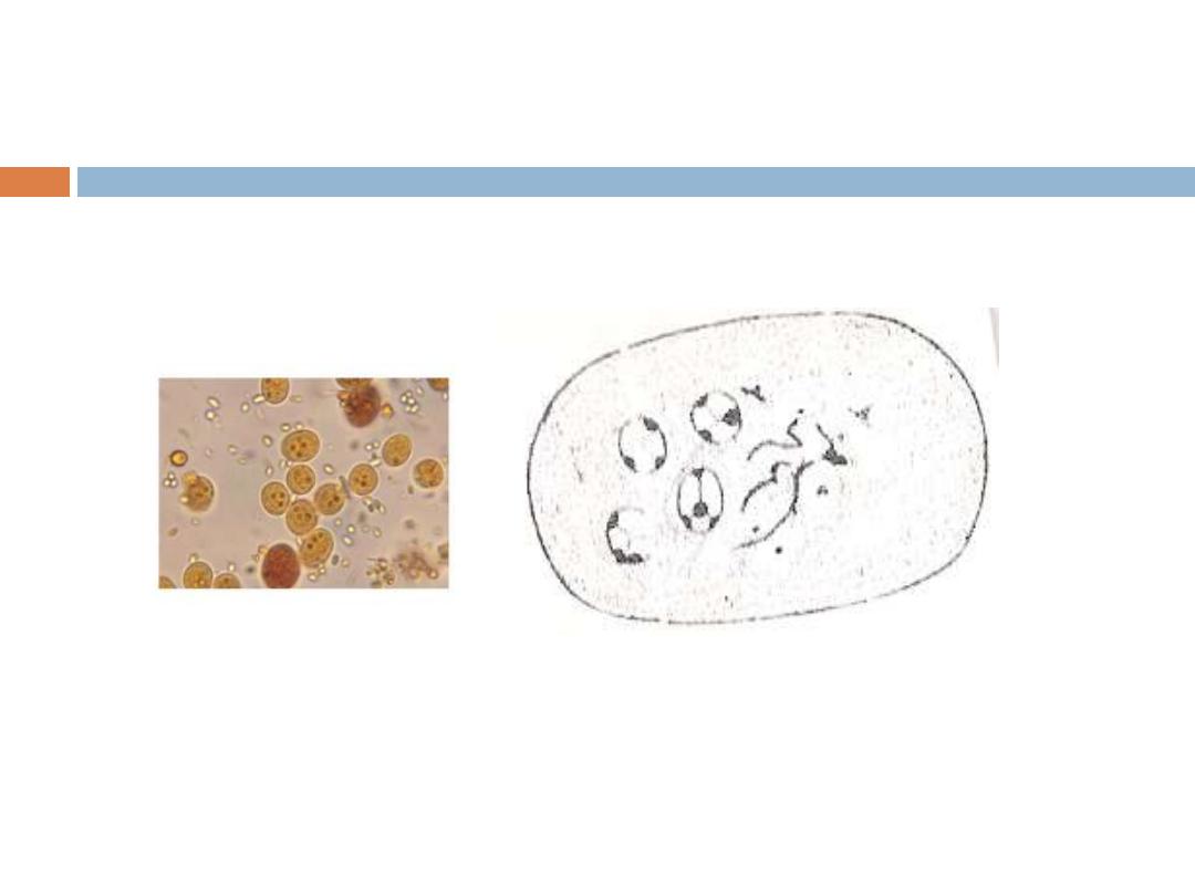
The cyst is irregular in shape with single nucleus only, cyst
contain well distinct glycogen mass which stain golden
brown with iodine solution so this amoeba called
Iodamoeba. The nucleus has large compact eccentric
karyosome and around this karyosome there are achromatic
granules.
Cyst of I.butschlii
Cyst of
I.buschlii
Lab.Diagnosis:
confused with N.fowleri troph. Both
have eye bull's appearance .The use od iodine stained
preparation will aid to differentiate between them.
4.Endolimax nana
This is the commonest small intestinal amoeba(nan:small)
Cosmopolitan amoebae, found in trophozoite and cyst stage. It is
commensal, living in
cecum
and
lower levels of large
intestine
.
Trophozoite: small, sluggishly motile, ectoplasm differentiated
from endoplasm. It has single nucleus with large pleomorphic or
lobulated karyosome.
Trophozoite of E.nana
Cyst: Spherical or oval in shape, mature cyst
contain 4 nuclei.
Diagnosis: by demonstrate troph. or cyst stage in stool specimen.
Cyst of E.nana
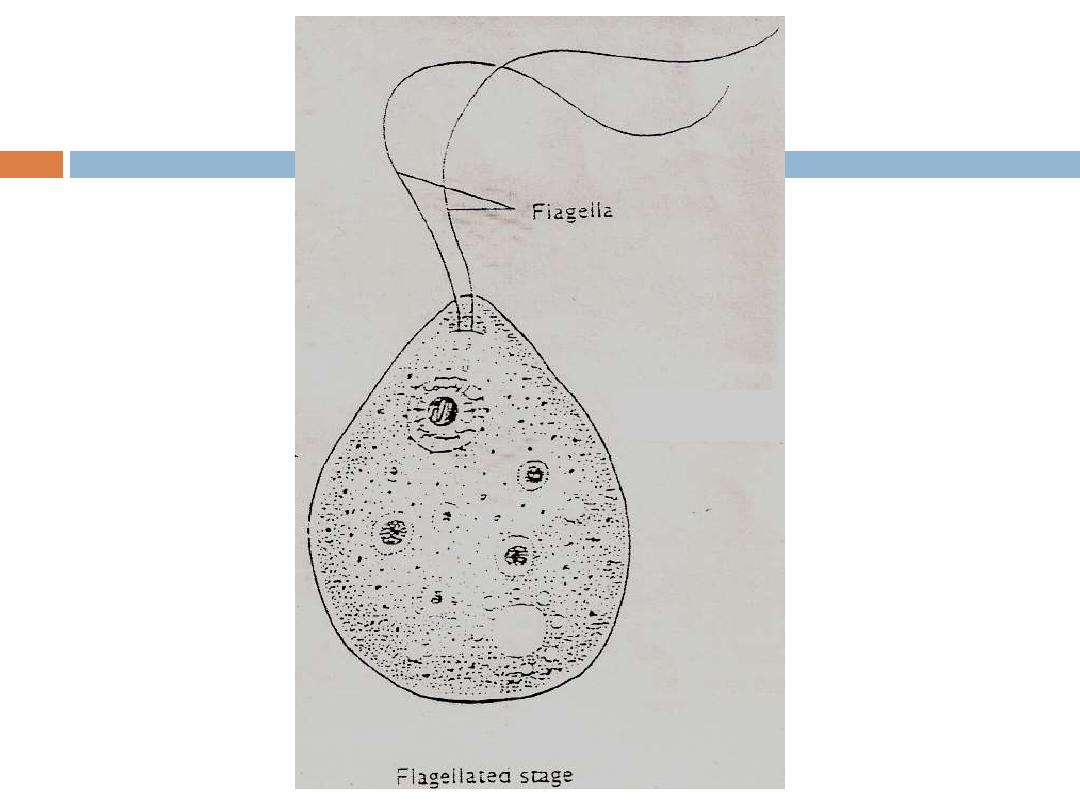
Dientamoeba fragilis
It was generally regarded as an amoeba, but it is placed
with flagellates because it has some characters of
Trichomonads. It is cosmopolitan parasite, but because of
its small size, it might sometimes overlook in fresh
Preparation.
Trophozoite of D.fragilis
Angular
pseudopodia
It is only known in
trophozoite stage
, it lives in
mucosal crypts of cecum and rectum.
Trophozoite
is small, sluggishly motile, it has 2 nuclei,
the nucleus characterized by having central
karyosome consisting of 4 – 8 granules.
Troph. Of D.fragilis

The mode of transmission :
is unknown, possibly
may be achieved by direct or indirect contact with the
trophozoite. Also it has been suggested that this
amoeba may be transmitted through the ova of certain
helminthes like entrobiasis.
Pathogenicity:
In most persons it causes no harmful
effects. Pathogenicity has been suspected when it has
been the only organism identified in case of anorexia,
abdominal pain and diarrhea.
Diagnosis:
by demonstrate trophozoites in formed or
diarrhic stool.

Thank you for your listen

Free
– Living Pathogenic
amoebae
Naegleria fowleri (brain
– eating amoeba)
It causes primary amoebic meningoencephalitis
(PAM) in humans. It is cosmopolitan, mainly in
North America, Western Europe, Africa, Japan and
Australia.
The amoebae found in warm fresh water of ponds,
lakes, pools and moist soil.
Morphology: It is the only amoebae which is found
in 3 forms; flagellate trophozoite, amoeboid
trophozoite and cyst stages.
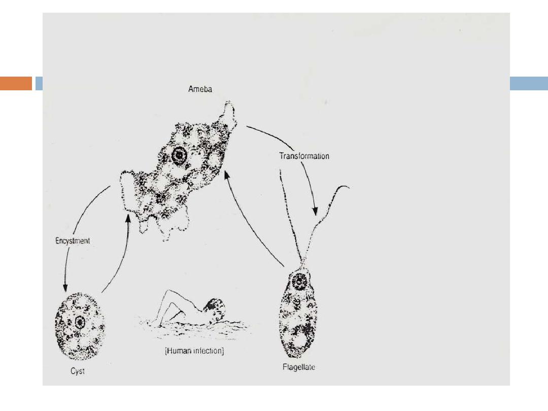

Pathogenicity and
Symptomatology:
The amoebae enter nasal passages when the persons
swimming or has contact with warm water, moving
along olfactory nerve, through cribriform plate into the
cranial cavity. In the brain the amoebae remain
associated with membranous covering of the brain, the
meninges, where they evoke an inflammatory
response.
Early in the infection the patient complains of upper
respiratory tract symptoms e.g. raning nose, sore
throat, fever and headache. Within 2
– 3 days the
headache becomes more sever and there may be
vomiting, stiff neck, mental confusion, coma as a result
of intracranial pressure. Death usually with 10 days of
the onset of symptoms.

Diagnosis:
Direct demonstration of motile amoebae in
unstained CSF or nasal discharge.
Stained smear of CSF.
Stained section of brain tissue at autopsy.
Culture on non-nutrient agar medium coated
with E. coli bacteria.
Serological tests.

Acanthamoeba Spp.:
It causes granulomatous amoebic encephalitis
and amoebic keratitis. It is cosmopolitan but
are not necessarily associated with warm
water, it is found in moist soil and in the air and
water. It is found in only two form; the
trophozoite and cyst, and either of these can
be a source of infection.
Amoeboid trophozoite has spikey
pseudopodia and a nucleus with large, central
karyosome similar to the nucleus of Naegleria.
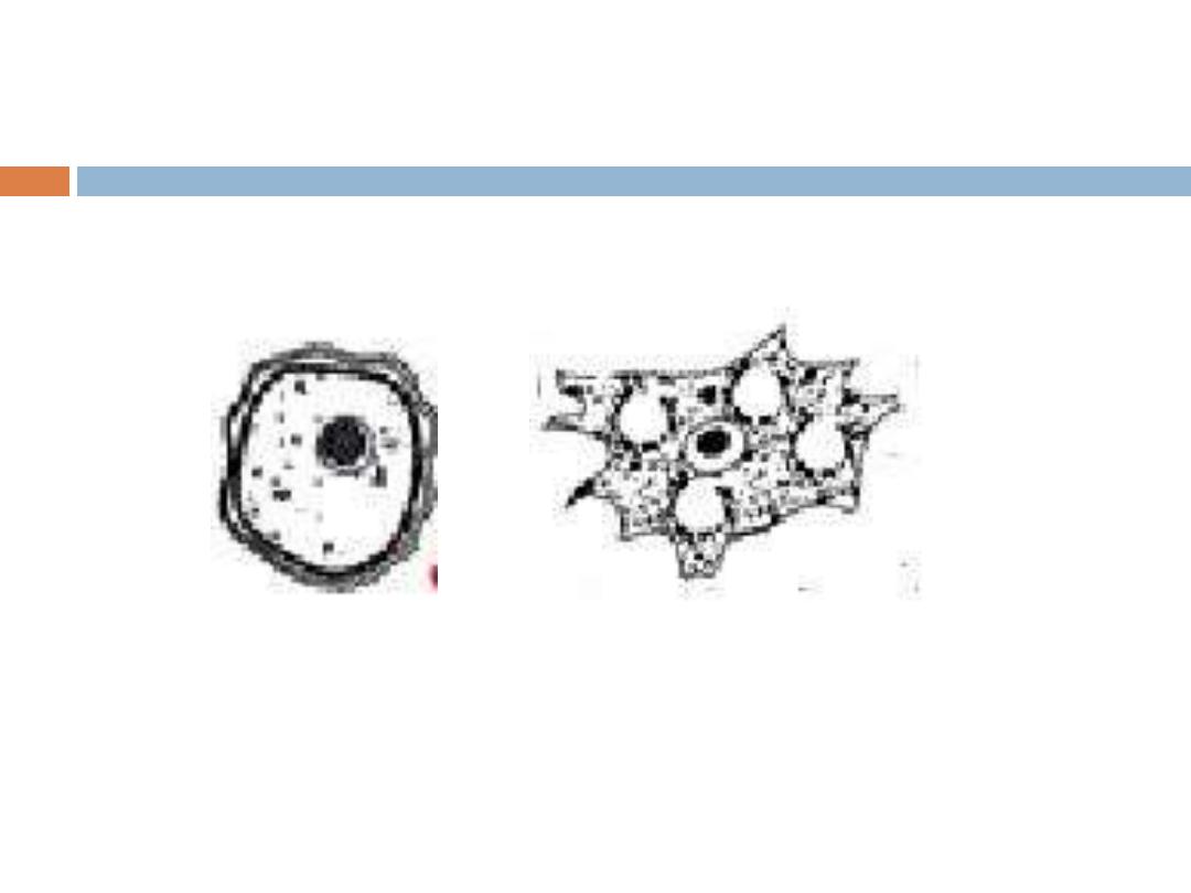
Trophozoite
Cyst

Host
– Parasite Interactions
The amoebae probably enter respiratory system,
or perhaps the skin, then migrate to CNS via the
blood.
Once in the brain, it cause granulomatous
encephalitis, which means that a more or less
discrete mass of inflammatory cells and amoebae
are found in the meninges and superficial layers
of the brain. The lesion develop slowly .if
treatment is not administered the progress of
disease is inexorable leading to death of the
patient. Most patients with GAE do not have
normal immune system. GAE has been seen in
AIDS patients.

Amoebic Keratitis:
Acanthamoeba spp. Caused ulcers of the cornea
of eye in humans. Cornea is invaded when there
is trauma in the eye or the presence of amoebae
in water. In most instances, there is an association
with wearing contact lenses and a failure to clean
them properly. Corneal lesions are painful and
differentiation must be made from herpes simplex
virus.
Diagnosis:
By finding amoebae in wet mounte (10% KOH) of
corneal ulcer scraping or in stained smear.
By isolation of amoebae form contact lenses or
washing solutions.