
The Immune System &
Lymphoid Organs
18
th
& 19
th
lecture
May 3 &5, 2016

The body has a system of cells—the immune system—
that has the ability to distinguish "self" (the organism's own
molecules) from "non-self" (foreign substances).
This system has the ability to neutralize or inactivate foreign
molecules (such as soluble molecules as well as those present in
viruses, bacteria, and parasites) and to destroy microorganisms or
other cells (such as virus-infected cells, cells of transplanted
organs, and cancer cells).
On occasion, the immune system of an individual reacts against its
own normal body tissues or molecules, causing autoimmune
diseases.
Immune System
• functional system
rather than organ
system
– Hematopoetic
– Vasculature
– Lymphatic
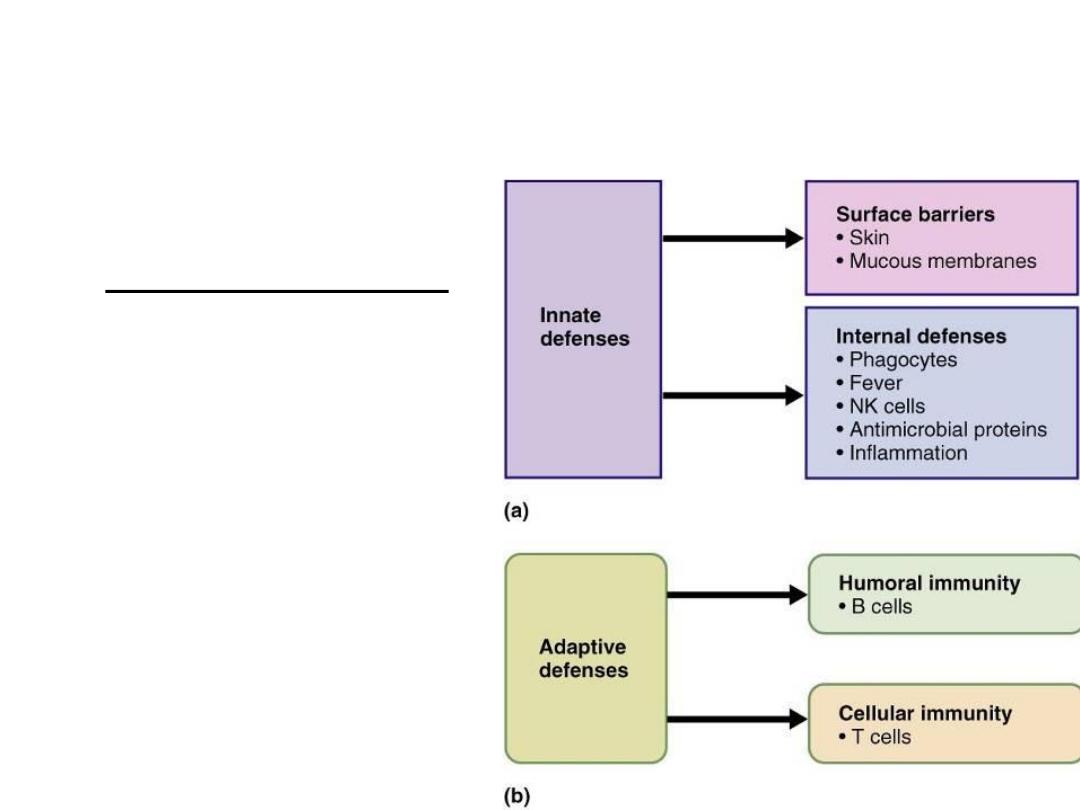
Innate vs. Adaptive Immune System –
Introduction
• Innate: structural defenses; responds to nonspecific
foreign substances
– First line: external surface epithelium & membranes
– Second line: inflammatory processes – antimicrobial
proteins, phagocytes, etc.
Innate vs. Adaptive Immune System –
Introduction
• Adaptive: responds to specific foreign substances
• Innate & adaptive mechanisms work together
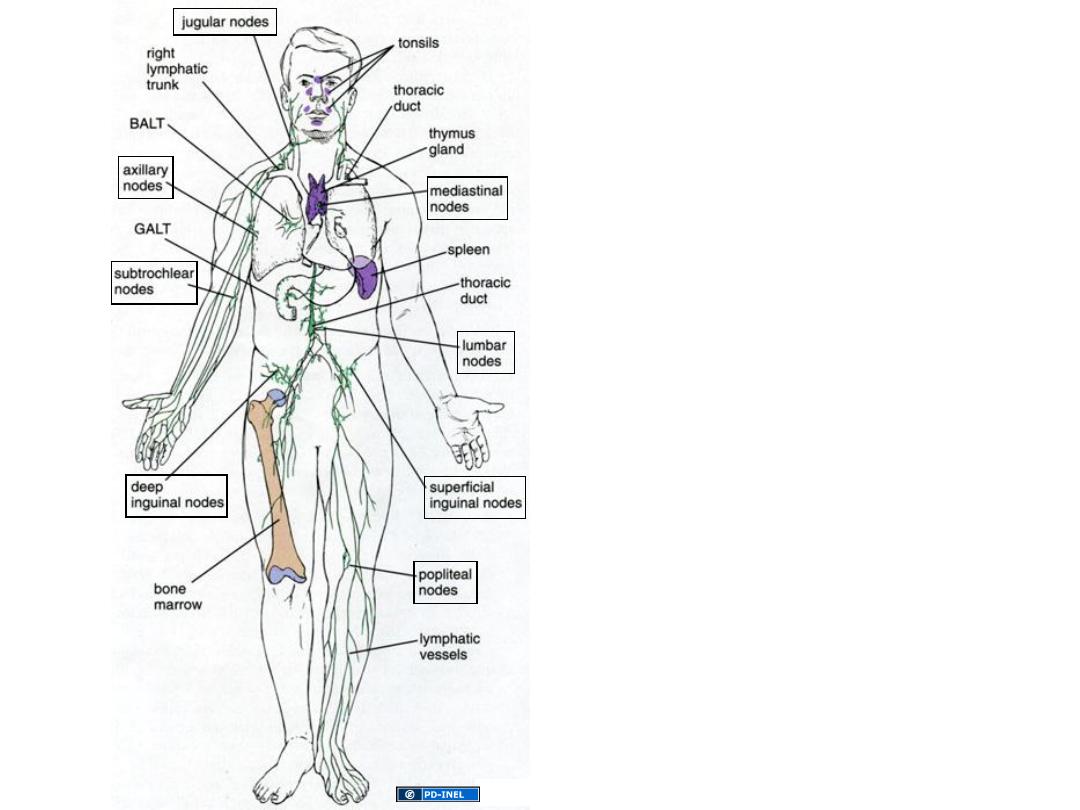
The lymphatic system is comprised of
1- lymphatic vessels, which transport
interstitial fluid (lymph) back to the blood
circulation
2- the lymphoid organs which house
lymphocytes and other cells of the body's
immune defense system .
-Primary lymphoid organs are the bone
marrow and thymus, where B and T
lymphocytes are formed respectively .
-The secondary lymphoid organs include
the lymph nodes, mucosa-associated
lymphoid tissue (MALT), and spleen
.

Cells of the Immune System
• (1) are distributed throughout the body in the blood,
lymph, and epithelial and connective tissues;
• (2) are arranged in small spherical nodules called lymphoid
nodules found in connective tissues and inside several
organs; and
• (3) are organized in larger lymphoid organs—the lymph
nodes, the spleen, the thymus, and the bone marrow.
• The wide distribution of immune system cells and the
constant traffic of lymphocytes through the blood, lymph,
connective tissues, and lymphoid organs provide the body
with an elaborate and efficient system defense

Cells of the Immune System
• The primary cells that participate in the immune response are
• Lymphocytes are classified as B, T, or natural killer (NK) cells.
• The B and T cells are the only cells that have the ability to selectively
recognize a specific epitope among a vast number of different
epitopes.
• B and T cells differ based on their life history, surface receptors, and
behavior during an immune response.
• T lymphocyte precursors, on the other hand, leave the bone marrow,
and through the blood circulation reach the thymus where they
undergo intense proliferation and differentiation or die by apoptosis.
After their final maturation, T cells leave the thymus and are
distributed throughout the body in connective tissues and lymphoid
organs.
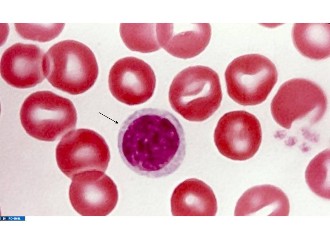
Lymphocytes in peripheral blood smear
These are B and T-cells that have undergone antigen-INDEPENDENT differentiation and are trafficking through
the bloodstream on their way to lymphoid organs/tissue.
lymphocyte
Mizobuti histology slide set

Cells of the Immune System
• Natural Killer Cells
• The natural killer lymphocytes lack the marker
molecules characteristic of B and T cells. They
comprise about 10–15% of the lymphocytes of
circulating blood. Their name derives from the
fact that they attack virus-infected cells,
transplanted cells, and cancer cells without
previous stimulation; for this reason they are
involved in what is called an innate immune
response.

B -Lymphocytes
• There are c.10 million different B-
lymphocytes, each of which make a different
antibody.
• The huge variety is caused by genes coding for
antibodies changing slightly during
development.
• There are a small group of clones of each type
of B-lymphocyte

B -Lymphocytes
• At the clone stage antibodies do not leave the B-
cells.
• The antibodies are embedded in the plasma
membrane of the cell and are called antibody
receptors.
• When the receptors in the membrane recognise and
antigen on the surface of the pathogen the B-cell
divides rapidly.

B -Lymphocytes
• Some activated B cells
PLASMA CELLS
these produce lots of antibodies
• The antibodies travel to the blood, lymph,
lining of gut and lungs.
• The number of plasma cells goes down after a
few weeks
• Antibodies stay in the blood longer but
eventually their numbers go down too.
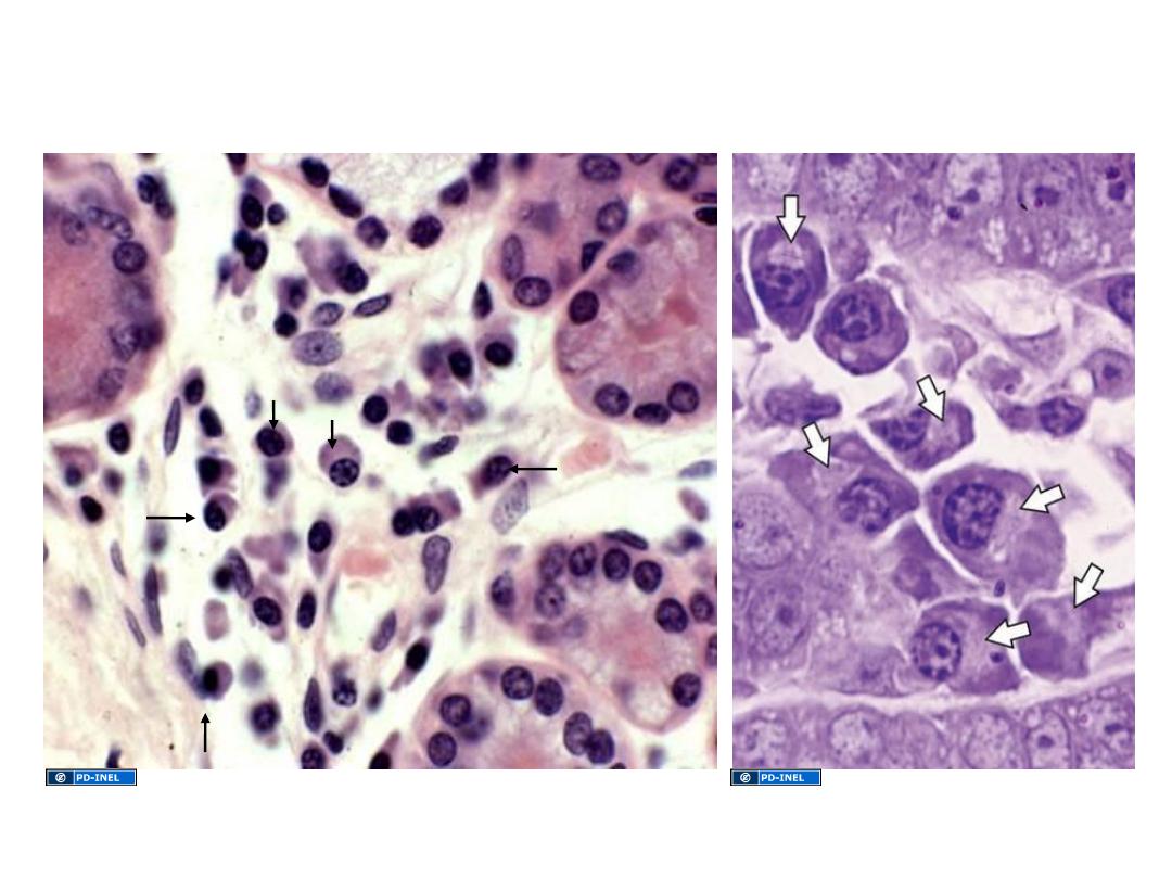
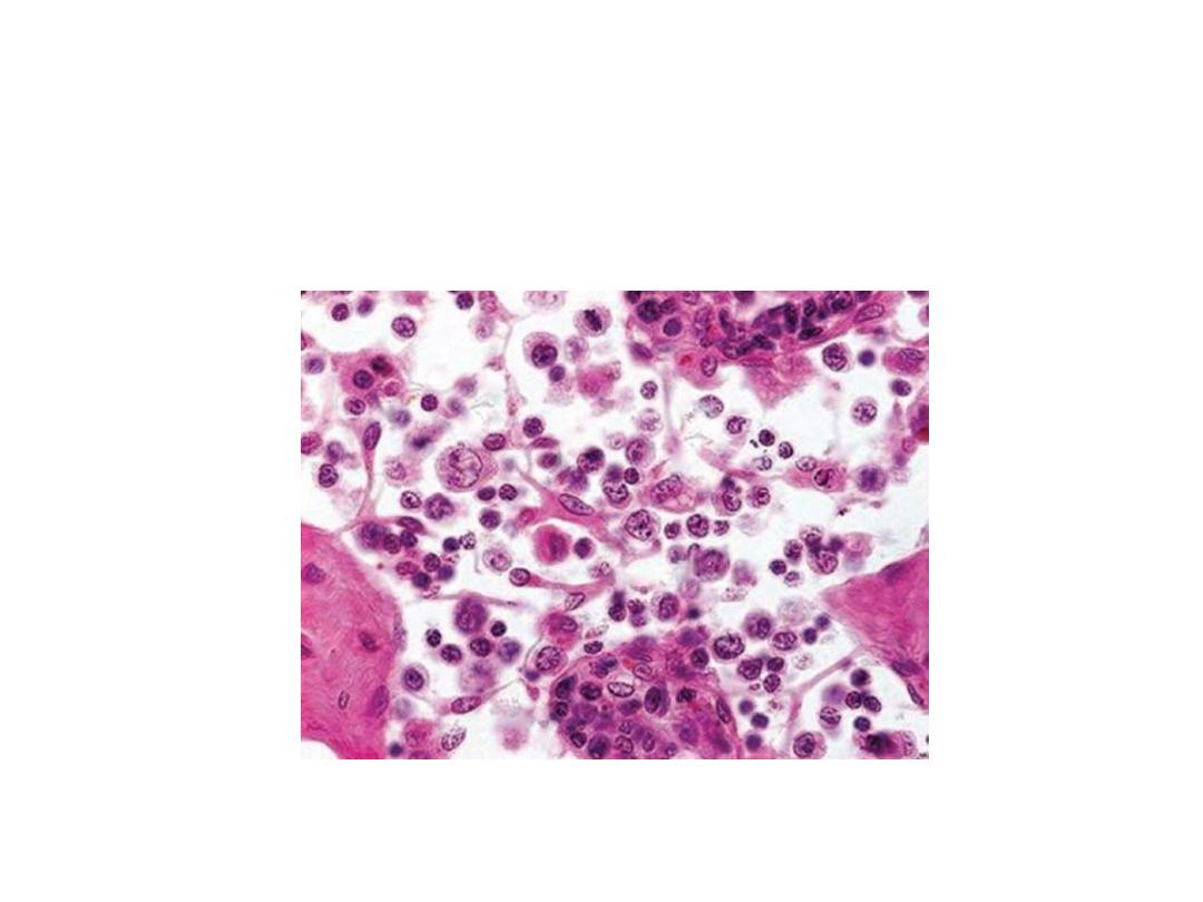
Plasma Cells
are mature B lymphocytes
White arrows = Golgi regions
Black arrows indicate several plasma cells
U-M Histology Collection
Junquiera and Carneiro. Basic Histology. Tenth Ed. 2003

B -Lymphocytes
• Some activated B cells
MEMORY CELLS
.
• Memory cells divide rapidly as soon as the
antigen is reintroduced.
• When the pathogen/infection infects again it
is destroyed before any symptoms show.

T Lymphocytes
T cells constitute 65-75 % of blood lymphocytes. To recognize epitopes, all
T cells have on their surfaces a molecule called a T cell receptor (TCR). T
seditpep llams yltsom( sepotipe ylno ezingocer setycohpmyl
(
Three important subpopulations of T cells are the following:
§
Helper cells, which produce cytokines that promote differentiation of B cells
into plasma cells Helper cells have a marker called CD4 on their surfaces and are,
hence, called CD4
+
T cells.
§
Cytotoxic T cells are CD8
+
and act directly against foreign cells or virus-
infected cells by two main mechanisms. By perforins or apoptosis.
§
Regulatory T cells are CD4
+
CD25
+
and play crucial roles in allowing immune
tolerance.
Neutrophils
• 60% of WBCs
• ‘Patrol tissues’ as they squeeze out of the
capillaries.
• Large numbers are released during infections
• Short lived – die after digesting bacteria
• Dead neutrophils make up a large proportion
of puss.

Macrophages
• Larger than neutrophils.
• Found in the organs, not the blood.
• Made in bone marrow as
monocytes
, called
macrophages once they reach organs.
• Long lived
• Initiate
immune responses as they display
antigens from the pathogens to the
lymphocytes.

Cytokines
• The functions of cells in the immune system are
regulated by a large number of molecules, mainly
cytokines, which are peptides or glycoproteins.
• They are primarily produced by cells of the
immune
system,
mainly
lymphocytes,
macrophages, and other leukocytes, but may also
be synthesized by other cell types, such as
endothelial cells and fibroblasts.
• Chemotaxins, or chemokines, are cytokines that
induce diapedesis of leukocytes and migration to
sites of inflammation
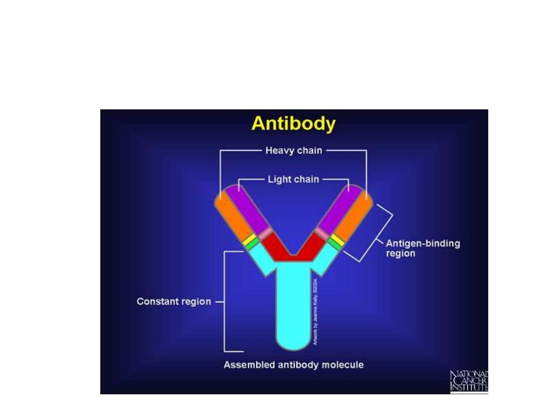
Antibody: a molecule that is recognized by cells of
the immune system is called an antigen and may
elicit a response from these cells

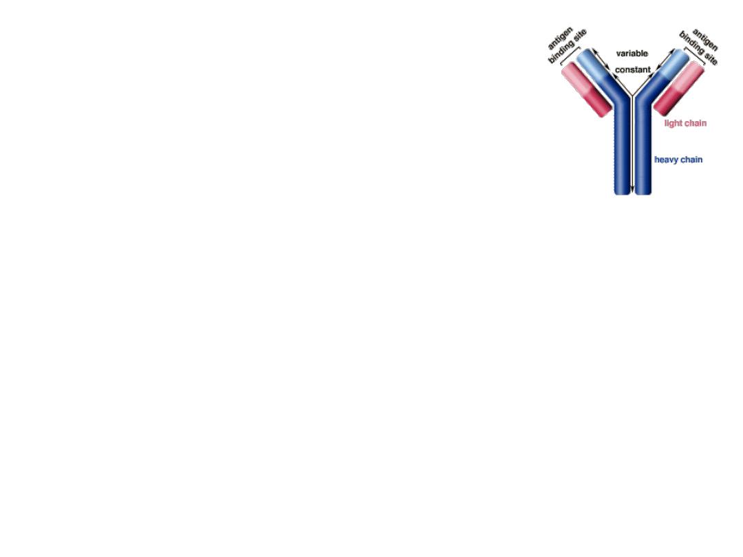
Antibodies
• Also known as
immunoglobulins
• Globular glycoproteins
• The heavy and light chains are polypeptides
• The chains are held together by disulphide
bridges
• Each antibody has 2 identical antigen binding
sites – variable regions.
• The order of amino acids in the variable region
determines the shape of the binding site
How Abs work
• Some act as
labels
to identify
antigens for phagocytes
• Some work as
antitoxins
i.e. they block toxins for
e.g. those causing diphtheria and tetanus
• Some attach to bacterial flagella making them
less active and easier for phagocytes to engulf
• Some cause
agglutination (clumping together)
of
bacteria making them less likely to spread
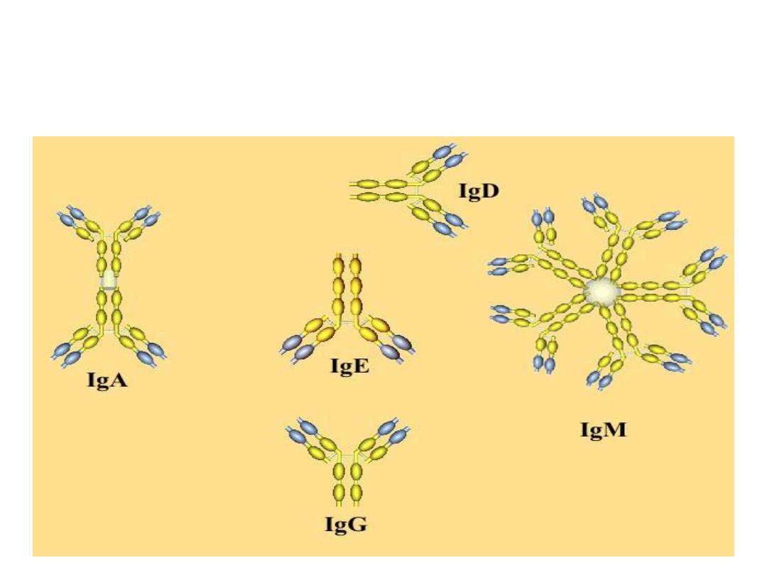
Different Immunoglobulins
Lymphoid Tissue
Lymphoid tissue is connective tissue characterized by a rich supply of
lymphocytes. It exists free within the regular connective tissue or is surrounded
by capsules, forming the lymphoid organs .
lymphocytes don’t just stay in one place
From the MALT, lymphocytes can squeeze into lymph vessels…
S.K. Kim. U-M Histology Collection
..go through larger lymphatic channels in the mesentery…
U-M Histology Collection
..and end up at a LYMPH NODE.
U-M Histology Collection
Lymph Nodes
Main functions:
1.
Filter lymph, thereby promoting
lymphocyte contact with antigen
2.
Provides necessary microenvironment
for antigen-dependent differentiation
Ross, Fig. 14.1

Role of Lymph Nodes in the Immune Response
• Lymph nodes are distributed throughout the body and lymph formed in
tissues must pass through at least one node before entering the
bloodstream.
• The lymph node is an important site of lymphocyte proliferation
(especially of B cells in the germinal centers) as well as of transformation
of B lymphocytes into plasma cells. Because of this, the lymph that leaves
a lymph node may be enriched in antibodies. When the lymph is returned
to the blood circulation, these antibodies will be delivered to the entire
body.

Thymus
The thymus is a bilateral organ located in the mediastinum; it attains its peak
development during youth. Like bone marrow and B cells, the thymus is
considered a central or primary lymphoid organ because T lymphocytes form
there .
The thymus has a connective tissue capsule that penetrates the parenchyma
and divides it into incomplete lobules, with continuity between the cortex and
medulla of adjoining lobules Each lobule has a peripheral darkly stained zone
known as the cortex
dna
a
lartnec
thgil
enoz
dellac
eht
medulla.
ehT
xetroc
si
rehcir
ni
llams
setycohpmyl
naht
eht
alludem
dna
erofereht
ti
sniats
erom
ylkrad
.
The thymus, an encapsulated,
bilateral
organ
in
the
mediastinum, is subdivided by
connective tissue (CT) septa into
connected lobes. Lobes of an
active
thymus
shown
have
peripheral regions of cortex (C),
where basophilic lymphocytes
are fairly dense, and more
central medulla (M) regions
with fewer lymphocytes
Mucosa-Associated Lymphoidt issue (MALT)
The digestive, respiratory, and genitourinary tracts are common sites of invasion by
pathogens because their lumens are open to the external environment. To protect the
organism, the mucosal connective tissue of these tracts contains large and diffuse
collections of dendritic cells, lymphocytes, IgA-secreting plasma cells, APCs, and lymphoid
nodules.
Intraepithelial lymphocytes
Shown here in resp. epith.
Homing mediated by
“addressins” (a sort of
lymphocyte “GPS”)
U-M Histology Collection
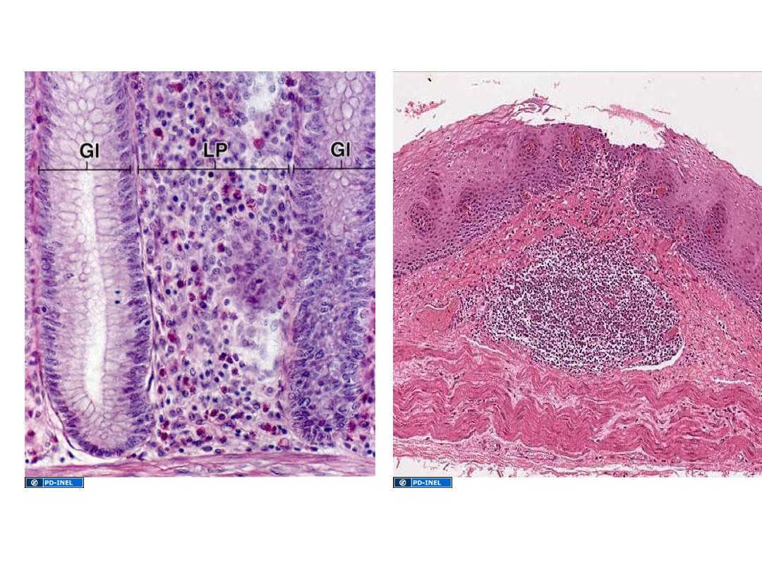
Diffuse lymphoid tissue
Lamina propria (LP) of gut shown here, but can be found
associated with mucosae anywhere in the gut,
respiratory, and genitourinary tracts.
Primary lymphatic nodule/follicle (LN)
Aggregation of lymphocytes in lamina propria or submucosa
LYMPHOCYTES IN CONNECTIVE TISSUE:
MALT = mucosa-associated lymphoid tissue
LN
Ross and Pawlina, Histology: A Text and Atlas
U-M Histology Collection
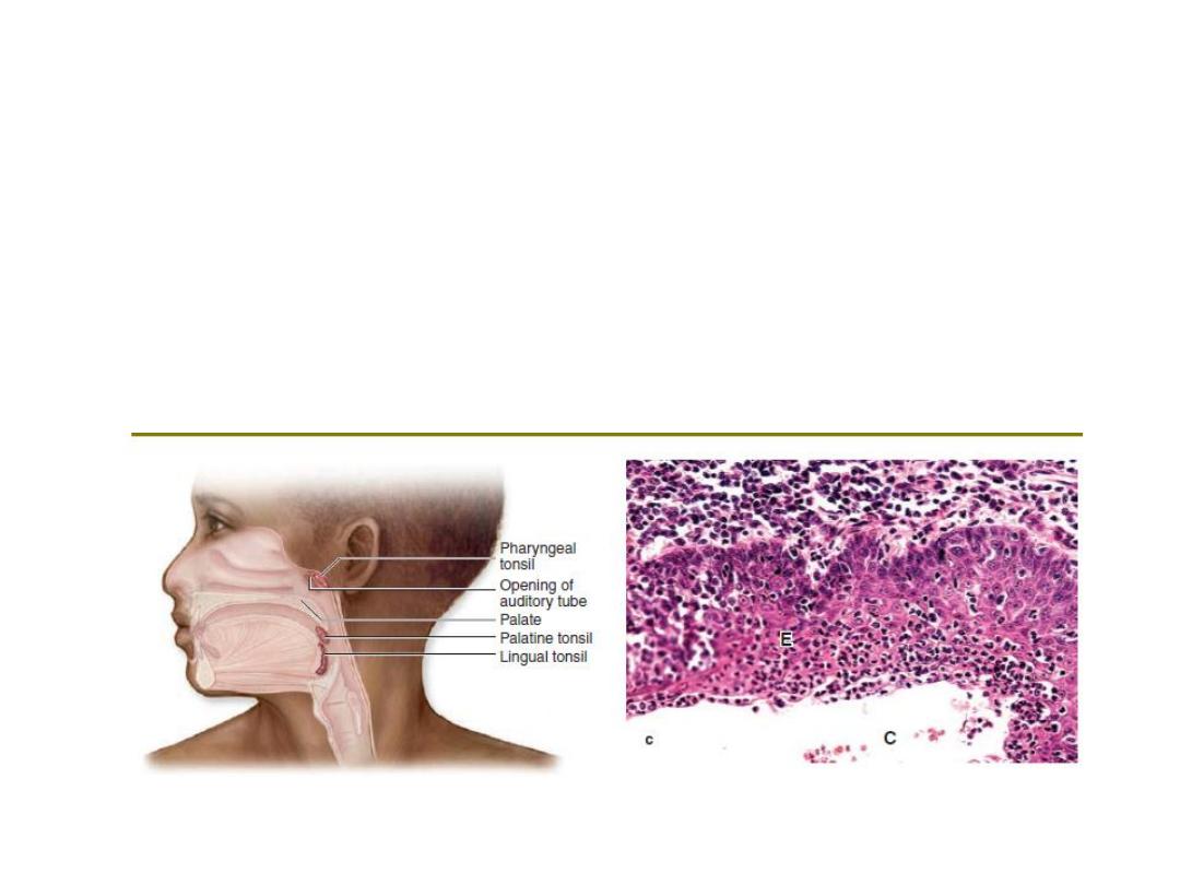
Tonsils
.
Masses of lymphoid nodules comprising the tonsils are collected in three general
locations in the wall of the pharynx. Palatine tonsils are located in the posterior lateral
walls of the oral cavity and lingual tonsils are situated along the surface of the posterior
third of the tongue. Both are covered with stratified squamous epithelium. The
pharyngeal tonsil is a single tonsil situated in the posterior wall of the nasopharynx. It is
usually covered by ciliated pseudostratified columnar epithelium typical of the upper
respiratory tract, but areas of stratified epithelium can also be observed. Hypertrophied
pharyngeal tonsils resulting from chronic inflammation are called adenoids.
TONSILS
Secondary follicles/nodules
• Contain germinal centers
• Arise when B-lymphocytes are
presented with appropriate
antigen, receive T-cell help, and
then begin proliferating as
lymphoblasts
• Lymphoblasts differentiate into
plasma cells or memory cells;
aberrant lymphoblasts undergo
apoptosis.
Ross and Pawlina, Histology: A Text and Atlas
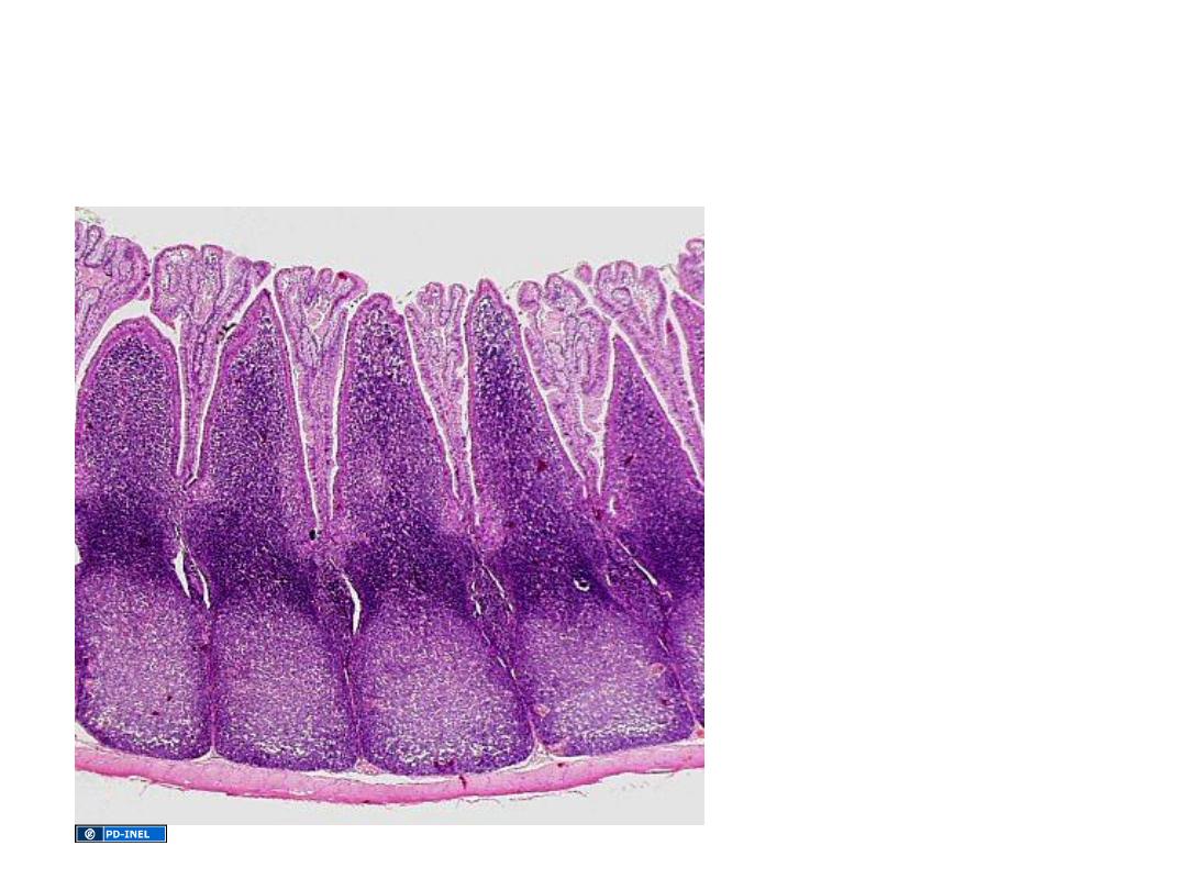
Aggregates of
lymphoid follicles in
the ileum.
Regions of extensive lymphoid infiltration:
Peyer’s patches
Source Undetermined
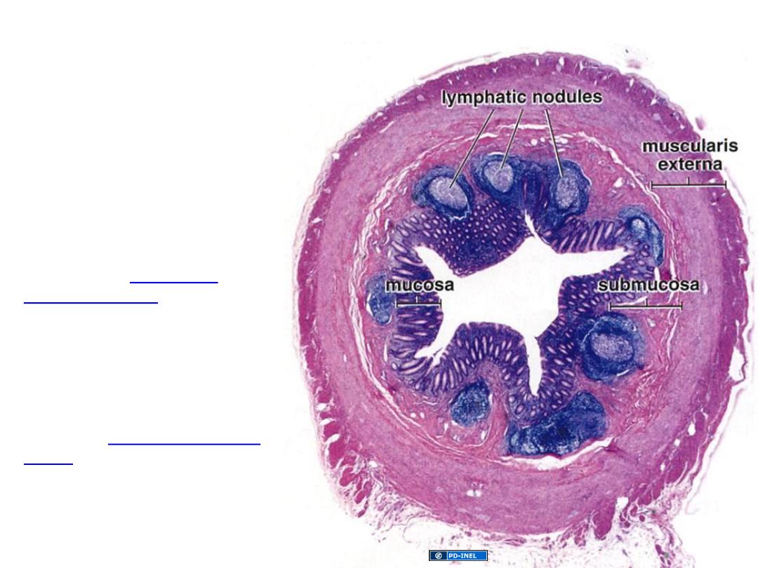
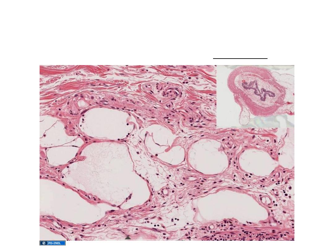
Appendix
Blind sac extending
from the caecum
• primary and secondary follicles
in lamina propria and
submucosa
• However, also a site of antigen-
INDEPENDENT differentiation
(similar to Bursa of Fabriscus is
birds)
Sorry about the various “primary” and “secondary”
nomenclature; that’s just the way it is…
Ross and Pawlina, Histology: A Text and Atlas
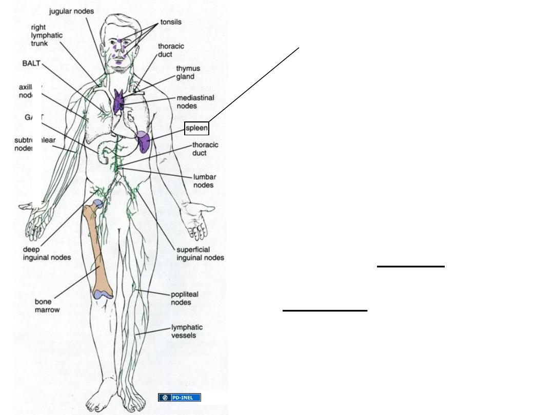
The Spleen
Filters the blood
Destroys old red blood cells
Serves as an immune organ
Divided into Red Pulp (RBC/
hemoglobin recycling)
White Pulp (responsible for
immune functions)
Ross, Fig. 14.1

•Monitoring antigens in blood
•Proliferation of lymphocytes
•Production of humoral antibodies
• Formation of blood cells in fetal life
• Removal and destruction of RBCs & platelets
• Retrieval of iron from RBC hemoglobin
• Storage of RBCs and platelets (more so in non-
human species)
Immune Functions
Of the Spleen
Hematopoietic
Functions
Of the Spleen
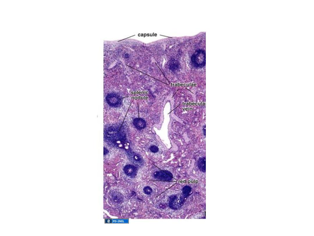
ORGANIZATION OF THE SPLEEN
Ross, 14.29
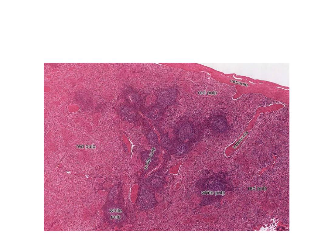
Organization of the spleen: white pulp and red pulp
White pulp: lymphatic aggregations around “central” arteries
Red pulp: cords and sinuses
As the body is exposed to antigens and the immune system rises an
immune response in the form of antibody production, lymph nodules
appear in the white pulp of the spleen.
U-M Histology Collection