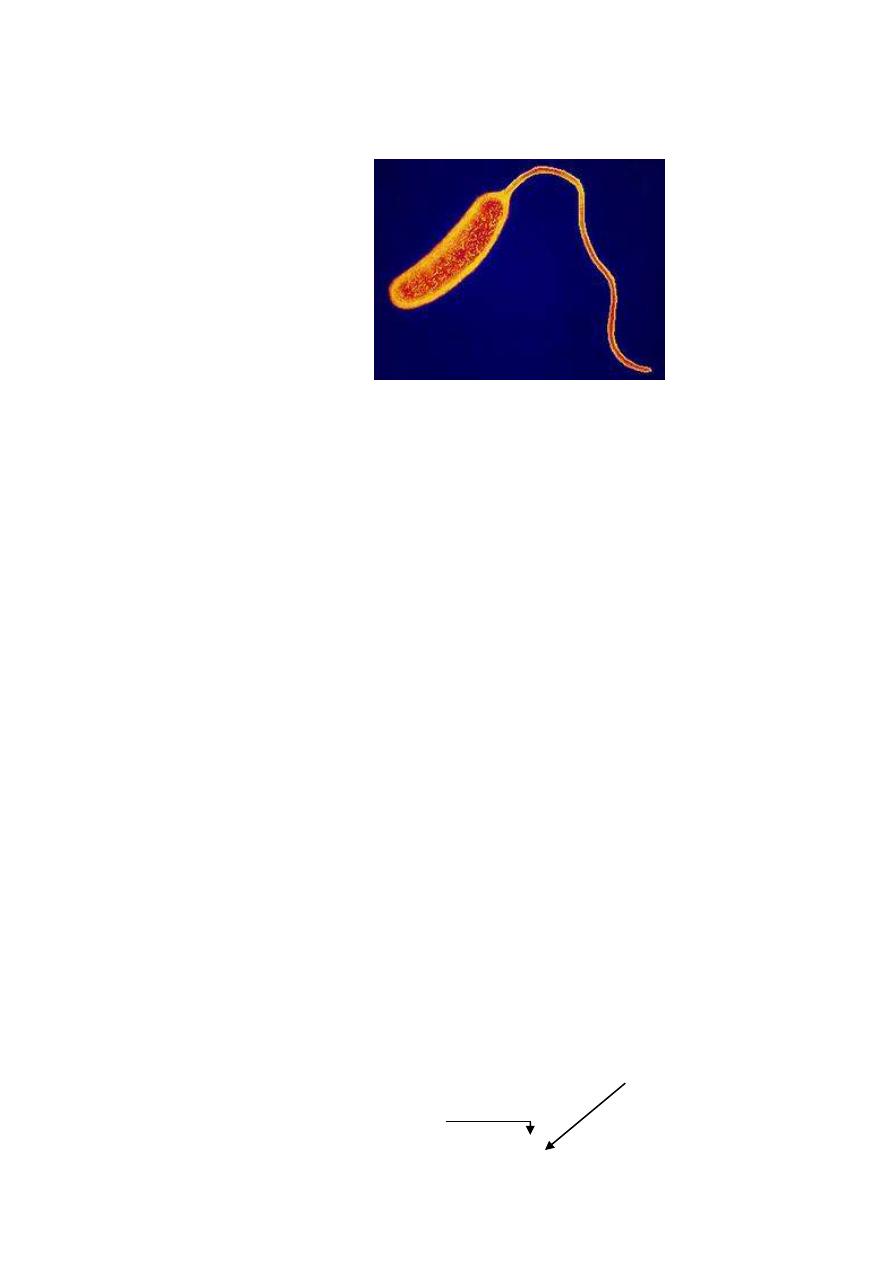
Genus Vibrio:-
Curved Gram negative rods (coma shaped), motile by polar
flagellum, they are either:
1-Halophilic
, require 8.5% and the isolated human pathogens
included are:-
-V.parahaemolyticum
self
limited
enteritis
from
contaminated seafood.
-V.vulnificus
wound infection
2-
Non - halophilic
, the most important pathogens are:-
-V. cholera
cholera
-Non – cholera Vibrio
(Non – agglutinable vibrio)
sever
enteritis
General characteristics of V. cholera
-actively motile Gram negative, curved rod.
-ferment glucose, sucrose & mannose but require few days to
ferment manitol (N L F)
-All are oxidase positive
-optimum PH required (8.5)
Indicator
bromothymol
Blue
Green
yellow
Acid production
PH
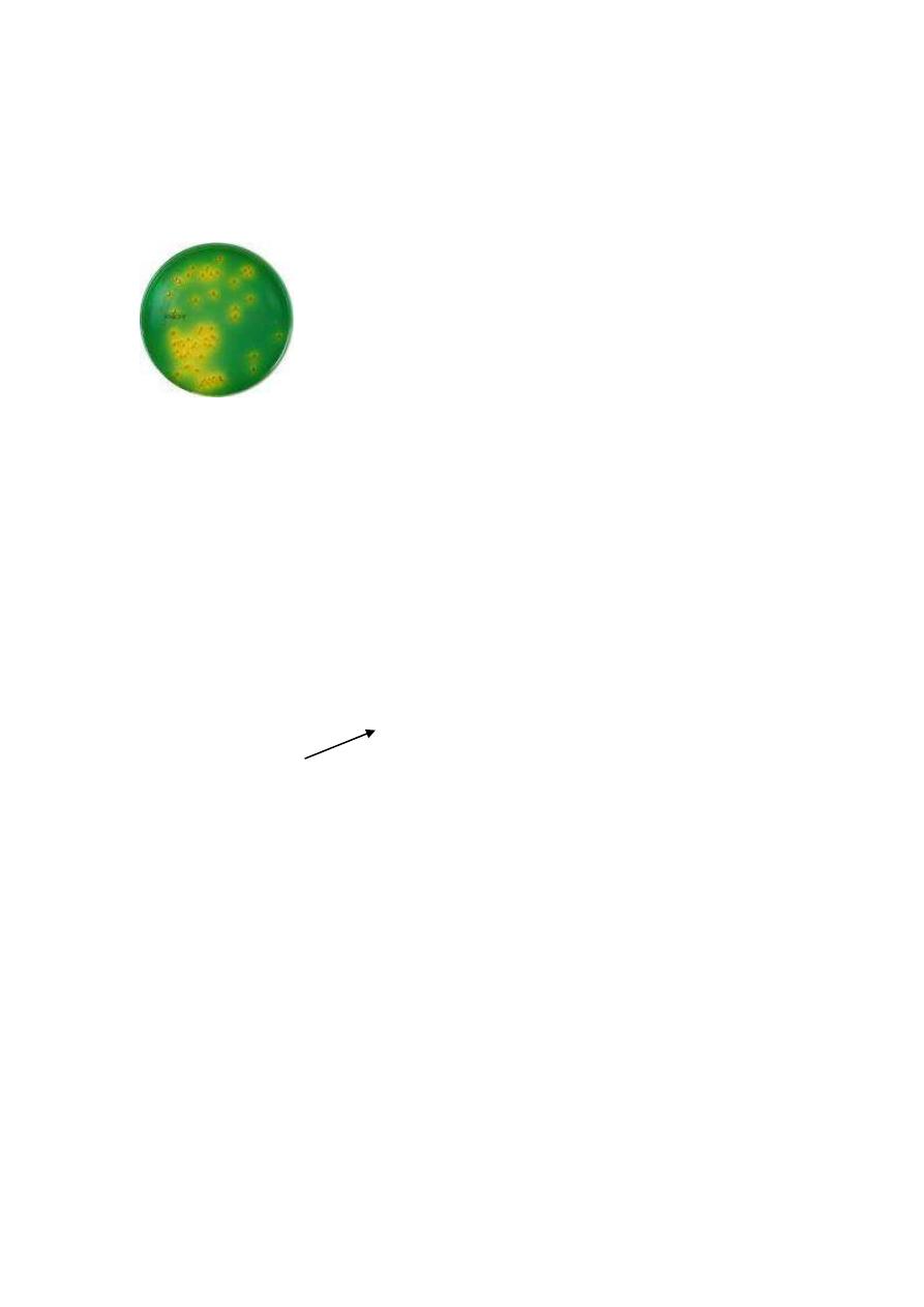
( T . C . B . S .)
alkaline media
This is selective & differential between sucrose fermenters
(V.cholera) & non fermenters (V.parahemolyticum)
-V.cholera killed by acidity and destroyed by 55
C
15 min. &
0.5%phanol.
-It reduce nitrates
nitrite with production of
Indole←
used for diagnosis of v.cholera
by (
Nitrose indol reaction
)
or (
cholera red reaction
).
This is done by isolating suspected micro – organism on
alkaline peptone water with nitrate, then after incubation
add
few drops of concentrated H
2
SO
4
if red color appear means
the presence of indole.
Antigenic structure of V.cholera
-All strains share a common, heat labile H, flagellar Ag that is of
no value.
-The O, lipopolysaccharide, there are 139 O Ag.
Thiosulfate Citrate
bile
Sucrose

The most important (O
1
), but O
139
also cause classical cholera,
and this strain shares the other non- O
1
cholera strains the
presence of a polysaccharide capsule.
The O
1
organisms can be subdivided into:
-Two biological types
Eltor
and
classical
biotype.
The Eltor show certain biologic criteria:
1-produce hemolysis.
2-positive vogas- proskauer test.
3-Resistance to polymyxin –B
-Three serological sub types
Inaba
,
Ogawa
and every rare
Hikojima
.
* Vibrio which lack O Ag called NAG vibrio which cause
cholera like disease.
V. cholerae Enterotoxin
Called (choleragen) because of its high antigenicity, but the
protective role of neutralizing Ab is not clear. This toxin
composed of 2 subunits (A&B), Epithelial ganglioside G
m1
serves as a receptor for B subunit ,and this will help entery of A
subunit, then activation of A subunit
increase level of
intracellular CAMP
Prolong hypersecretion of water &
electrolyte
causing watery diarrhea without inflammatory
cells (non– invasive) as much as 20-30 L
DAY
sever
dehydration and electrolyte imbalance.

Pathogenesis of V. cholera
Cholera is transmitted by fecal contamination of water and food
from human sources (infected or carriers), large infective dose
must be ingested (about 1 billion bacteria) because they are
sensitive to stomach acid.
The micro – organism adhere to epithelial cells of brush border
of the gut, multiply and secret enterotoxin and mucinase enzyme
which dissolves the protective glycoprotein coating the
intestinal cells.
Clinical findings
After IP (1-4) days, a sudden onset of nausea, vomiting and
sever profuse diarrhea resemble "rice water" contain mucus,
epithelial cells and large number of vibrios leading to marked
dehydration. The loss of fluid & electrolytes → acidosis &
hypokalemia → cardiac & renal failure. The mortality rate
without treatment reach 40% .
Laboratory diagnosis:-
Specimen must be collected in sterile containers (without
disinfectant) including:
- Stool or mucus flecks.
- Rectal swab or catheter.
- Vomitus (unusual).

1-Microscopical examination:
- Cram’s stain → G negative curved rods.
2-Motility test.
a-by stabbing the micro-organism with needle into semisolid
media (incubation 24hr at 37c̊ ) → the micro-organism grow
forming brush-like growth around the needle.
b-Hanging drop preparation by using slide with central
depression and put a drop of stool on cover slip and turn it over
the depression, the drop will be hanged and if examined under
dark field microscope ,the vibrio will be motile.
3-Cholera immobilization test
: a serological test done by mixing
stool specimen with specific antisera → under dark field
agglutination will appear, causing non-motile micro-organism.
4-culture
: the specimen must be inoculated at first into
enrichment media (alkaline peptone water) for 6-8hr to facilitate
their growth, then subculture on selective media (TCBS).
5-Biochmical tests
*Cholera red test.
*Oxidase test.
6-Typing of the isolated micro-organism with specific antisera
7-Serological test
could be done as retrospective diagnosis by
demonstrating the rising titer of agglutinins (one done in the first
3 days and the other after 7-10 days).
Antitoxin can be detected by ELISA.

Treatment of cholera:
-Adequate replacement of water and electrolyte to either orally
or intravenous.
-Antibiotics such as tetracycline are not necessary but they
shorten the duration of illness.
Prevention of cholera:
-Mainly by public health measures that insure a clean water &
food supply.
-The vaccine, composed of killed organisms with limited
usefulness (only 50% effective in preventing disease for 3-6
months).
-The use of protective tetracycline is effective in close contact.
-Detection of carriers is important in limiting outbreaks.