Dr. Abdul Ameer M.
Hussein
Lec. 1
DISEASE OF THE VEINS
Tues. 2 / 12 / 2014
Published by : Ali Kareem
مكتب اشور لالستنساخ
5102
-
5102
Disease of The Veins Dr. Abdul Ameer M. Hussein
2–12-2014
2
DISEASE OF THE VEINS
Venous disease refers to all conditions related to or caused by veins that
become diseased or abnormal.
Venous disease is quite common.
Mild venous disease is usually not a problem for patients, but as venous
disease worsens, it can become crippling chronic venous insufficiency.
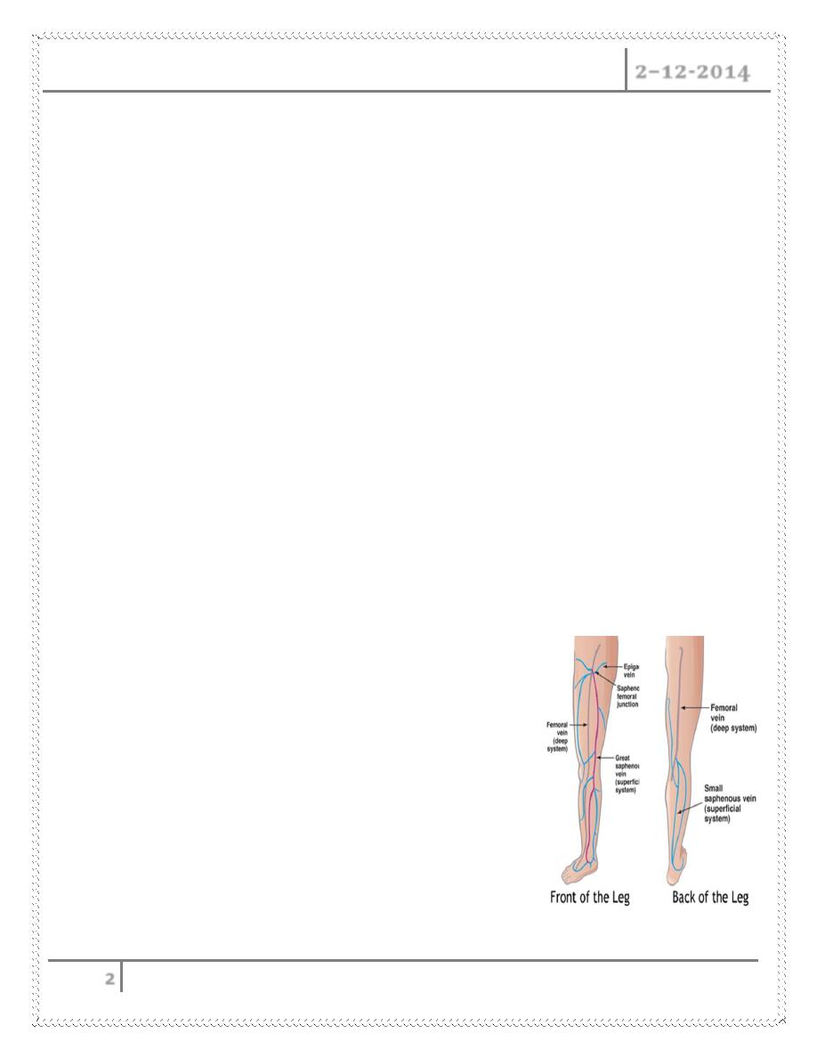
VEINS
Divided into Superficial and Deep System
Deep System
Named for by associated arteries
Found running along the arteries
Predictable anatomy
Causes most of the Morbidity
1- DVT
2- PE
3- Severe Leg Swelling
4- Ulcerations
Little Surgical interventions (IVC Filter)
Medical Management
Superficial Venous System
These are the veins we see
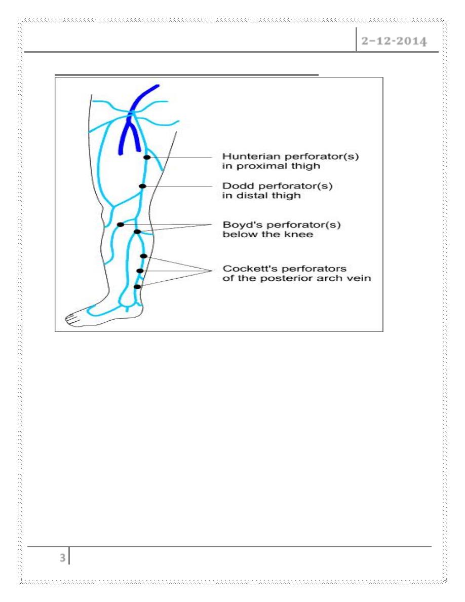
Perforators connect superficial and deep systems
Highly variable anatomy
Many unnamed branches and Tributaries
Disease of The Veins Dr. Abdul Ameer M. Hussein
2–12-2014
3
Named perforators along the greater saphenous distribution
Venous Disease
- Superficial System
Varicose Veins
Spider Veins
Venous Malformation
Venous Reflux
Leg Swelling
Venous Ulceration
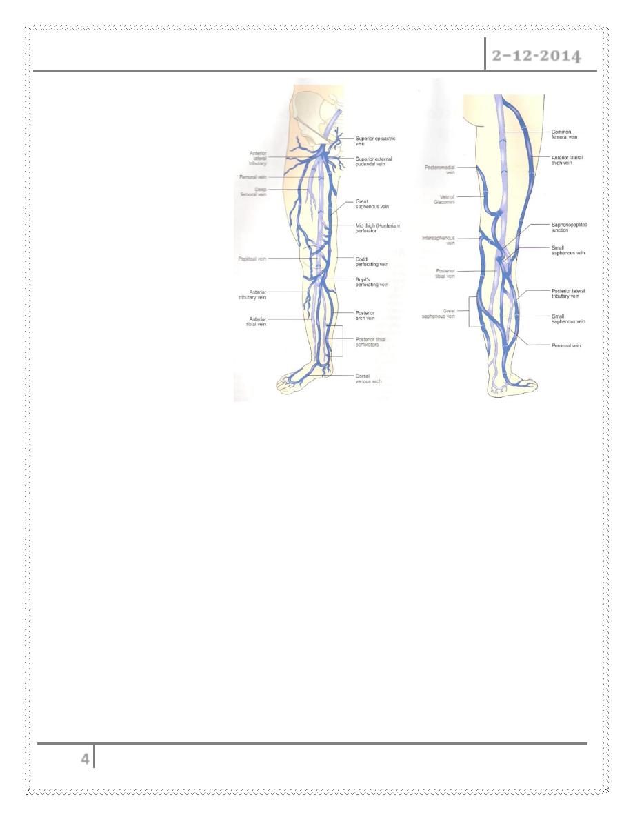
Superficial Anatomy
Disease of The Veins Dr. Abdul Ameer M. Hussein
2–12-2014
4
Deep System = Light
blue
Superficial System =
Dark blue
Complex and variable
anatomy
Physiology
Arteries deliver blood to tissue
Veins return blood to the heart
Heart is the arterial pump
o What pumps the venous blood back to the heart ?
Venous pressure is about 25mmHg at the foot
Pressure needed 80mmHg to return blood
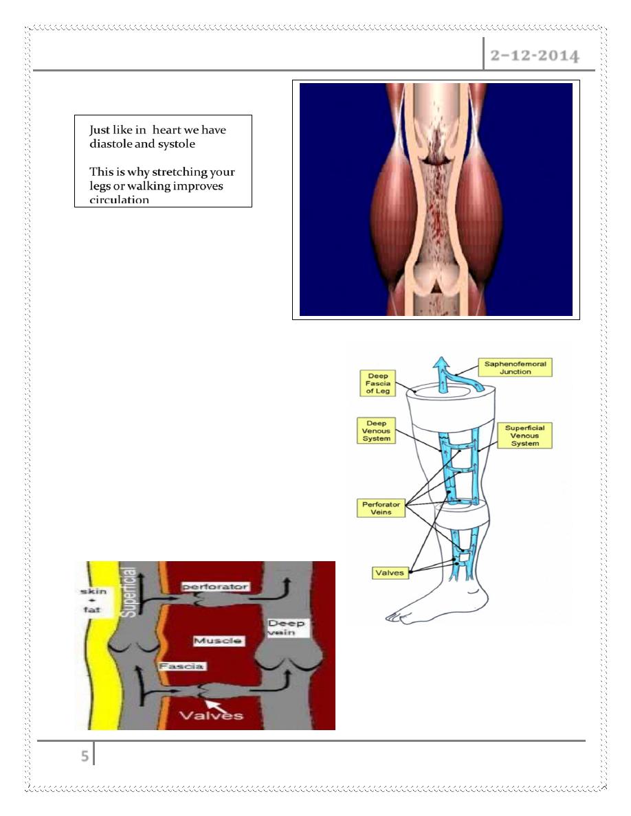
o Two unique features of veins accomplish this
Most important is one-way Venous Valves
Easily compressible by surrounding muscle (calf pump)
Disease of The Veins Dr. Abdul Ameer M. Hussein
2–12-2014
5
Calf Muscle Pump
Normal venous flow in the Leg
Normal Flow
Superficial veins drain into the deep
veins
From the foot up to the heart
Superficial vein disease always starts with
abnormal valves and interruption to normal
flow called venous reflux
Disease of The Veins Dr. Abdul Ameer M. Hussein
2–12-2014
6
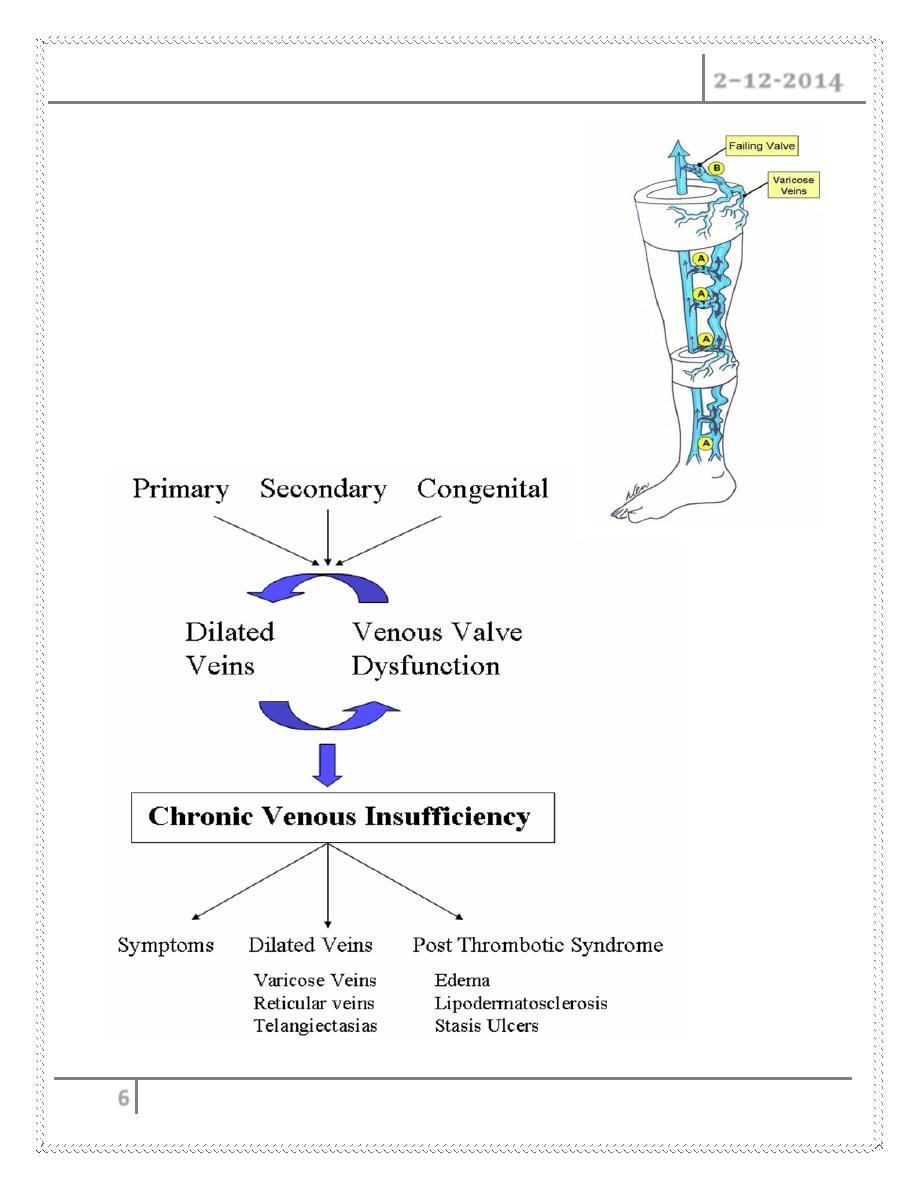
Abnormal flow = Venous Reflux
Damaged Valves
1- Blood flows to the skin
2- Blood is pushed distally and proximally
3- Close loop recirculation
4- Blood is retained in the leg
Increased volume of blood (heaviness Fatigue)
Increased venous pressure
Veins Dilate (varicose veins)
Causes of Venous Reflux
Disease of The Veins Dr. Abdul Ameer M. Hussein
2–12-2014
7
Symptoms of venous reflux
Leg Fatigue
Leg Heaviness
Itching and pain along veins
Varicose Veins
Spider veins (not always 2
nd
to reflux)
Leg swelling( think DVT 1
st
)
Skin Discoloration (lipo dermatosclerosis)
Venous ulceration
Varicose Veins
Definition: Swollen and enlarged veins that you can see just under the surface of
the skin. These veins usually occur in the legs, but they also can form in other parts
of the body.
Located in the Subcutaneous(between skin and fascia)
Remember this is only a manifestation of the underlying disease
Mild Disease is cosmetic issue
Advanced Disease significant medical problem
i) -
Pain
ii) Swelling
iii) Ulcerations
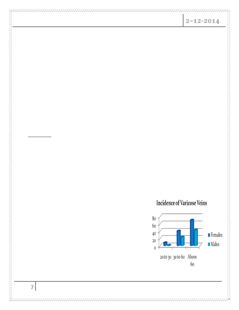
Incidence Increases with age
Females to male 3 to 1
50% of the population will affected in their
life time

Disease of The Veins Dr. Abdul Ameer M. Hussein
2–12-2014
8
Spider Veins
The proper term is Telangiectasia
These are non raised dilated veins located in the dermis (deep layer of the
skin)
Single layer endothelium, minimal muscle
Can be Red or Blue in color depending on the origin
Do not cause major medical complications
Appears earlier than varicose veins
More common in females
Reticular Veins are lager feeding veins
Etiology : Multifactorial
Venous Hypertension associated with varicose veins
Congenital: vascular nevi, neonatal hemangiomatosis.
Collagen Vascular Disease: lupus
Hormonal factors: pregnancy, estrogen therapy, topical steroids
Trauma: contusion, incisions
Infections
Venous Stasis Ulcers
Etiology
1- Venous Hypertension
Venous reflux
DVT
Varicose veins
2- Edema
3- Biological factors
Leakage of proteins impedes O2 diffusion

Disease of The Veins Dr. Abdul Ameer M. Hussein
2–12-2014
9
Aggregation of white cells
Block capillary flow
Release on inflammatory proteins
Symptoms :
Itching
Discoloration
Redness or swelling in the affected area
Pain or a feeling of heaviness in the affected leg
More common in elderly population
Venous ulcerations 50% are non healing ulcers
Differential Diagnosis
1- Arterial ulcers
2- Malignancy : basal and squamous cell, lymphoma
3- Infections: HIV, fungal
4- Collagen vascular disorders
5- Lymphatic obstruction
Diagnosis of venous disease
Physical exam
Appearance
Trendelenburg test
Palpation
Hand Doppler
Duplex Examination
DVT
Size of veins
Map out superficial veins
Locate the site of reflux
Find refluxing perforators
Disease of The Veins Dr. Abdul Ameer M. Hussein
2–12-2014
10
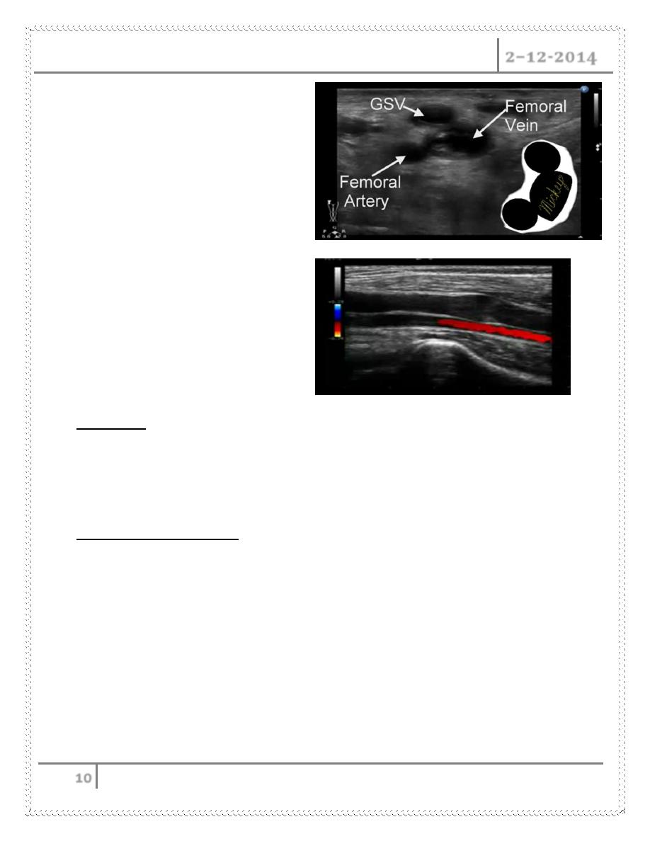
Duplex Anatomy
Locate GSV Junction(FSJ)
Look for Mickey's
Normal venous flow
Look at valve
Venous flow is opposite the artery
Treatments
Conservative management techniques
Ultrasound guided Sclerotherapy with foam
Conservative management
This can offer relief from burning, itching, and general discomfort caused by
large, unhealthy veins.
requires minimal lifestyle changes and can alleviate symptoms without
requiring a medical procedure
Common techniques include :
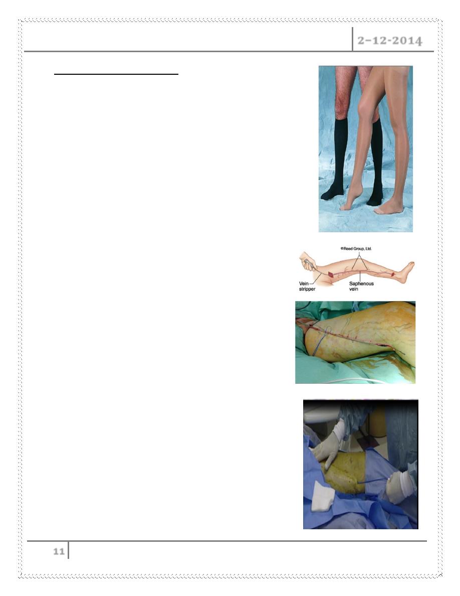
o Compression stockings.
o Periodic elevation of legs during the day.
o Medication.
Disease of The Veins Dr. Abdul Ameer M. Hussein
2–12-2014
11
Treatment of Varicose Veins
Conservative management
o Exercise
o Leg elevation
o Compression stocking
Surgical treatment
o Standard Ligation and stripping
o Phlebectomies
Minimally invasive procedures
o Laser Ablation
o Radio Frequency ablation
o Sclerotherapy
Surgical ligation and Stripping
Standard treatment for a century
General anesthesia
Pain
Long recovery
Some complications
Good cosmetic results
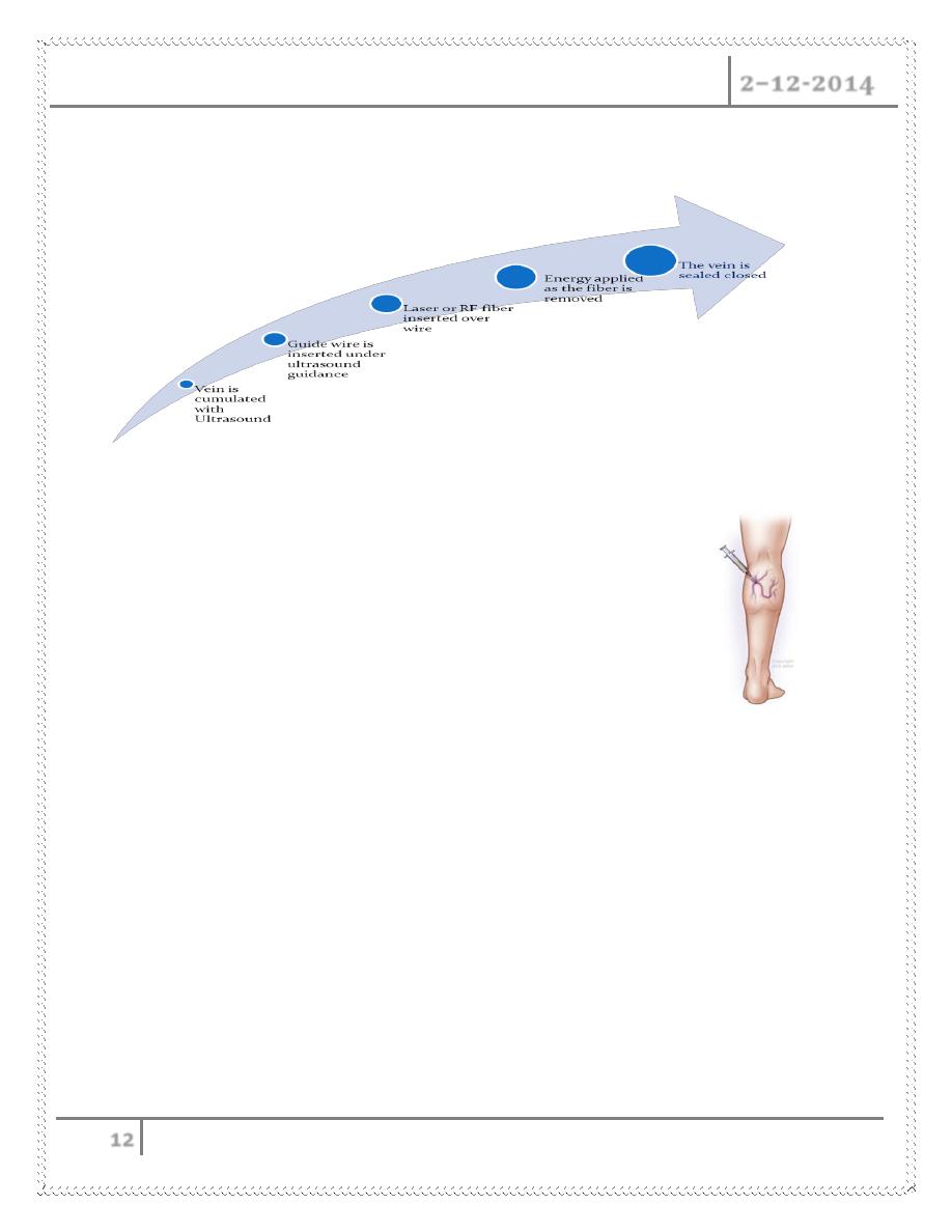
Vein Ablation
Laser Ablation (EVLA or EVLT)
Uses light to heat the vein
Radio Frequency
Uses radio frequency to heat the vein
Office based procedure
Done under local anesthesia
One needle puncture at the level of the knee
Takes about 1 hour
Disease of The Veins Dr. Abdul Ameer M. Hussein
2–12-2014
12
Patient resumes normal activity same day
Sclerotherapy
Cumulate vein with needle
Inject Sclerosing Solution
o Sotradecol (Sodium tetradecyl sulfate)
o Pilodocanol
o Hyper tonic Saline
o Foam
Intravenous injection causes intima inflammation and thrombus formation
Sclerotherapy Use
Neovascularization
Perforators
Spider veins
Reticular veins
GSV : has high recurrence rate
Done By
Ali Kareem