
1
PHYSIOLOGY
Dr. Basim Mohamad Awan Lecture 8
CELLS AND LAMINAE
OF THE CEREBRAL CORTEX
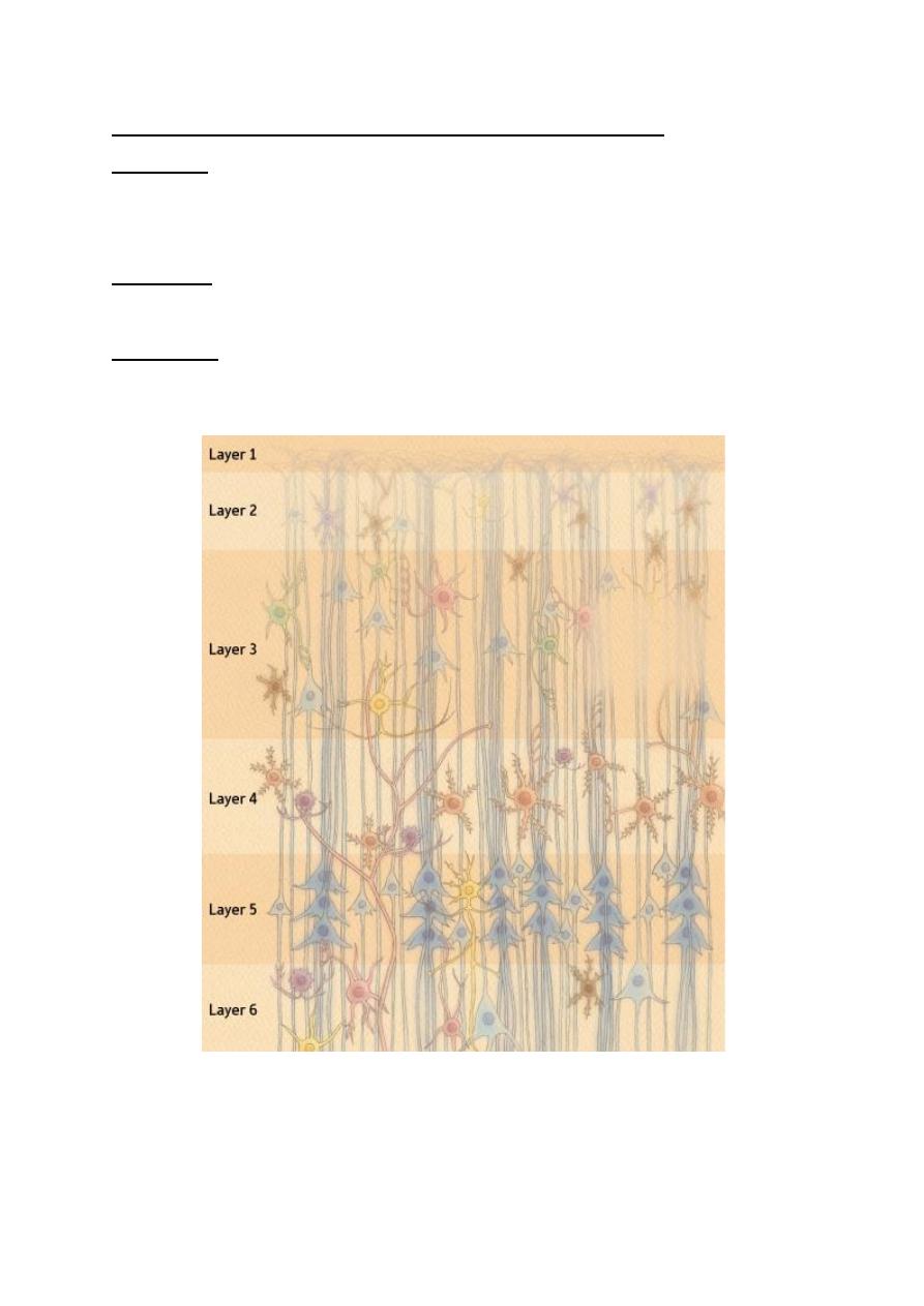
The cerebral cortex is arranged in six distinct laminae according to the
histological architecture of each lamina (Fig. 8-1). These laminae are labeled
from outside inwards as lamina I to lamina VI. There are generally two types of
cells in the cerebral cortex.
1. The pyramidal cells; which have a pyramidal shape and are found in layers
II, III, V and VI. The special feature of these cells is that their axons leave the
cortex and these axons terminate in one of the following destinations:
a. Association fibers: which originate from lamina II and terminate in other
cortical areas on the same side?
b. Commissural fibers; which originate from lamina III and terminate in
cortical areas on the opposite side.
c. Pyramidal tracts; which originate from lamina V and terminate in
motor nuclei in the brainstem and spinal cord.
d. Cortical projection fibers; which originate from lamina VI and
terminate in subcortical structures; e.g. corticothalamic projections.
2. The stellate cells: which have a star-like shape and are found mainly in layer
IV? Their axons terminate within the cortex. They are the final cortical sensory
neurons which are responsible for conscious perception of different sensations.
2
FUNCTIONS OF THE DIFFERENT CORTICAL LAMINAE
Lamina I: It consists of interconnecting nerve fibers which connect different
areas of the cortex. These fibers arise from all other laminae in the cortex. It also
contains fibers from the nonspecific thalamic nuclei.
Lamina II: Contains pyramidal cells which send association fibers to other areas
of the cortex on the same side.
Lamina III: Contains pyramidal cells which send commissural fibers to the cortex
on the opposite side.
Figure 8-1: The types of cells in different layers of the cerebral cortex and
their connection.

3
Lamina IV: Consists of stellate cells which receive input fibers from the specific
sensory nuclei of the thalamus. These cells are the final sensory neurons which are
responsible for the conscious perception of different sensations.
Lamina V: Consists of pyramidal cells whose axons descend as the motor
pyramidal tracts.
Lamina VI: Consists of pyramidal cells whose axons form corticofugal fibers
that project to subcortical structures (e.g. corticothalamic projections).
4
MOTOR FUNCTIONS OF THE CEREBRAL CORTEX
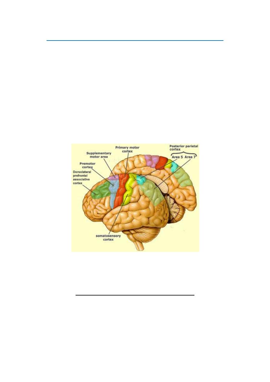
All voluntary movements involve the conscious activity of the "motor
cerebral cortex of the brain. The motor cortex lies in front of the central
sulcus and occupies most of the frontal lobe (Fig. 8-2). It is divided into 4
separate areas:
[I] THE PRIMARY MOTOR AREA
[II] THE PREMOTOR AREA
[III] THE SUPPLEMENTARY MOTOR AREA
[IV] THE MOTOR ASSOCIATION AREA
Figure 8 - 2: The lateral and medial surfaces of the left cerebral
hemisphere showing the motor cortex
[I] THE PRIMARY MOTOR AREA (Area 4)
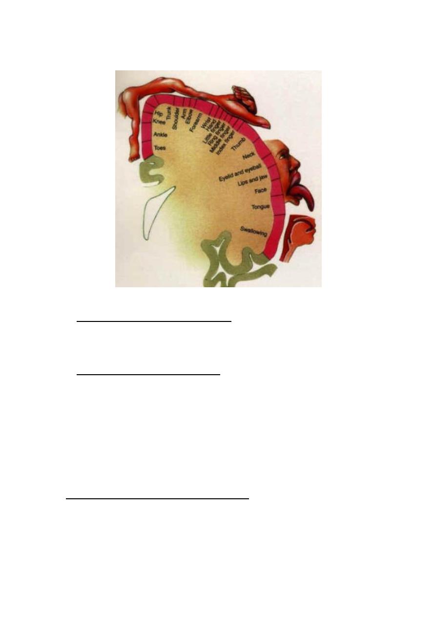
The primary motor area of the cerebral cortex (area 4 in Brodmann
classification) occupies the precentral gyrus in the frontal lobe. The body is
topographically represented in an inverted and crossed manner (Fig. 8-
1). The upper part of the face is bilaterally represented and the area of

5
representation of each part is proportionate to the degree of fine
movements in this part, e.g. hands and muscles of speech are represented
by large areas, whilst the trunk is represented by a small area. The
primary motor area contains two types of neurons:
1. Dynamic neurons: Which discharge at high frequency for a short time
at the beginning of contraction causing the initial development of force?
2. Static neurons: Which discharge at a much slower frequency but for
much longer time to maintain contraction for as long as required.
FUNCTIONS OF THE PRIMARY MOTOR AREA
1. Initiation of voluntary, fine, discrete (separate) movements of the distal
parts of the body e.g. hands and fingers.
2. Facilitation of stretch reflex; i.e. facilitation of skeletal muscle tone and
tendon jerks.
[II] THE PREMOTOR AREA (Areas 6, 8 and 44)
The premotor area of the cerebral cortex (mainly area 6 of Brodmann
classification, but it also includes areas 8 and 44) lies immediately
anterior to the primary motor area. The topographic representation of the
body is nearly the same as in area 4. The premotor area includes some
specialized areas with specific functions (Fig. 8-1):
1. Broca's area of speech (area 44):
This area lies at the upper border of the lateral sulcus in front of the
primary motor cortex. It stores the motor programs for verbalization.
Its damage leads to inability to speak whole words except simple ones as
"yes" or "no".
2. Eye field area (area 8):
It lies above Broca's area. It directs the eyes voluntarily towards any
desired object. It also controls the blinking movements of the eye lids.
Its damage leads to locking of the eye on objects.
6
Figure 8-3: Body representation in the primary motor cortex
3. Head rotation area (part of area 6):
Lies immediately above area 8 and works in close association with it. It
directs the head towards different objects.
4. Hand skills area (part of area 6):
This area lies immediately anterior to the primary motor area for hands
and fingers. It stores the motor programs for skilled hand movements; e.g.
sharpening a pencil or peeling a potato. Damage of this area leads to
"motor apraxia"; i.e. inability to do skilled hand movements. This area
includes the "Exner center" for writing skill.
FUNCTIONS OF THE PREMOTOR AREA
1. Initiation of gross movements that involve groups of muscles to
support and' facilitate fine movement, e.g. fix the shoulders and arms at a
certain position so that the hands and fingers can do skilled movements
7
(e.g. threading a needle).
2. Weak inhibition of the stretch reflex. It tends to decrease the skeletal
muscle tone.
3. A center for rotation of the head towards objects.
4. A center for skilled hand movements.
5. A center for verbalization of words (area 44).
6. A center for voluntary eye and lid movements (area 8),
7. Inhibition of the grasp reflex.
THE MECHANISM OF ACTION OF THE PREMOTOR AREA
The premotor area acts by activating the corresponding nearby motor
cells in the primary motor area. This occurs in two ways:
First: directly through direct projection fibers to area 4.
Second: indirectly through circuits starting from the premotor area to the
corpus striatum of the basal ganglia, to the thalamus, then back to
terminate in area 4 of the cerebral cortex.
[III] THE SUPPLEMENTARY MOTOR AREA
The supplementary motor area (Fig. 8-1) is an extension of area 6 in the
medial surface of the cerebral hemisphere. Accordingly, it is also called
"the medial area 6". Topographic representation of the body is bilateral
and in a horizontal position; head anteriorly and legs posteriorly. This
area is connected by projection fibers to the premotor and the primary
motor areas.
The supplementary motor area supplements the functions of the premotor
area in producing positioning and fixation of the different parts of the
body as a background for finer hand or feet movements; e.g. the
coordinated movement of the trunk with the hand and feet during boxing.
This area is involved in preparation for movements before they start. It

8
shows electrical potentials shortly before the start of the movement
(readiness potential) and increase in metabolism and local blood flow on
the mere intention to move a muscle,
[IV] THE MOTOR ASSOCIATION AREA
This area occupies the frontal lobe in front of the premotor area (Fig. 8-
1). It receives input signals from the parieto-tempro-occipital
association area and projects output signals to the motor and premotor
areas through the caudate and putamen circuits.
FUNCTIONS OF THE MOTOR ASSOCIATION AREA
1. Setting off goals and aims of movement and then taking the
decision to start the movement. Once the decision is taken, signals are
sent to the basal ganglia (the motor consultant of the cerebral cortex) to
activate programs or set plans for the movement. The motor plans and
programs are then fed to the motor and premotor areas.
2. Elaboration of thoughts; i.e. carrying out prolonged thought
processes which involve setting of plans and developing new constructive
ideas. This is the area of the brain which is concerned with deep quiet
thinking during rest. A lesion in this area abolishes creativity and
planning for the future.

9
CONNECTIONS OF THE MOTOR CORTEX
AFFERENT CONNECTIONS
A. FROM OTHER CORTICAL AREAS
• Of the same side: from somatic sensory areas, visual and auditory
areas.
• Of the opposite side: from the contralateral motor cortex to connect
corresponding points on both sides.
B. FROM THE THALAMUS
• The ventrobasal complex (VPL and VPM nuclei); it receives specific
sensory signals.
• The intralaminar nuclei; it receives nonspecific signals to arouse the
cortex.
• The ventral and medial nuclei; it receives impulses coming from the
cerebellum and basal ganglia.
EFFERENT CONNECTIONS
A. Pyramidal tract fibers to motor nuclei in the brainstem and spinal
cord.
B. To the basal ganglia (the caudate and putamen circuits).
C. To the red nucleus of the midbrain.
D. To the cerebellum through the middle cerebellar peduncle
(cortico-ponto-cerebellar fibers).
E. To adjacent cortical areas to inhibit any unwanted discharge
(lateral inhibition). These inhibitory fibers are collaterals from the axons
of the giant Betz cells. This helps to sharpen the outgoing signals.