
1
Physiology
Dr. Basim Mohamad Alwan Lecture (2)
PROPERTIES OF SYNAPTIC TRANSMISSION
Transmission of signals across the synapses is characterized by:
1. FORWARD DIRECTION
Transmission in synapses is unidirectional, i.e. from the presynaptic to
the postsynaptic neuron, not the reverse. This is because the
postsynaptic neuron cannot release a chemical transmitter at the
synapse. So, the synapse acts as a
unidirectional "valve"
to keep the
flow of signals between neurons always in the right direction.
2. SYNAPTIC DELAY
When an impulse reaches a nerve terminal, it takes a delay time of
0.5-1.0 ms to pass across the synapse to the postsynaptic neuron. This
time is taken for the release of the chemical transmitter, its diffusion in
the synaptic extracellular fluid and then synaptic cleft, activation of
receptors, induction and summation of postsynaptic potentials.
3. SYNAPTIC AFTERDISCHARGE
After discharge is the persistence of output signals after stoppage of
the input signals. Synaptic afterdischarge occurs at some synapses
because of the delay of inactivation of the chemical transmitter. So, an
impulse conducted by a presynaptic neuron may produce more than
one impulse in the postsynaptic neuron. The duration of the synaptic
afterdischarge is longer if the chemical transmitter released by the

2
presynaptic neuron is a long acting one (substance P).
4. FATIGUE
Fatigue is the decline in response caused by prolonged activity. For a
synapse, fatigue is the decline in the response of the postsynaptic
neuron after a long period of high frequency stimulation of the
synapse (> 60 Hz). It is manifested by prolongation of the synaptic
delay, then failure to transmit some or all of the impulses across the
synapse.
The synapse is an early site of fatigue in the reflex arc and the fatigue of
the neural synapses is caused by:
i. Exhaustion of the chemical transmitter in the presynaptic terminals
which is the main cause.
ii. Inactivation of some postsynaptic receptors due to accumulation of
metabolites.
iii. Marked increase of the intracellular Ca
2+
in the postsynaptic
neuron. This high Ca
2+
level opens K
+
channels so K
+
efflux and
hyperpolarization of the postsynaptic membrane decreasing the
excitability of postsynaptic neuron.
Fatigue is a protective mechanism against excess neuronal activity
Fatigue is the most important means by which the excess excitability
of an epileptic circuit is cut off and stopped. This leads to spontaneous
ending of the epileptic fit (normal protective mechanism).

3
5. SYNAPTIC POTENTIATION (FACILITATION)
This is an increase in the postsynaptic response caused by previous
presynaptic stimulation. It may be a short-term or a long-term
potentiation.
i. SHORT TERM (POST TETANIC) POTENTIATION
This occurs after a short period of low frequency stimulation of the
synapse (< 60 Hz). It is caused by an increase in the intracellular Ca
2+
level in the presynaptic neuron, which increases the release of the
transmitter. Short-term potentiation lasts for few seconds up to few
minutes.
ii. LONG TERM POTENTIATION (LTP)
This occurs after a short period of high frequency stimulation (>60
Hz). Long term potentiation is caused by the release of arachidonic
acid from the postsynaptic neuron which acts on the presynaptic
neuron to release more of the transmitter (Glutamate).
Long-term potentiation occurs in several parts of the CNS,
particularly in the hippocampus and it plays an important role in
memory and learning.
6. SYNAPTIC DEPRESSION (HABITUATION)
Habituation is the gradual decrease in the postsynaptic response
when stimulation of the presynaptic neuron is frequently repeated.
With complete habituation, the postsynaptic response may disappear
altogether.
Habituation is due to inactivation of Ca
2+
channels in the presynaptic
neuron which decrease in intracellular Ca
2+
so release of smaller
amount of transmitter from the presynaptic terminals.
4
Habituation could be short-term or long-term depending on how many
times the stimulus is applied. It is an important mechanism of
learning, as it enables the subject to ignore insignificant stimuli.
Habituation of synapses is different from adaptation which occurs in
excitable tissues. Adaptation is the decline in response to a constant
maintained stimulus.
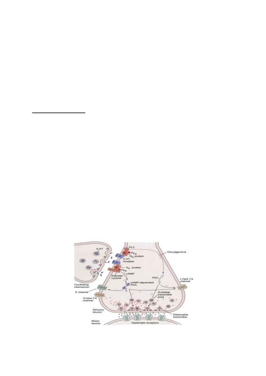
7. SENSITIZATION
Sensitization of a synapse is the potentiation of the postsynaptic
response to a certain stimulus by coupling the stimulus to another
intense (usually painful) stimulus (fig.2-1).
The terminal which conducts the intense or painful stimulus is called
a facilitator terminal which relays on the presynaptic sensory
terminal. The facilitator terminal stimulates the presynaptic sensory
terminal lead to prolonged action potential in the sensory terminal and
more Ca
2+
influx into the sensory terminal so release of more
transmitter and potentiated postsynaptic response result.
Sensitization is an important mechanism in memory and learning.
Figure 2-1: The mechanism of synaptic sensitization.

5
8. EFFECT OF pH
Alkalosis enhances synaptic transmission. A rise of arterial blood pH
from 7.4 to 7.8 leads to increased cerebral excitability and
convulsions.
Acidosis depresses synaptic transmission. Breathing of air with high
Co
2
level will lead to hypercapnea and acidosis and then depression of
synaptic transmission in the brain resulting in drowsiness and sleep or
even anesthesia. A drop of arterial pH down to 7.0 produces coma
because of failure of synaptic transmission between various neurons in
the brain.
9. EFFECT OF HYPOXIA
Hypoxia depresses synaptic transmission and prolongs reflex time due
to accumulation of acidic metabolites.
10. EFFECT OF DRUGS
Caffeine, theophylline and theobromine which are found in coffee,
tea enhance synaptic transmission. They increase neuronal excitability
by lowering the threshold of excitation.
Strychnine enhances synaptic transmission by blocking the action of
central inhibitory transmitters (e.g. glycine).
Hypnotics and anesthetics depress synaptic transmission by
decreasing neuronal excitability. They stabilize the cell membrane by
increasing the resting membrane potential (hyperpolarization).
6
PROCESSING OF SIGNALS IN THE CNS
Nerve signals (impulses) enter the CNS to be directed to various
neuronal pools
(collection of neurons). In the neuronal pools, input
signals are processed, and output signals emerge out to proceed to
specific destinations.
THE DISCHARGE ZONE AND THE FACILITATED FRINGE
(THE LIMINAL ZONE AND THE SUBLIMINAL FRINGE)
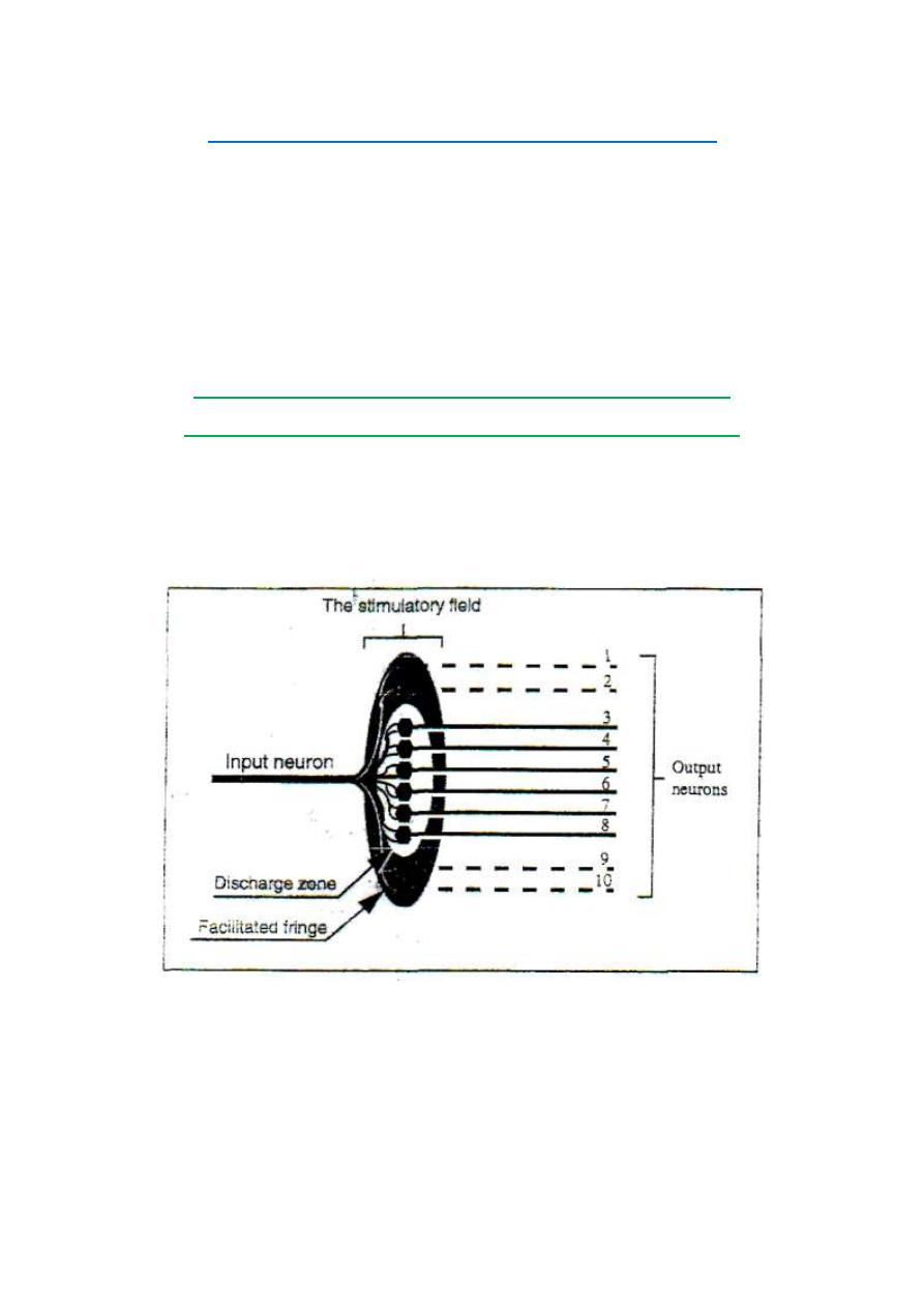
When an impulse in an excitatory input neuron reaches the neuronal pool,
it stimulates a group of neurons which form the "stimulatory field'' of
this neuron (fig.2-3).
Figure 2-3: The stimulatory field of an input neuron.
At the middle of the field, stimulation reaches a liminal level
(threshold) and the neurons in this zone discharge impulse. The zone
where neurons discharge impulse is called the discharge zone or the

7
liminal zone of the input neuron.
Around the discharge zone, there is a circular zone (a fringe) in which
the neurons are only facilitated without reaching the
liminal
firing
level. This zone is called "the facilitated fringe" or "the subliminal
fringe" of the input neuron.
An impulse in an inhibitory input neuron produces an "inhibitory
field'' with maximum inhibition at its center.
FORMS OF SIGNAL PROCESSING IN THE NEURONAL POOLS
Signal processing in the neuronal pools takes one of the following
forms:
[I] Convergence.
[II] Divergence.
[III] Prolongation.
[IV] Shortening.
[V] Sharpening.
[I] CONVERGENCE OF SIGNALS
Convergence is the direction of signals from several input neurons to
excite a single output neuron. There are two main types of
convergence in the neuronal pools.
1. CONVERGENCE FROM A SINGLE SOURCE
This is important because no neuron can be excited by a single input
terminal. So, convergence must occur on neurons to excite those
neurons (fig.2-4). The spatial and the temporal summation of
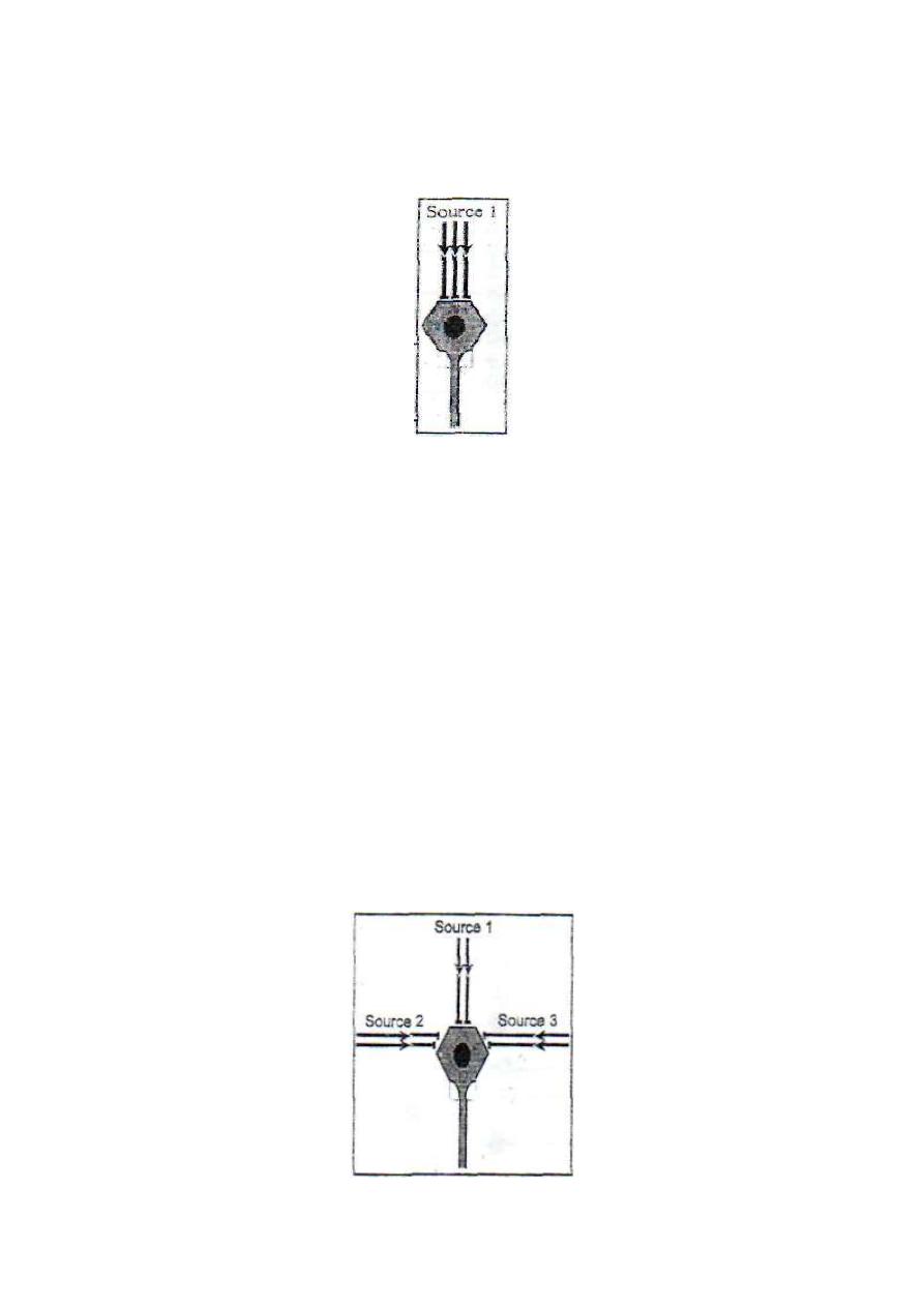
8
postsynaptic potentials from the multiple input terminals build up a
threshold membrane potential to excite the neuron.
Figure 2-4: convergence from single
source
2. CONVERGENCE FROM .MULTIPLE SOURCES
This is important because it enables neurons of the neuronal pool to
receive signals from different sources (fig. 2-5).
The effect produced will be the resultant of all the inputs whether
excitatory or inhibitory; e.g. motor neurons of the ventral horn of the
spinal gray matter receive inputs from the pyramidal and extra
pyramidal tracts and from the afferent fibers of the stretch reflex and
several intermediate neurons. All these input neurons influence the
contraction and relaxation of the skeletal muscles.
Figure 2-5: Convergence from multiple sources.
9
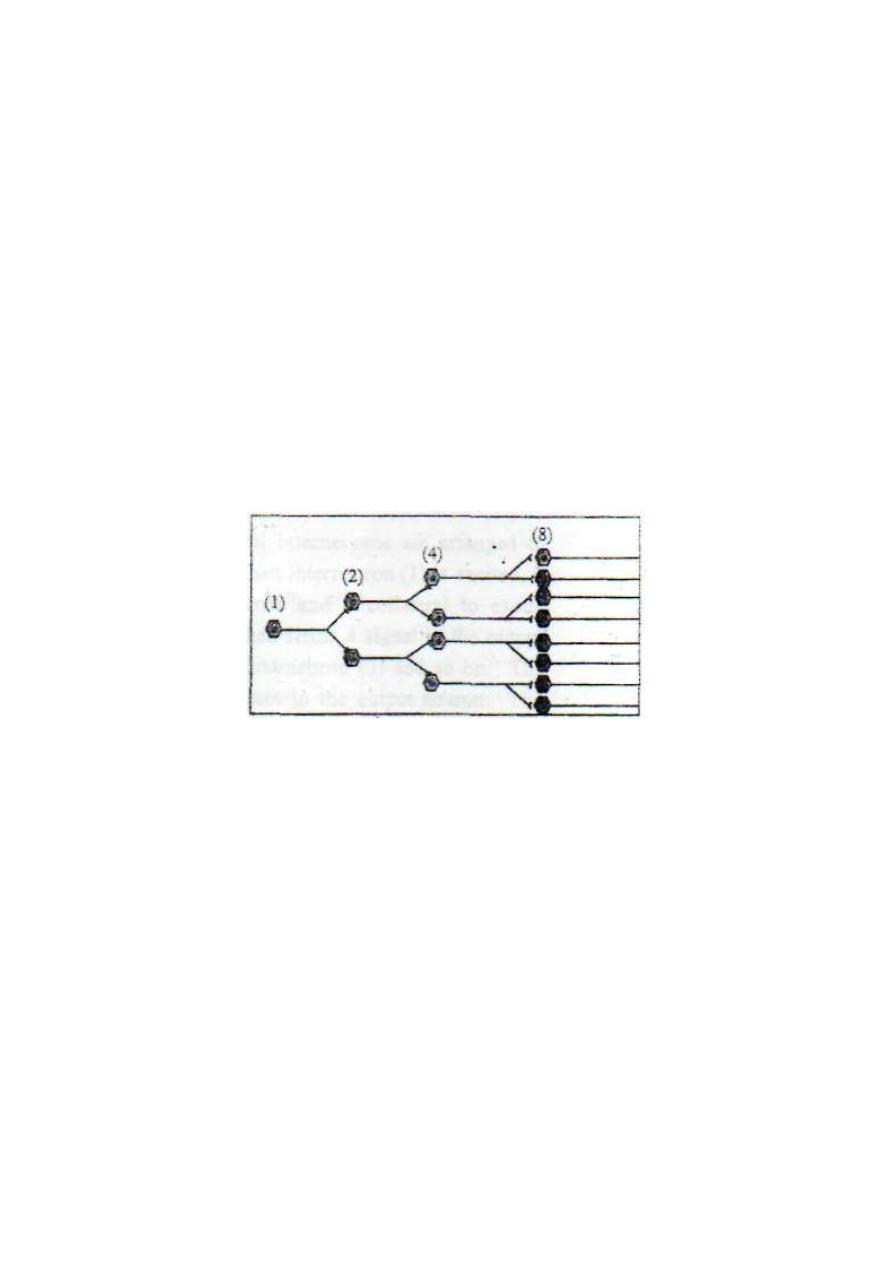
[II] DIVERGENCE OF SIGNALS
Divergence is the spread of a signal from one input neuron into more
than one output neuron. There are two main types of divergence in the
neuronal pools:
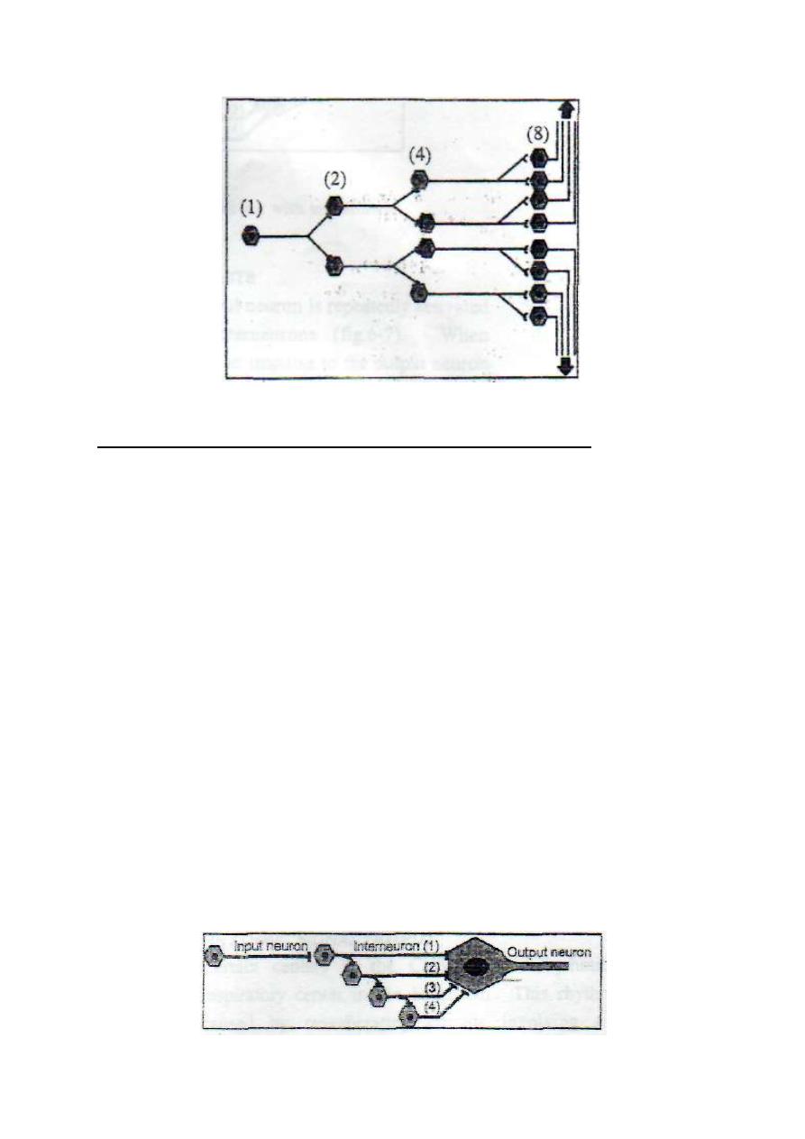
1. DIVERGENCE IN THE SAME BATHWAY
This leads to spread of the signal into an increasing number of
neurons as it passes from one order of neurons into another (fig. 2-6).
It may be called an "amplifying divergence". It occurs, for example,
in the pyramidal tract where a single pyramidal neuron in the motor
cerebral cortex can excite up to 10,000 muscle fibers.
Figure 2 - 6: Amplifying divergence.
2. DIVERGENCE INTO MULTIPLE PATHWAYS
This leads to spread of the signal into two or more separate directions
from the pool (fig .2-7). It may be called a "diversifying
divergence". It occurs, for example, in the paleospinothalamic tract
where some signals proceed directly to the thalamus and others enter
the spinoreticular tract.
10
Figure 2-7: Diversifying divergence.
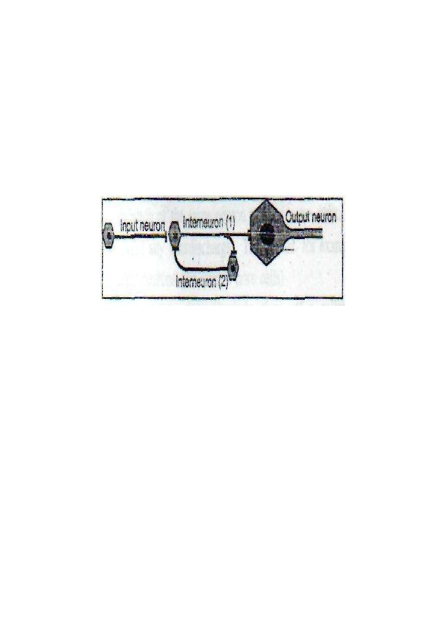
[III] PROLONGATION OF SIGNALS (AFTERDISCHARGE)
Afterdischarge is the persistence of output signals after stoppage of the
input signals. This is possible through the following mechanisms:
1. SYNAPTIC AFTERDISCHARGE:
2. OPEN-OHAIN CIRCUITS
These are circuits in which several interneuron's are arranged to form
an open circuit (fig. 2-7). When interneuron (1) is excited, it sends a
signal to the output neuron and collateral to excite interneuron (2).
Interneuron (2) then sends a signal to the output neuron and collateral
to excite interneuron (3) and so on. The result will be a barrage
(train
of impulses follows each other)
of impulses in the output neuron. The
interneurons of the open-chain circuits are called "the interneuronal
barrages.
Figure 2 - 8: An open chain circuit with interneuronal barrages
11
3. CLOSED {REVERBERATING} CIRCUITS
These are circuits in which the output neuron is repeatedly activated
through a closed circuit of interneurons (fig.2-8). When interneuron
(1) is excited, it sends an impulse to the output neuron and collateral
to excite interneuron (2). Interneuron (2) then re-excites interneuron
(1), and so on.
Figure 2 - 8: A closed chain (reverberating) circuit of neurons.
Reverberating circuits can be facilitated or inhibited by other input fibers.
When facilitated the frequency and duration of discharge in output fibers
increase. When inhibited, the frequency and duration decrease.
The cycle of reverberation stops by either fatigue of synapses or
inhibition by other input fibers. The frequency and duration of
discharge from a reverberating circuit depends on the number of
neurons (i.e. the number of synapses) in the circuit. The larger the
number the lower is the frequency and the longer is the duration.
RHYTHMIC SIGNAL OUTPUT
Certain centers in the CNS produce rhythmic signals, e.g. the
respiratory center in the brainstem. This rhythmic signal output is

12
caused by reverberating circuits involving a large number of
interneurons. The large number of synapses slows down the frequency
of output discharge and delays fatigue of synapses
[IV] SHORTENING OF SIGNALS
Shortening of signals means suppression of afterdischarge in the output
neurons. This is done by either feedback or feed forward inhibition.
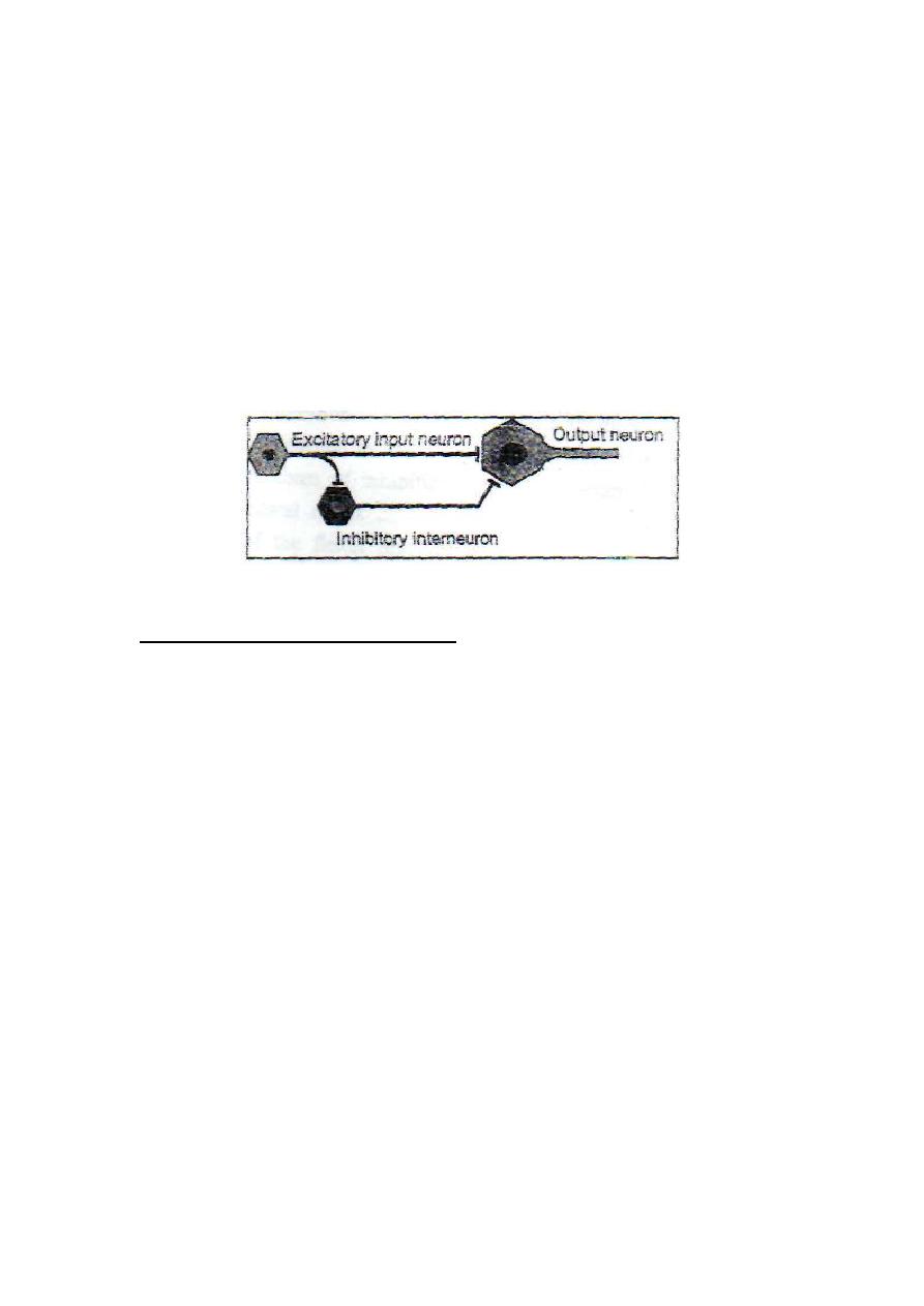
1. FEEDBACK INHIBITION
This occurs when an excitatory neuron stimulates an inhibitory neuron
then the inhibitory neuron turns back to inhibit the initial excitatory
neuron. In this case, stimulation of a neuron results in feedback
inhibition of the same neuron to shorten the duration of discharge and
prevent any afterdischarge. This occurs, for example, with the spinal
motor neurons (the ventral horn cells). Each spinal motor neuron
regularly gives off a collateral branch which synapses with an
inhibitory interneuron called "Renshaw cell".
Renshaw cell sends inhibitory signals to the cell body of the original
spinal motor neuron (feedback inhibition). The inhibition of the original
motor neuron suppresses any synaptic afterdischarge to prevent
undesired prolonged activity of the motor nerve.
2. FEED FORWARD INHIBITION
This occurs when an input neuron stimulates an output neuron plus an
inhibitory interneuron then the inhibitory interneuron inhibits the
output neuron (fig. 2-9).
13
In this case, stimulation of an input neuron results in stimulation
then rapid inhibition of the output neuron. This prevents any
undesired prolonged discharge from the output neuron.
Feed forward inhibition occurs in the cerebellum where a single
input neuron stimulates an output neuron and a Purkinje cell.
The Purkinje cell then inhibits the output neuron, cutting short
any undesired afterdischarge.
Figure 2 - 9: Feed forward inhibition.
[V] SHARPENING OF SIGNALS
Sharpening of signals means the limitation of signals to the
target neurons only. This requires the inhibition of any undesired
activity in the nearby neurons. This is achieved by either lateral
or reciprocal inhibition mechanisms.
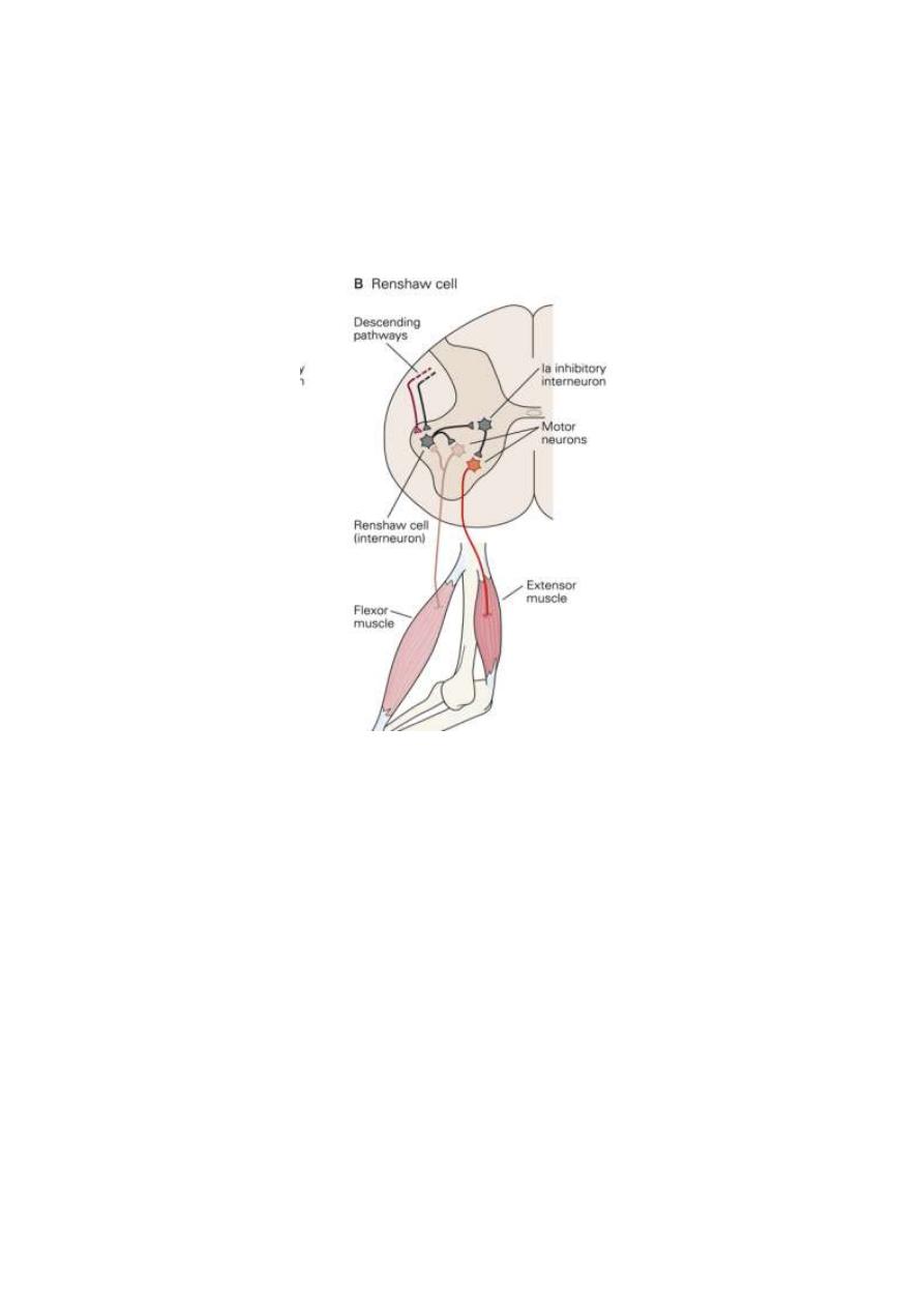
1. LATERAL INHIBITION
Lateral inhibition occurs when a neuron sends collaterals to
inhibit the nearby neurons through intermediate inhibitory
neurons. This helps to focus the activity to the original neuron
and eliminate any undesired discharge from the nearby neurons.
The function of Renshaw" cell shows an example of both
feedback inhibition (of the original motor neuron) and lateral
inhibition (of the nearby neurons). Fig. 2-10
Lateral inhibition occurs also in sensory neurons. Sensory fibers
14
conducting touch (scratching) laterally inhibit itch and pain
conducting fibers at the dorsal horn of the spinal gray matter. In
this way scratching relieves itch and pain sensations.
Fig. 2-10 Lateral and reciprocal inhibition
2. RECIPROCAL INHIBITION
In reciprocal inhibition the activation of one output neuron is
accompanied by simultaneous inhibition of another output
neuron (Fig.2-10). This form of inhibition occurs, for example,
during the flexor withdrawal reflex. In this reflex, contraction of
the flexor muscles is accompanied by concomitant reflex
relaxation of the extensor muscles. Reciprocal inhibition helps to
optimize the reflex response by inhibiting any antagonistic
contraction.