1
Physiology
Dr. Basim Mohamad Alwan
Lecture (1)
INTRODUCTION
The nervous system is unique in the vast complexity of thought
processes and control actions it can perform. It receives each minute
literally millions of bits of information from the different sensory
nerves and sensory organs and then integrates all these to determine
responses to be made by the body. The nervous system contains more
than 100 billion neurons and consists of the central nervous system,
the peripheral nerves and autonomic nervous system (fig. 1-1).
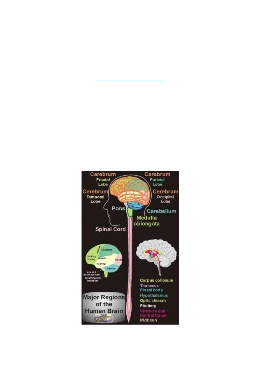
Figure 1-1: General view of the brain, spinal cord and spinal nerves

2
The central nervous system (the neural axis or neuraxis) consists of
the brain and the spinal cord. Anatomically, the brain comprises the
cerebrum which consists of two cerebral hemispheres, the cerebellum,
and the brain stem. The brainstem consists of the midbrain, the pons
and the medulla oblongata. The CNS contains the nerve centers which
receive and process the nervous signals, then formulate the response
to these signals.
The peripheral nerves are divided into cranial and spinal nerves. The
cranial nerves are twelve pairs of nerves, which arise from the brain
and emerge out through foramina in the bones of the cranium (skull).
The spinal nerves are 31 pairs of nerves that arise from the spinal cord
and emerge out through foramina in the vertebral column.
Anatomically, the spinal nerves are sorted out into 5 groups according
to their site of origin from the spinal cord which are 8 cervical pair, 12
dorsal pairs, 5 lumbar pairs, 5 sacral pairs and one cooccygeal pair.
DIVISIONS OF THE NERVOUS SYSTEM
The nervous system comprises three major systems;
I. THE AUTONOMIC NERVOUS SYSTEM
: Is the part of the nervous
system which is concerned with the involuntary control of the visceral
activity. It includes sympathetic, parasympathetic and enteric
divisions.
II. THE SOMATIC NERVOUS SYSTEM:
Is the part of the nervous
system which is concerned with conscious perception of different
sensations, and voluntary control of the muscular activity.

3
This system is divided into two divisions;
(i) SENSORY DIVISION:
Which is concerned with conscious
perception of somatic sensations? It includes the sensory (afferent)
nerves, the sensory (ascending) tracts inside the CNS, the sensory
reticular formation, the thalamus and the sensory cerebral cortex.
(ii) MOTOR DIVISION:
Which is concerned with voluntary control of
muscular activity? It includes the motor cerebral cortex, the basal
ganglia, the cerebellum, the motor reticular formation, the motor
(descending) tracts inside the CNS and the motor (efferent) nerves.
III. THE INTEGRATIVE NERVOUS SYSTEM: Is the part of the
nervous system which is concerned with the sophisticated functions of
the brain. These functions include memory, thinking, learning,
language, speech, emotions and general behavior. The main parts of
the integrative division are the cortical association areas and the
limbic system.
All these three systems and divisions are interconnected and their
functions are integrated together and with other systems in the body.
The basic structural unit of the nervous system which is capable of
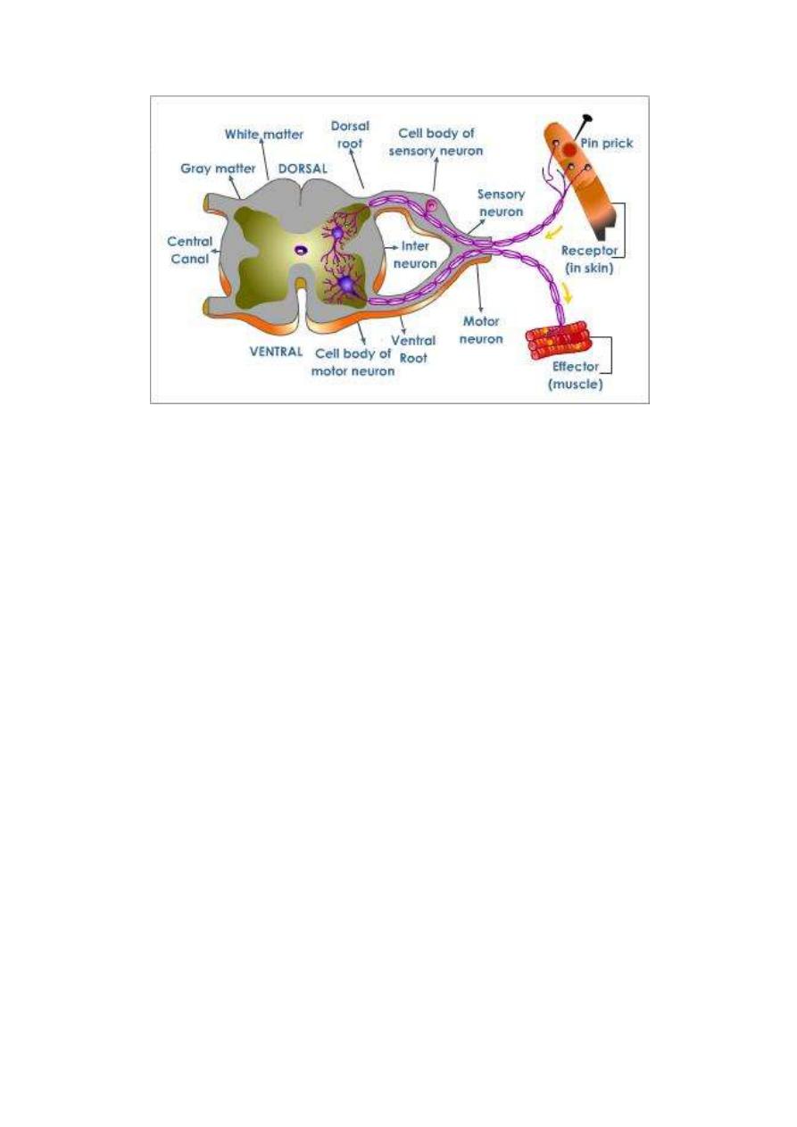
conducting a reflex action is the reflex arc (fig. 1-2). A reflex arc;
consists of 5 components:
4
Figure 1-2: The five basic components of a reflex arc
1. Receptor: A sensor which is excited by the stimulus.
2. Afferent nerve: Which conveys input signals to the CNS? The
afferent nerve is also called the sensory nerve,
3. Center: A collection of neurons that receive the sensory
information and issue the order for proper response.
4. Efferent nerve: A nerve that conveys output signals from the CNS
to the effectors organ. The efferent nerve is either a motor nerve to a
muscle or a secretary nerve to a gland.
5. Effectors organ: A muscular or glandular structure which receives
the final order and executes the reflex response.
The basic functional unit in the nervous system is the reflex action.
A reflex action is an involuntary action in response to a stimulus e.g.
a. painful stimulus applied to the hand leads to reflex withdrawal of
the arm (the withdrawal reflex).

5
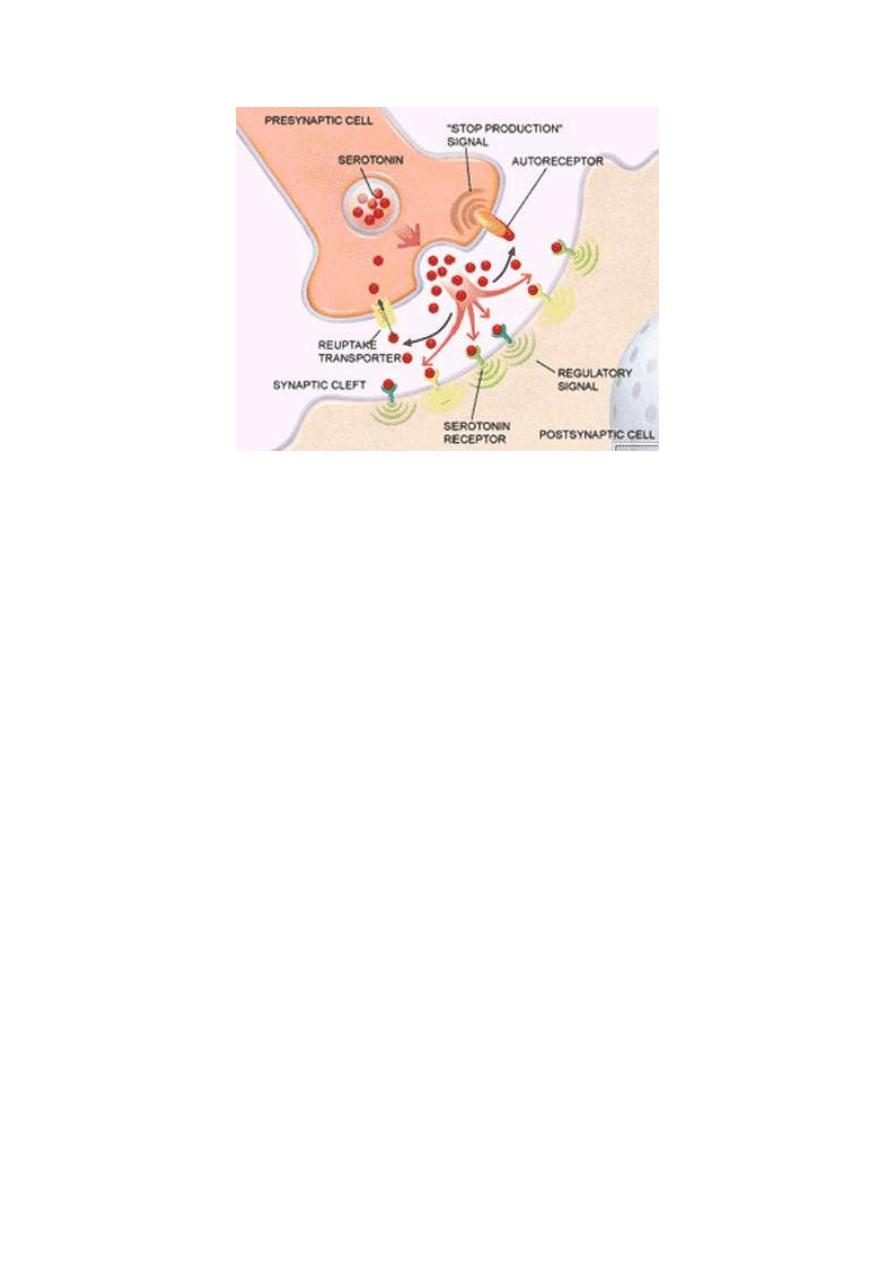
NEURAL SYNAPSES
A synapse is the junction area between a nerve terminal and another
cell. If the second cell is a neuron the synapse is then called a "neural
or neuronal synapse".
The axon of a neuron conducts impulses away from the cell body to
relay onto another cell at the synapses (fig. 1.3). The axon branches
extensively near its end, giving off 1000 branches on the average.
Each branch ends in a nerve terminal. This terminal is a disc-like
expansion called the synaptic knob (the terminal button or the end
foot). There is a gap between the nerve terminal and the adjacent
neuron 30-50 nm wide called the synaptic cleft. So, at the neural
synapse there is contiguity but no continuity of the two adjacent
neurons.
The neuron which conducts impulses to the synapse is called the
"presynaptic neuron'' or "input neuron" and that which conducts
impulses away from the synapse is called the "postsynaptic neuron"
or "output neuron". The synaptic knobs of the presynaptic neuron
contain vesicles called synaptic or transmitter vesicles which contain
the chemical transmitter of the neuron. A polypeptide called synapsin
is found in the walls of the vesicles which bind the transmitter
vesicles to the cytoskeleton keeping them in the cytoplasm away from
the release sites on the presynaptic membrane.
6
Figure 1-3: Neural synapse
THE IMPORTANCE OF SYNAPSES IN THE NERVOUS
SYSTEM
Synapses act as "unidirectional valves" in the nervous
pathways, i.e. they allow the flow of impulses from the pre to
the postsynaptic neurons only. This ensures the flow of impulses
in the nervous pathways in the forward "orthodromic" direction
only. Any impulse that travels along a neuron in the opposite
"antidromic" direction cannot be transmitted to the next neuron
because it dies off at the first set of synapses it meets.
Also, synapses are the sites in the nervous pathways at which
transmission of impulses can be most easily influenced.
At the synapse, transmission of impulses can be accelerated,
slowed down, or blocked by physiological, pathological or
pharmacological influences.
7
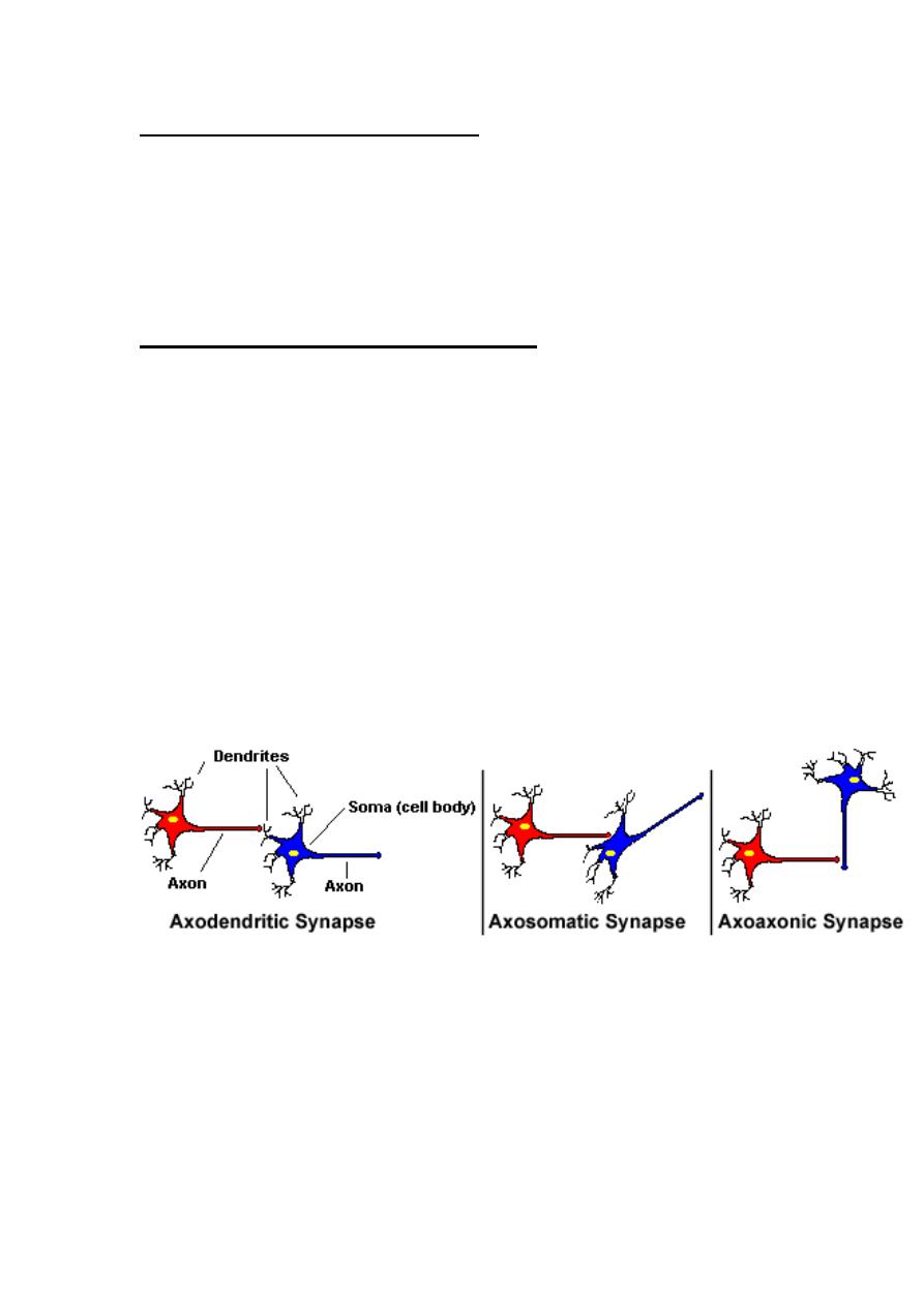
CLASSIFICATION OF SYNAPSES
Synapses could be classified according to either their location
between the pre and postsynaptic neurons (histological
classification), or the mechanism of transmission of impulses
across them (physiological classification),
HISTOLOGICAL CLASSIFICATION
According to this classification, synapses are classified into
three types (fig.1-4):
1. Axodendritic synapses: These are synapses between the
axon terminals of the presynaptic neuron and the dendrites of
the postsynaptic neuron.
2. Axosomatic synapses: These are synapses between the axon
terminals of the presynaptic neuron and the soma of the
postsynaptic neuron
Figure 1-4: histological classification of synapses
3. Axoaxonic synapses: These are synapses between the axon
terminals of the presynaptic neuron and the axon of the
postsynaptic neuron.

8
PHYSIOLOGICAL CLASSIFICATION
According to this classification, synapses are classified into
three types:
1. CHEMICAL SYNAPSES:
In these synapses, transmission of
signals occurs by releasing a ''chemical transmitter" from the
presynaptic terminal into the synaptic cleft. The transmitter then
acts on specific receptors on the postsynaptic membrane to
generate postsynaptic potential. There are more than
40
different synaptic transmitters in the CNS which are either small
molecule rapidly acting (acetylcholine) or large molecule slowly
acting (substance P).
Chemical synapses are the only type of synapses found in the
human nervous system.
2. ELECTRICAL SYNAPSES:
In these synapses, there are gap
junctions between the pre and postsynaptic membranes which
allow the transmission of the depolarization wave directly from
the pre to the postsynaptic membrane.
3.
CONJOINT SYNAPSES (ELECTROCHEMICAL):
In these
synapses, transmission of impulses occurs by both mechanisms
electrical and chemical. They are found in some fish and
invertebrates.
THE MECHANISM OF RELEASE OF TRANSMITTER AT THE
CHEMICAL SYNAPSES
When the action potential reaches the nerve terminal, it opens the
calcium gates allowing Ca
2+
influx from the extracellular fluid into

9
the cytoplasm. Ca
2+
induces the phosphorylation of synapsin.
This detaches the synaptic vesicles from their binding to the
cytoskeleton. The vesicles get attached and fused to specific release
sites on the presynaptic membrane. The release sites then rupture
and the chemical transmitter is released into the synaptic cleft. This
process is a passive process.
THE MECHANISM OF ACTION OF THE CHEMICAL
TRANSMITTER
The transmitter moves in the fluid in the synaptic cleft by simple
diffusion to the receptors on the postsynaptic membrane. It activates
these receptors to generate postsynaptic potential (PSP). There
are two types of PSPs; excitatory and inhibitory:
1. THE EXCITATORY POSTSYNAPTIC POTENTIALS (EPSPs)
EPSPs are produced by depolarization of the postsynaptic
membrane due to sodium influx. The action of the chemical
transmitter is to open the gated sodium channels leading to sodium
influx. Some transmitters produce EPSPs by closing K
+
channels
(membrane depolarization).
2. THE INHIBITORY POSTSYNAPTIC POTENTIALS (IPSPs
)
IPSPs are produced by hyperpolarization of the postsynaptic
membrane due to C1
-
influx and / or K
+
efflux. The action of the
transmitter in this case is to open the gated chloride and / or
potassium channels. The IPSPs decrease the excitability of the
10
postsynaptic neuron and resist the development of any action
potential in it.
The excitatory and inhibitory postsynaptic potentials do not
obey the all or none rule. They can be summated either
temporally or spatially.
SUMMATION OF THE POSTSYNAPTIC POTENTIALS
There are two ways of summation of the postsynaptic potentials;
temporal and spatial summation.
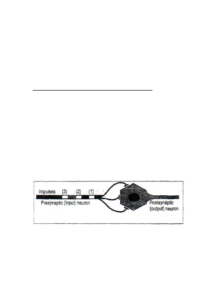
1. TEMPORAL SUMMATION
This is summation of the postsynaptic potentials produced by a
train of impulses on one presynaptic terminal, reaching the same
synapse one shortly after the other (fig. 1-5). Each time an
impulse reaches the synapse it creates a PSP which is summated
with other PSPs in its magnitude.
Figure 1-5: The mechanism of temporal summation.
Temporal summation is possible because the opening of a
ligand-gated channel lasts for about one ms (millisecond) whilst
the PSP produced by this opening lasts for about 15 ms. In this
way any other opening of the same channel within 15 ms would
11
produce another PSP that will potentiate the previous one.
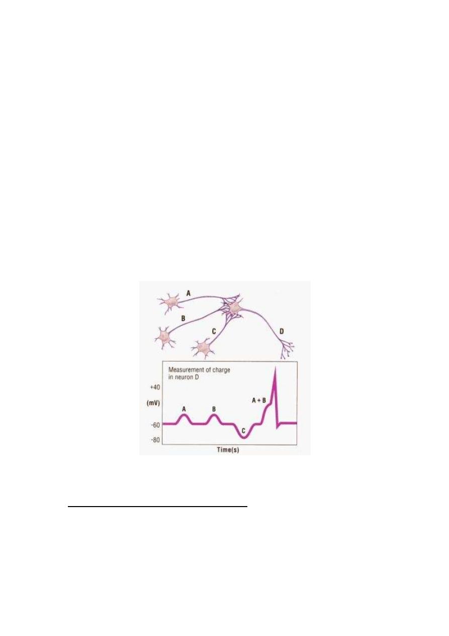
2. SPATIAL SUMMATION
This is summation of postsynaptic potentials produced by multiple
impulses in several presynaptic terminals which reach several synapses
at the same moment (fig. 1-6). A large number of postsynaptic spots
are stimulated simultaneously. This increases the stimulated surface
area of the postsynaptic neuron. The simultaneously generated
postsynaptic potentials potentiate each other.
In most cases in vivo, both types of summation occur at the same
time (tempro-spatial summation) where multiple impulses arrive
one shortly after another at several presynaptic terminals.
Figure 1-6: The mechanism of spatial summation
THE RESULT OF SUMMATION OF PSPs
An average neuron in the CNS receives about 1000 terminals from
different presynaptic neurons. Some of these terminals are
excitatory and some are inhibitory. Excitatory and inhibitory

12
postsynaptic potentials may occur at the same time. Summation of
PSPs results in one of two conditions in the postsynaptic neuron:
1. INHIBITORY STATE
This occurs when the inhibitory input is greater than the excitatory
input so IPSPs are produced and the cell membrane becomes
hyperpolarized and its excitability decreases.
2. EXCITATORY STATE
This occurs when the excitatory input is greater than the inhibitory
input so more EPSPs are produced and the cell membrane
becomes depolarized. When depolarization reaches a critical
threshold level (the firing level) a propagated action potential (nerve
impulse) is produced.
The propagated action potential starts at the initial segment of
the neuronal axon not at the soma of the neuron because:
i. The density of the voltage-gated Na
+
channels at the initial
segment is seven times as much as those at the membrane of
the soma. This allows greater and faster Na
+
influx which
creates more rapid depolarization up to the firing level.
ii. The depolarization required to open the voltage gated Na
+
channels at "the initial segment is only +15 mV, whilst the
required depolarization at the soma is +30 mV.
This explains why the axoaxonic synapses are the most
effective in exciting the postsynaptic neuron. This is because

13
they are the nearest to the initial segment. The least effective
synapses are the axodendretic synapses.
THE EXCITATORY AND INHIBITORY NEURONS
The presynaptic neuron is either excitatory or inhibitory to the
postsynaptic neurons. This is because neurons can release only
one type of transmitter which is either excitatory or inhibitory to
the postsynaptic neuron.
A co-transmitter may be released with the primary
transmitter. It is, however, always a potentiator of the
primary one.
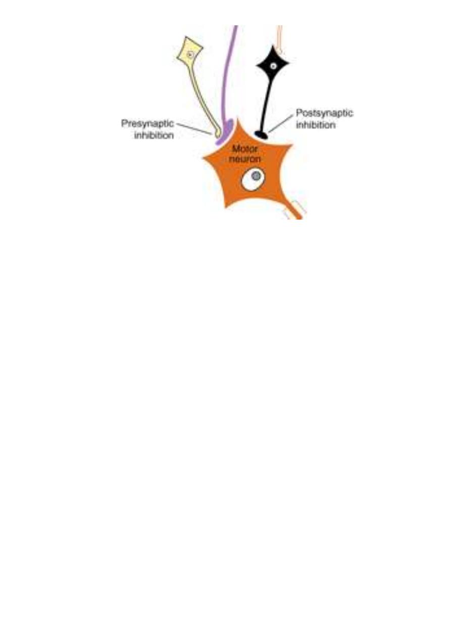
PRESYNAPTIC AND POSTSYNAPTIC INHIBITION
Transmission of impulses across the synapse can be inhibited or
blocked in two ways (fig. 1-6):
1. PRESYNAPTIC INHIBITION
Presynaptic inhibition is the inhibition of synaptic transmission
by inhibiting the release of the transmitter from the presynaptic
nerve terminal. It can be induced by certain drugs (botulinum,
toxin) or by certain inhibitory neurons (attenuators) which
synapse on presynaptic terminals. The attenuator neuron inhibits
the synapsin phosphorylase enzyme of the excitatory neuron so
the transmitter vesicles remain attached to the cytoskeleton and
no release of the transmitter.
14
Figure 1-7: Presynaptic and postsynaptic inhibition.
2. POSTSYNAPTIC INHIBITION
Postsynaptic inhibition is inhibition of synaptic transmission by
induction of an inhibitory state in the postsynaptic neuron. It is
induced either by drugs or by inhibitory neurons.