Respiratory Physiology
Pulmonary ventilationDiffusion of gases
Transport of gases
Control of respiration
Respiration
Ventilation: Movement of air into and out of lungsExternal respiration: Gas exchange between air in lungs and blood
Transport of oxygen and carbon dioxide in the blood
Internal respiration: Gas exchange between the blood and tissues
Respiratory System Functions
Gas exchange: Oxygen enters blood and carbon dioxide leavesRegulation of blood pH: Altered by changing blood carbon dioxide levels
Voice production: Movement of air past vocal folds makes sound and speech
Olfaction: Smell occurs when airborne molecules drawn into nasal cavity
Protection: Against microorganisms by preventing entry and removing them
Respiratory System Divisions
Upper tract
Nose, pharynx and associated structures
Lower tract
Larynx, trachea, bronchi, lungs
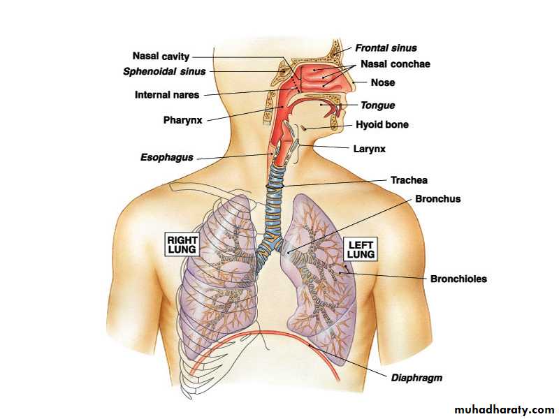
Nasal Cavity and Pharynx
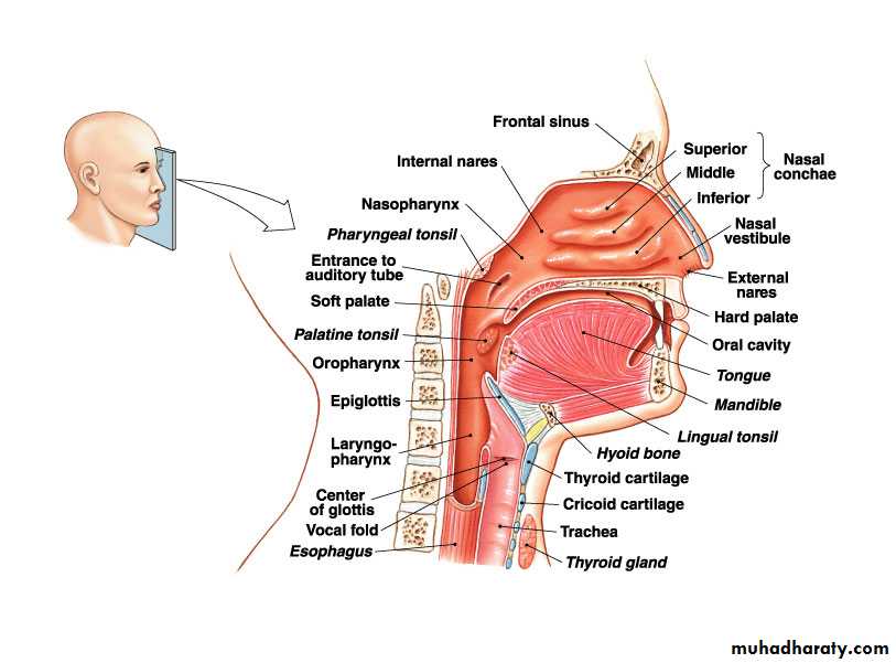
Nose and Pharynx
NoseExternal nose
Nasal cavity
Functions
Passageway for air
Cleans the air
Humidifies, warms air
Smell
Along with paranasal sinuses are resonating chambers for speech
Pharynx
Common opening for digestive and respiratory systems
Three regions
Nasopharynx
Oropharynx
Laryngopharynx
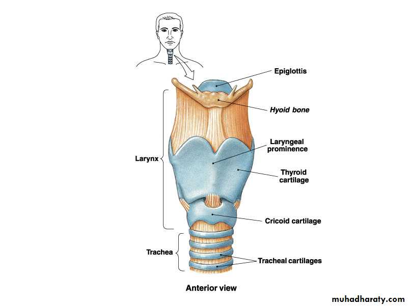
Larynx
Functions
Maintain an open passageway for air movement
Epiglottis and vestibular folds prevent swallowed material from moving into larynx
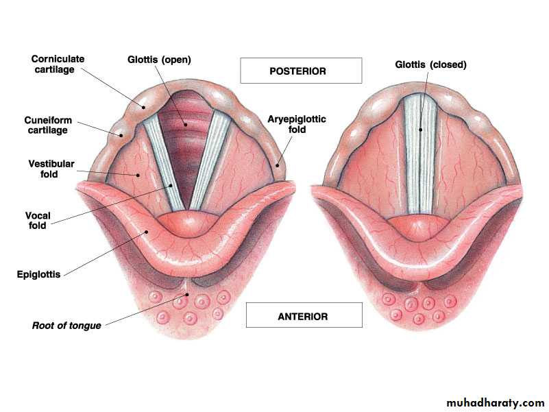
Vocal folds are primary source of sound production
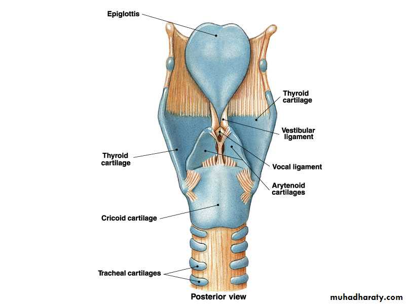
Vocal Folds
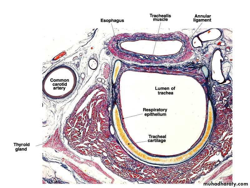
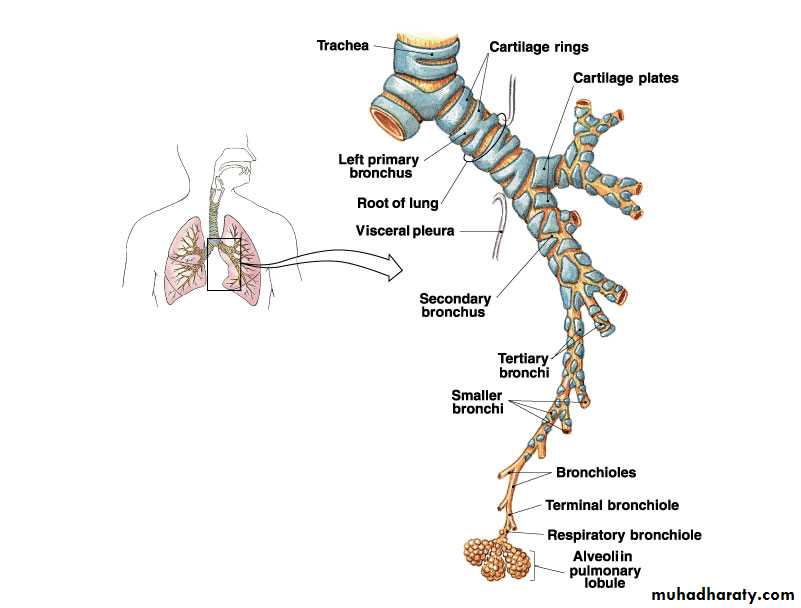
Trachea
WindpipeDivides to form
Primary bronchi
Carina: Cough reflex
Tracheobronchial Tree
Conducting zoneTrachea to terminal bronchioles which is ciliated for removal of debris
Passageway for air movement
Cartilage holds tube system open and smooth muscle controls tube diameter
Respiratory zone
Respiratory bronchioles to alveoli
Site for gas exchange
Tracheobronchial Tree
Bronchioles and Alveoli
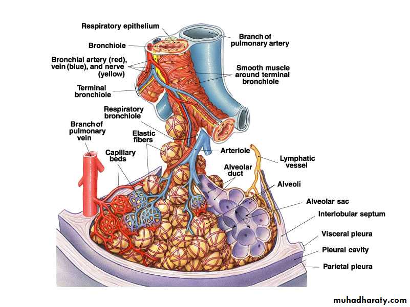
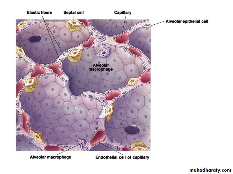
Alveolus and Respiratory Membrane
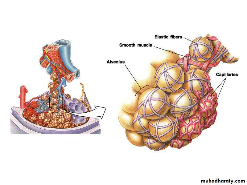
The normal adult human lung weighs about 1000g and consists of about 50% blood and 50% tissue by weight. About 10% of the total lung volume is composed of various types of conducting airways and some connective tissue. The remaining 90% is the respiratory or gas exchange portion of the lung, composed of alveoli and supporting capillaries.
Thoracic WallsMuscles of Respiration
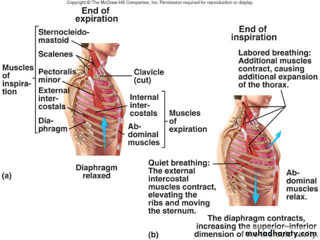
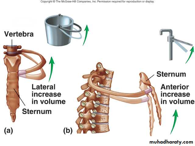
Thoracic Volume
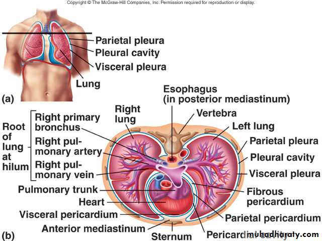
Pleura
Pleural fluid produced by pleural membranesActs as lubricant
Helps hold parietal and visceral pleural membranes together
Ventilation
Movement of air into and out of lungsAir moves from area of higher pressure to area of lower pressure
Pressure is inversely related to volume
Alveolar Pressure Changes
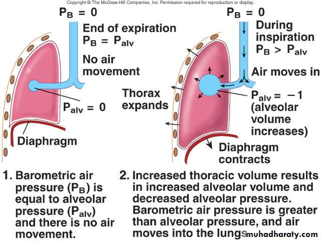
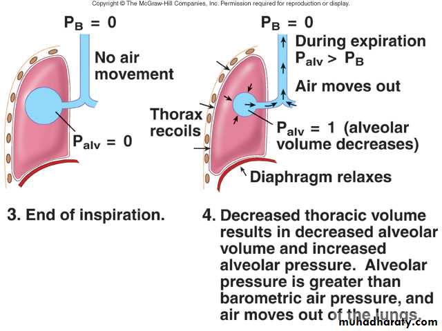
Changing Alveolar Volume
Lung recoilCauses alveoli to collapse resulting from
Elastic recoil and surface tension
Surfactant: Reduces tendency of lungs to collapse
Pleural pressure
Negative pressure can cause alveoli to expand
Pneumothorax is an opening between pleural cavity and air that causes a loss of pleural pressure
Normal Breathing Cycle
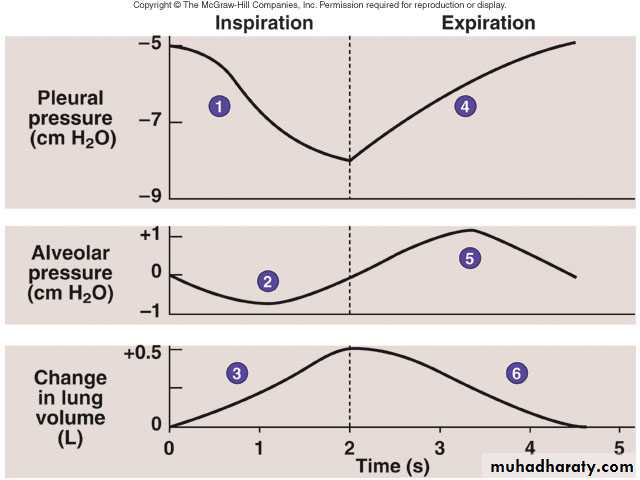
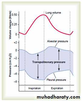
Transpulmonary pressure [pressure difference between the alveolar pressure and the pleural pressure]. It is the measure of the elastic forces that leads to collapse of the lung and it is called the recoil pressure.
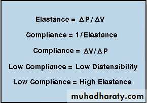
The Opposing Force of Pulmonary Elastance or Compliance
The lung is an elastic structure with an anatomical organization that promotes its collapse to essentially zero volume, much like an inflated balloon.The term elastic means a material deformed by a force tends to return to its initial shape or configuration when the force is removed. It oppose lung inflation. Elastance.
Compliance (distensibility) is the reciprocal of elastance, is a measure of the ease of deformation (inflation).
Compliance
Measure of the ease with which lungs and thorax expandThe greater the compliance, the easier it is for a change in pressure to cause expansion
A lower-than-normal compliance means the lungs and thorax are harder to expand
Conditions that decrease compliance
Pulmonary fibrosis
Pulmonary edema
Respiratory distress syndrome
Lung compliance: Which equals to change in volume divided by change in pressure (1 cm = 200 ml). That is, every time the transpulmonary pressure increases 1 centimeter of water, the lung volume, after 10 to 20 seconds, will expand 200 milliliters.
1/3 to overcome pleural pressure
2/3 to overcome surface tension
Effect of thoracic cage: Compliance of both lung + cage = 110 ml (instead of 200ml/cm)
Surfactant: The surface active agent in water and it consists of lipids, protein and ions.
Fetal lung surfactant also is not fully functional until about the seventh month of gestation. Respiratory Distress Syndrome (RDS) is related to non-functional alveolar surfactant.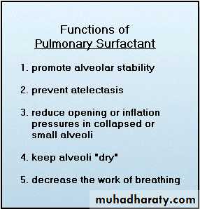
Work of breathing:
Compliance work: against elastic forces of lung + cageTissue resistance work: against viscosity of both lung and cage
Airway resistance work
During quite breathing, 3-5% of total energy of the body are spent for respiration, while in heavy exercise, it increases up to 50 folds
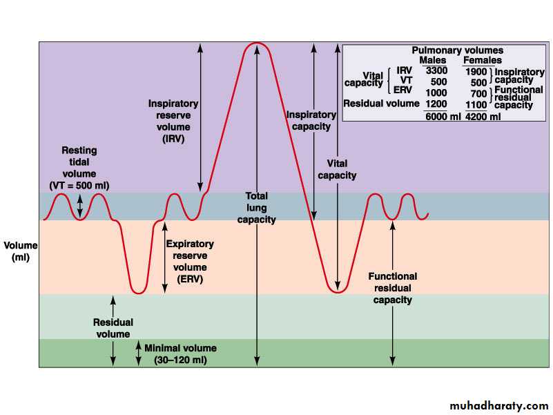
Pulmonary Volumes
Tidal volumeVolume of air inspired or expired during a normal inspiration or expiration
Inspiratory reserve volume
Amount of air inspired forcefully after inspiration of normal tidal volume
Expiratory reserve volume
Amount of air forcefully expired after expiration of normal tidal volume
Residual volume
Volume of air remaining in respiratory passages and lungs after the most forceful expiration
Pulmonary Capacities
Inspiratory capacity
Tidal volume plus inspiratory reserve volume
Functional residual capacity
Expiratory reserve volume plus the residual volume
Vital capacity
Sum of inspiratory reserve volume, tidal volume, and expiratory reserve volume
Total lung capacity
Sum of inspiratory and expiratory reserve volumes plus the tidal volume and residual volume
Volumes
Capacities1- Tidal vol. (500ml)
1- Inspiratory cap. (3500ml)
2- Inspiratory reserve vol. (3000ml)
2- Functional residual cap. (2300ml)
3- Expiratory reserve vol. (1100ml)
3- Vital cap. (4600ml)
4- Residual vol. (1200ml)
4- Total lung cap. (5800ml)
Spirometer and Lung Volumes/Capacities
Minute and Alveolar Ventilation
Minute ventilation: Total amount of air moved into and out of respiratory system per minuteRespiratory rate or frequency: Number of breaths taken per minute
Anatomic dead space: Part of respiratory system where gas exchange does not take place
Alveolar ventilation: How much air per minute enters the parts of the respiratory system in which gas exchange takes place
Respiratory dead space
Is the space where no gas exchange occurs. It is either anatomically (150 ml) (anatomical dead space (nose, pharynx, larynx, trachea, bronchi, bronchioles); or physiological dead space whereby some alveoli are not functional because of absent or partial blood supply (normally it should be zero).So the total dead space is the sum of anatomical and physiological dead spaces and so equals to 150 ml. So the alveolar ventilation per minute equals to pulmonary ventilation per minute minus dead space and equals to 500-150 = 350 ml/min X 12 = 4200 ml/min.
Nerve stimulation (sympathetic, i.e., adrenalin dilatation; parasympathetic, i.e., Ach. constriction).
Cough reflex: afferent vagus nerve medulla autonomic inspiration of 2.5 liters closure of epiglottis and vocal cords contraction of abdominal muscles sudden opening expel air at a velocity of 400 miles per hour + narrowing of trachea and bronchi.
Sneeze reflex: Similar except to nasal passages instead of lower airways. Afferent is fifth cranial medulla similar but depression of uvula so that large amounts of air pass through the nose.
Pulmonary circulation:
Blood supply to the lungs goes to bronchi (nutrition) and respiratory units (gaseous exchange).When O2 concentration drops to 70% (73mmHg), pulmonary blood vessels constricts (opposite to other capillaries) and this is important to shift the blood to more aerated areas.
Right atrial pressure is 25 mmHg systolic and 0 mmHg diastolic.
Pulmonary artery pressure is 25mmHg systolic and 8mmHg diastolic (mean arterial pressure equals 15 mmHg)
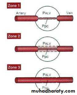
Lung Zones
In the normal, upright adult, the lowest point in the lungs is about 30 centimeters below the highest point. This represents a 23 mm Hg pressure difference, about 15 mm Hg of which is above the heart and 8 below.
For the whole lung, an ideal ventilation to perfusion ratio is between 0.8 to 1.0.
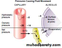
Perfusion across capillaries
Capillary pressure equals 7mmHg (while it is 17 in general circulation).Plasma colloid equals 28 mmHg.
Interstitial colloid 14 mmHg (7 in general circulation)
-ve interstitial pressure equals 8 mmHg
Total = 29, so 29-28 = 1mmHg which removed by lymphatics and evaporation
Physical Principles of Gas Exchange
Partial pressureThe pressure exerted by each type of gas in a mixture
Dalton’s law: Ptotal = P1 + P2 + P3 + ... + Pn
Water vapor pressure
Diffusion of gases through liquids
Concentration of a gas in a liquid is determined by its partial pressure and its solubility coefficient
Henry’s law: concentration of dissolved gas = pressure × SC
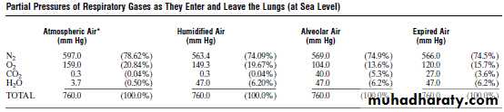
Gases Comprising the Earth's Atmosphere
The earth's atmosphere is a mixture of gases consisting of about 78% molecular nitrogen (N2), 20.9 % molecular oxygen (O2) and 1.0 % argon (Ar). Other gases, like carbon dioxide (0.03%), are also detectable, but only in trace amounts.
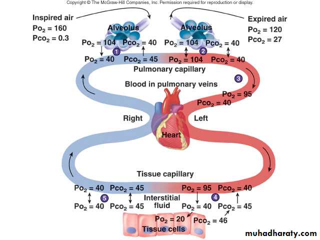
Only a portion of each tidal volume is delivered to the alveoli. The total air volume of all lung alveoli before inspiration (end-expiration) is by definition the Functional Residual Capacity. For a normal adult, the FRC is about 2500 ml. So, if the volume of fresh ambient air reaching the alveoli is 300 ml, it is added to an FRC of 2500 ml. As a result, the partial pressures of alveolar gases do not fluctuate markedly with each breath since only a portion of the FRC is exchanged.
Factors affecting the diffusion of gasses in air:
Pressure X Area X TemperatureD = ----------------------------------------------, distance X SQR(Molecular Weight)
diff. coef. = T/SQR(MW) (constant)
Solubility of O2 = 0.024
Solubility of CO2 = 0.57 (20 times of O2)
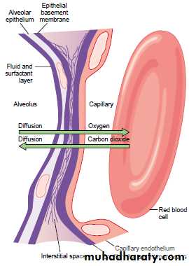
Diffusion of Gases through the Respiratory Membrane
The respiratory unit: respiratory bronchioles, alveolar ducts, atria, and alveoli.Blood flows as a sheet.
Respiratory membrane is 0.2 micrometer thickness and composed of: 1) fluid (surfactant), 2) epithelium, 3) epithelial basement membrane, 4) interstitial fluid, 5) capillary basement membrane, 6) endothelial cells
Physical Principles of Gas Exchange
Diffusion of gases through the respiratory membraneDepends on membrane’s thickness, the diffusion coefficient of gas, surface areas of membrane, partial pressure of gases in alveoli and blood
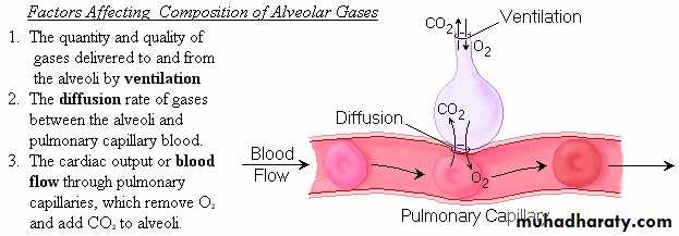
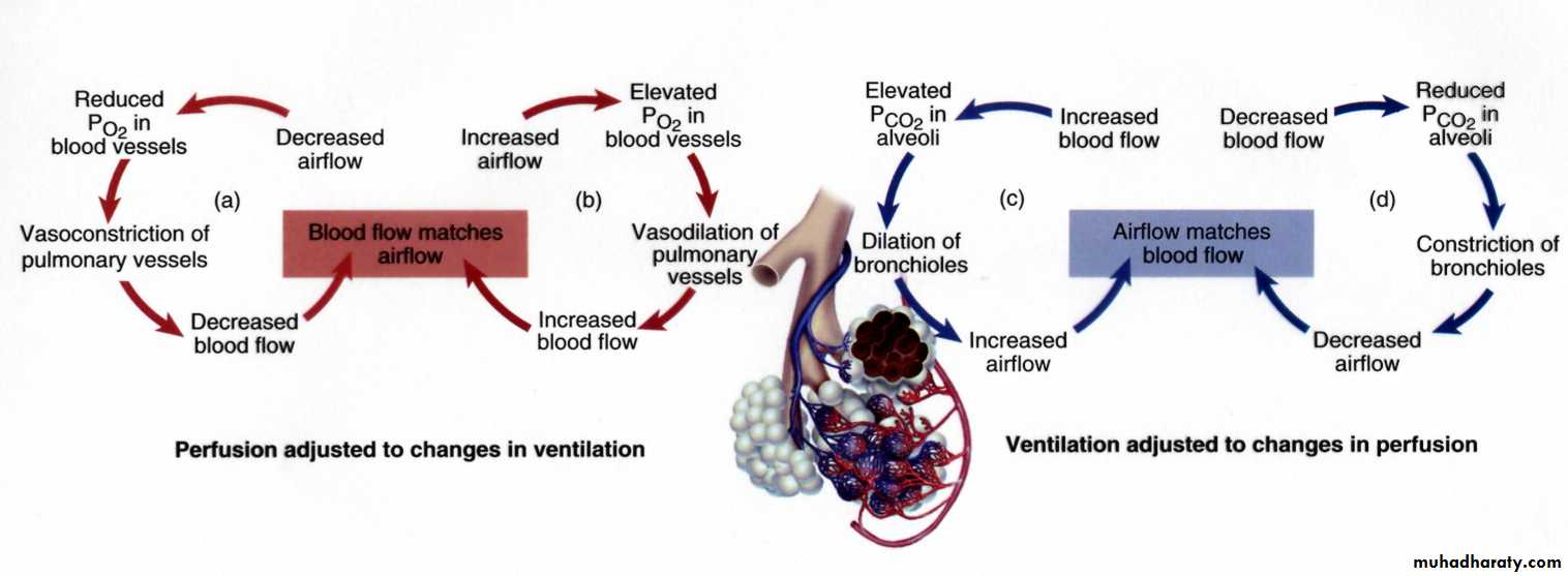
Relationship between ventilation and pulmonary capillary flow
Increased ventilation or increased pulmonary capillary blood flow increases gas exchange
Physiologic shunt is deoxygenated blood returning from lungs
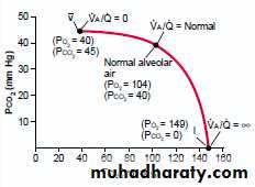
Effect of ventilation perfusion ratio on alveolar gas concentration
VA/Q = 0 O2 = 40, CO2 = 45mmHgVA/Q = infinity O2 = 149, CO2 = 0mmHg
VA/Q = normal O2 = 104, CO2 = 40mmHg
If less than normal then called physiological shunt
If more than normal then called physiological dead space
Normally at the tip of the lung, VA/Q is (2.5) times normal (phys. dead space), while at the base, it is (0.6) times normal (phys. shunt).
Normally, there are abnormal VA/Q ratios in the upper and lower portions of the lung. In the upper both ventilation and perfusion are low but VA is more than Q, so there is physiological dead space, but in the lower VA is less than Q, so there is physiological shunt.
Changes in Partial Pressures
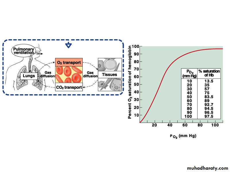
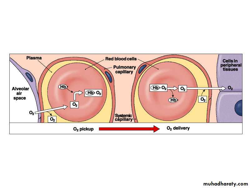
Hemoglobin and Oxygen Transport
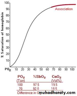
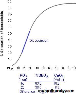
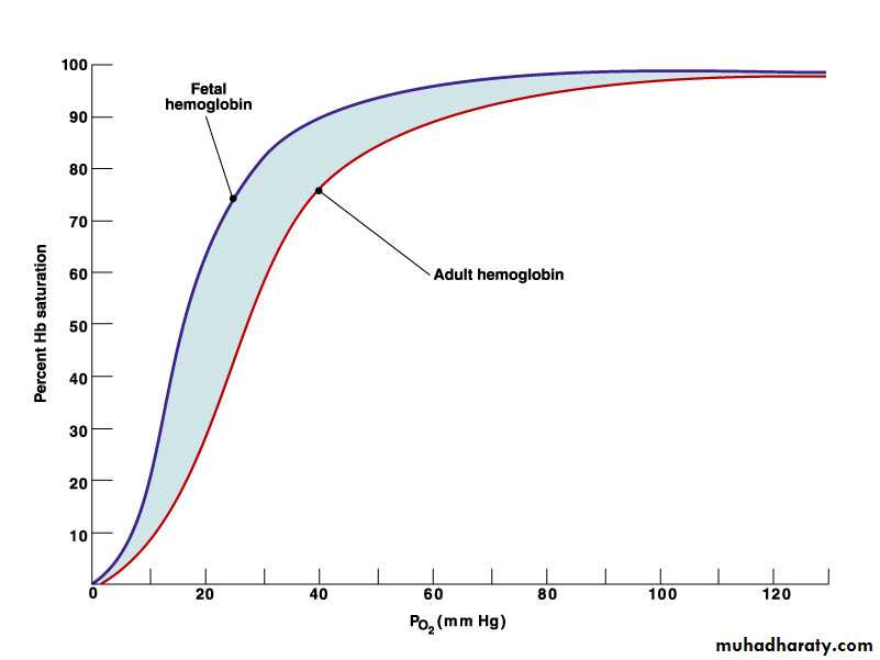
Oxygen is transported by hemoglobin (97%) and is dissolved in plasma (3%)Oxygen-hemoglobin dissociation curve shows that hemoglobin is almost completely saturated when P02 is 80 mm Hg or above. At lower partial pressures, the hemoglobin releases oxygen.
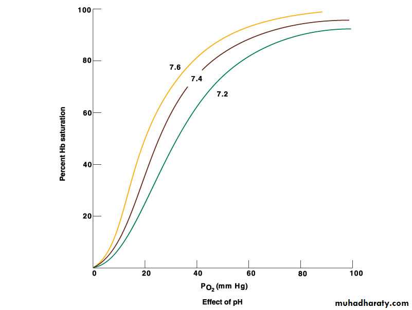
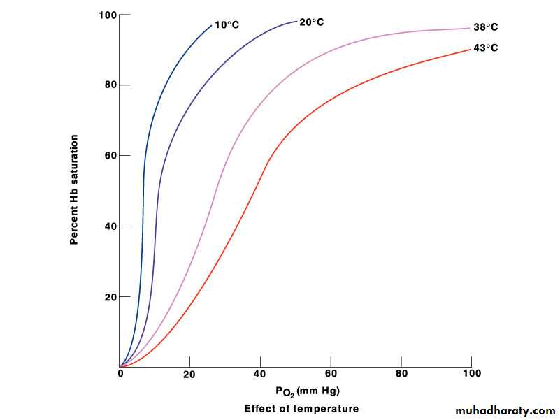
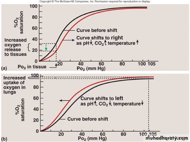
A shift of the curve to the left because of an increase in pH, a decrease in carbon dioxide, or a decrease in temperature results in an increase in the ability of hemoglobin to hold oxygen
Hemoglobin and Oxygen Transport
A shift of the curve to the right because of a decrease in pH, an increase in carbon dioxide, or an increase in temperature results in a decrease in the ability of hemoglobin to hold oxygenThe substance 2.3-bisphosphoglycerate increases the ability of hemoglobin to release oxygen
Fetal hemoglobin has a higher affinity for oxygen than does maternal
Oxygen-HemoglobinDissociation Curve at Rest
Bohr effect:
Temperature effects:
Shifting the Curve
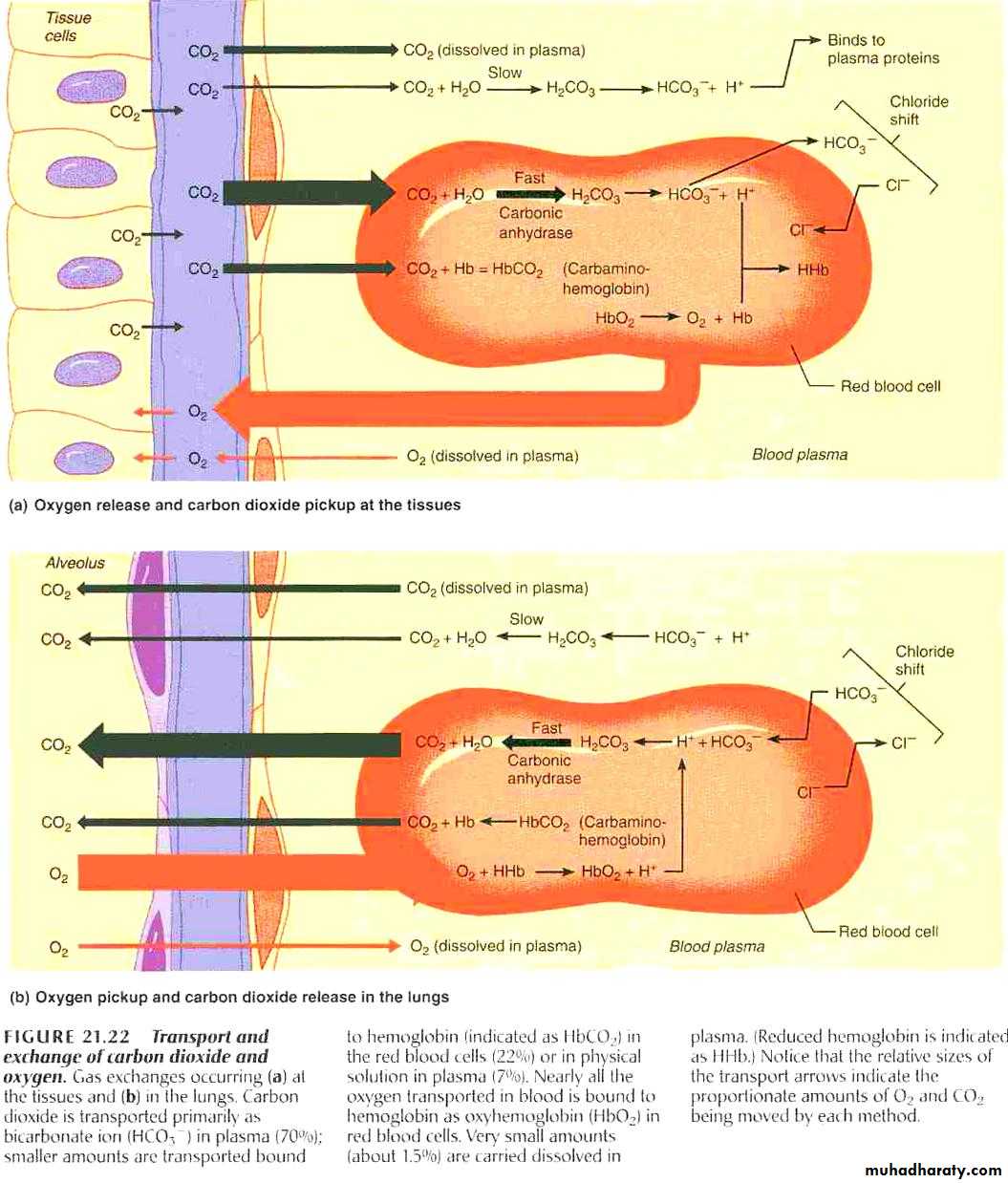
Transport of Carbon Dioxide
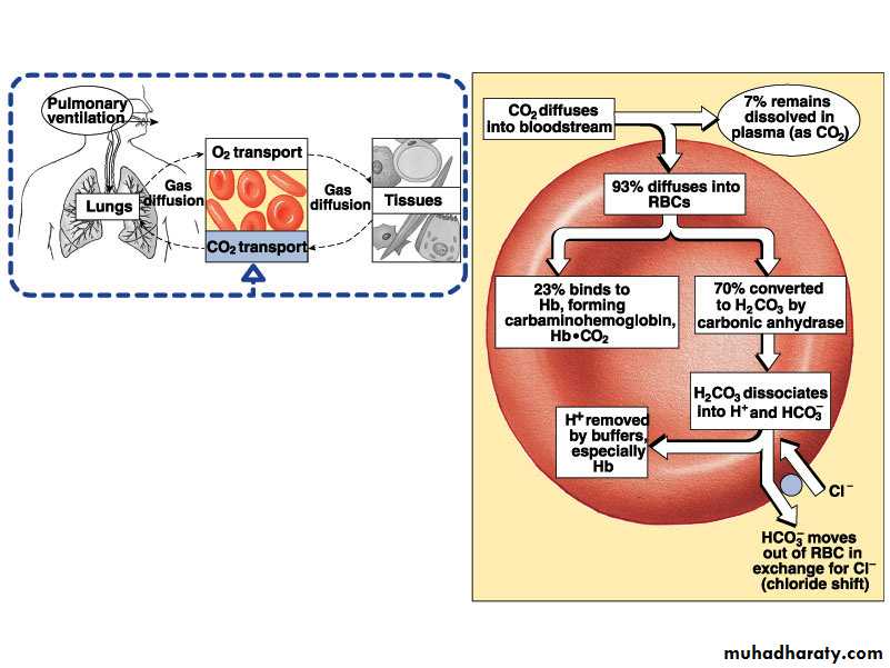
Carbon dioxide is transported as bicarbonate ions (70%) in combination with blood proteins (23%) and in solution with plasma (7%)Hemoglobin that has released oxygen binds more readily to carbon dioxide than hemoglobin that has oxygen bound to it (Haldane effect)
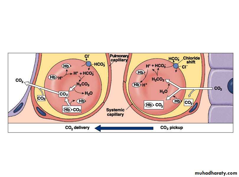
In tissue capillaries, carbon dioxide combines with water inside RBCs to form carbonic acid which dissociates to form bicarbonate ions and hydrogen ions
Transport of Carbon Dioxide
In lung capillaries, bicarbonate ions and hydrogen ions move into RBCs and chloride ions move out. Bicarbonate ions combine with hydrogen ions to form carbonic acid. The carbonic acid is converted to carbon dioxide and water. The carbon dioxide diffuses out of the RBCs.
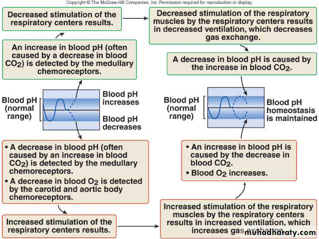
Increased plasma carbon dioxide lowers blood pH. The respiratory system regulates blood pH by regulating plasma carbon dioxide levels
Haldane effect
It was pointed out that an increase in carbon dioxide in the blood causes oxygen to be displaced from the hemoglobin (the Bohr effect), which is an important factor in increasing oxygen transport.The reverse is also true: binding of oxygen with hemoglobin tends to displace carbon dioxide from the blood.
The Haldane effect results from the simple fact that the combination of oxygen with hemoglobin in the lungs causes the hemoglobin to become a stronger acid.
This displaces carbon dioxide from the blood and into the alveoli in two ways:
(1) The more highly acidic hemoglobin has less tendency to combine with carbon dioxide to form carbaminohemoglobin, thus displacing much of the carbon dioxide that is present in the carbamino form from the blood.(2) The increased acidity of the hemoglobin also causes it to release an excess of hydrogen ions, and these bind with bicarbonate ions to form carbonic acid; this then dissociates into water and carbon dioxide, and the carbon dioxide is released from the blood into the alveoli and, finally, into the air.
CO2 Transport and Cl- Movement
Ventilation-perfusion coupling:
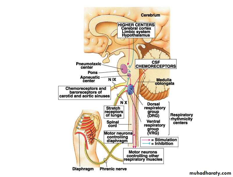
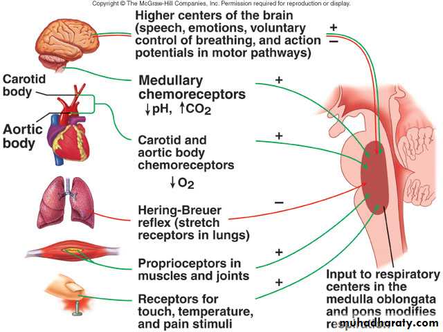
Respiratory Areas in Brainstem
Medullary respiratory centerDorsal groups stimulate the diaphragm
Ventral groups stimulate the intercostal and abdominal muscles
Pontine (pneumotaxic) respiratory group
Involved with switching between inspiration and expiration (respiratory ramp).
Pontine (apneuostic center) prevents the switch off of the respiratory ramp.
Respiratory Structures in Brainstem
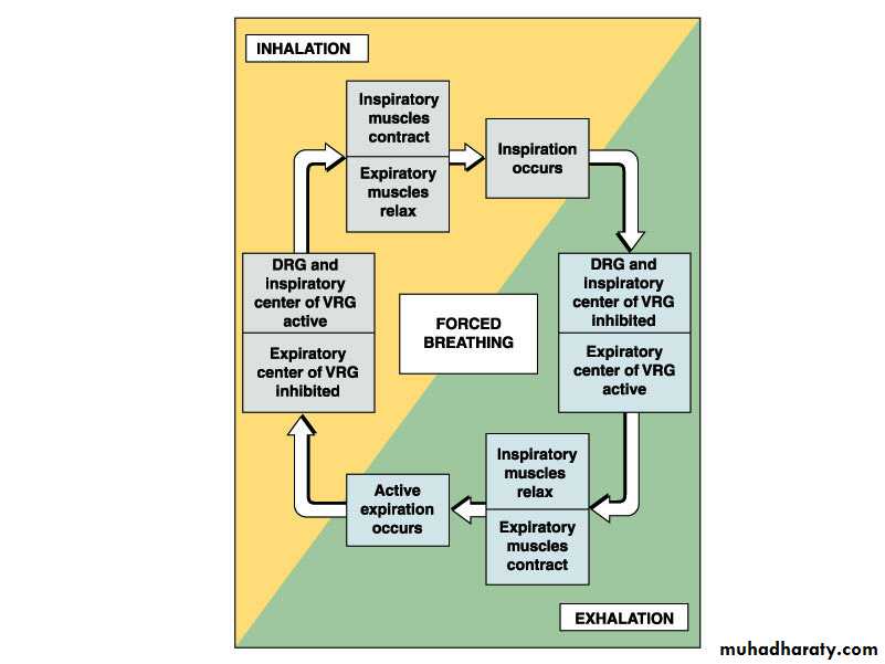
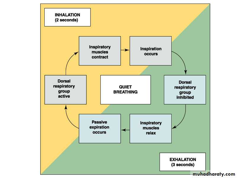
Rhythmic Ventilation
Starting inspiration
Medullary respiratory center neurons are continuously active
Center receives stimulation from receptors and simulation from parts of brain concerned with voluntary respiratory movements and emotion
Combined input from all sources causes action potentials to stimulate respiratory muscles
Increasing inspiration
More and more neurons are activated
Stopping inspiration
Neurons stimulating also responsible for stopping inspiration and receive input from pontine group and stretch receptors in lungs. Inhibitory neurons activated and relaxation of respiratory muscles results in expiration.
Modification of Ventilation
Cerebral and limbic systemRespiration can be voluntarily controlled and modified by emotions
Chemical control
Carbon dioxide is major regulator
Increase or decrease in pH can stimulate chemo- sensitive area, causing a greater rate and depth of respiration
Oxygen levels in blood affect respiration when a 50% or greater decrease from normal levels exists
Modifying Respiration
Regulation of Blood pH and Gases
Herring-Breuer Reflex
Limits the degree of inspiration and prevents overinflation of the lungsInfants
Reflex plays a role in regulating basic rhythm of breathing and preventing overinflation of lungs
Adults
Reflex important only when tidal volume large as in exercise
Ventilation in Exercise
Ventilation increases abruptlyAt onset of exercise
Movement of limbs has strong influence
Learned component
Ventilation increases gradually
After immediate increase, gradual increase occurs (4-6 minutes)
Anaerobic threshold is highest level of exercise without causing significant change in blood pH
If exceeded, lactic acid produced by skeletal muscles
Effects of Aging
Vital capacity and maximum minute ventilation decrease
Residual volume and dead space increase
Ability to remove mucus from respiratory passageways decreases
Gas exchange across respiratory membrane is reduced
Other types of respiratory control as:
Voluntary controlIrritant receptors of airways.
Lung “J” receptors
Brain edema
Anesthesia
Periodic breathing (normally damped).
Slow blood flow to the brain (heart failure)
Increased negative feedback gain (brain damage)
Sleep apnea
Loss of spontaneous breathingMay last for > 10 seconds
May recur 300-500 per night sleep
May be due to obstruction of pharynx
May be due to impaired CNS respiratory drive
Respiratory investigations
Blood pH
Blood gas determination
Respiratory function tests
Maximum expiratory flow
FCV
FEV1
FVC/FEV1 ratio
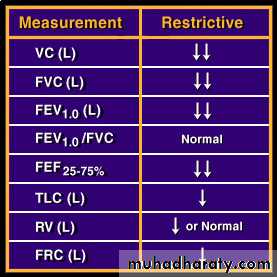
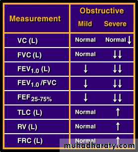
Types of respiratory abnormalities
ObstructiveRestrictive
Pulmonary emphysema
Infection, obstruction, alveolar damage, decrease diffusing capacity, and may lead to pulmonary hypertension.Pneumonia
Infection, filling of areas with fluid and consolidation leading to hypoxia and hypercapnia.
Atelectasis
Lung collapse
Asthma: Spastic contraction to bronchioles leading to hypoxia.
Hypoxia
Circulatory hypoxia:
Histotoxic hypoxia
Anemic hypoxia
Hypoxic hypoxia