
Granulopoiesis
Regulated by GM-CSF

At the end of this lecture, the 1
st
medical
student will be able to review maturation
stages of
1. granulocytes
2. lymphocytes
3. monocytes
4. thrombocytes
Granulopiosis
1.
pluripotential stem cells
2.
myeloid multipotential stem cells
3.
granulocyte colony forming cell
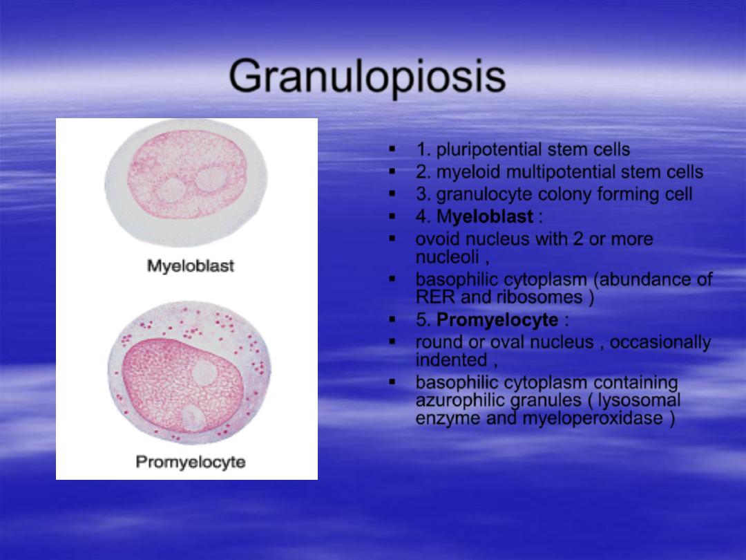
4.
Myeloblast
:
ovoid nucleus with 2 or more
nucleoli ,
basophilic cytoplasm (abundance of
RER and ribosomes )
5.
Promyelocyte
:
round or oval nucleus , occasionally
indented ,
basophilic cytoplasm containing
azurophilic granules ( lysosomal
enzyme and myeloperoxidase )
Granulopiosis
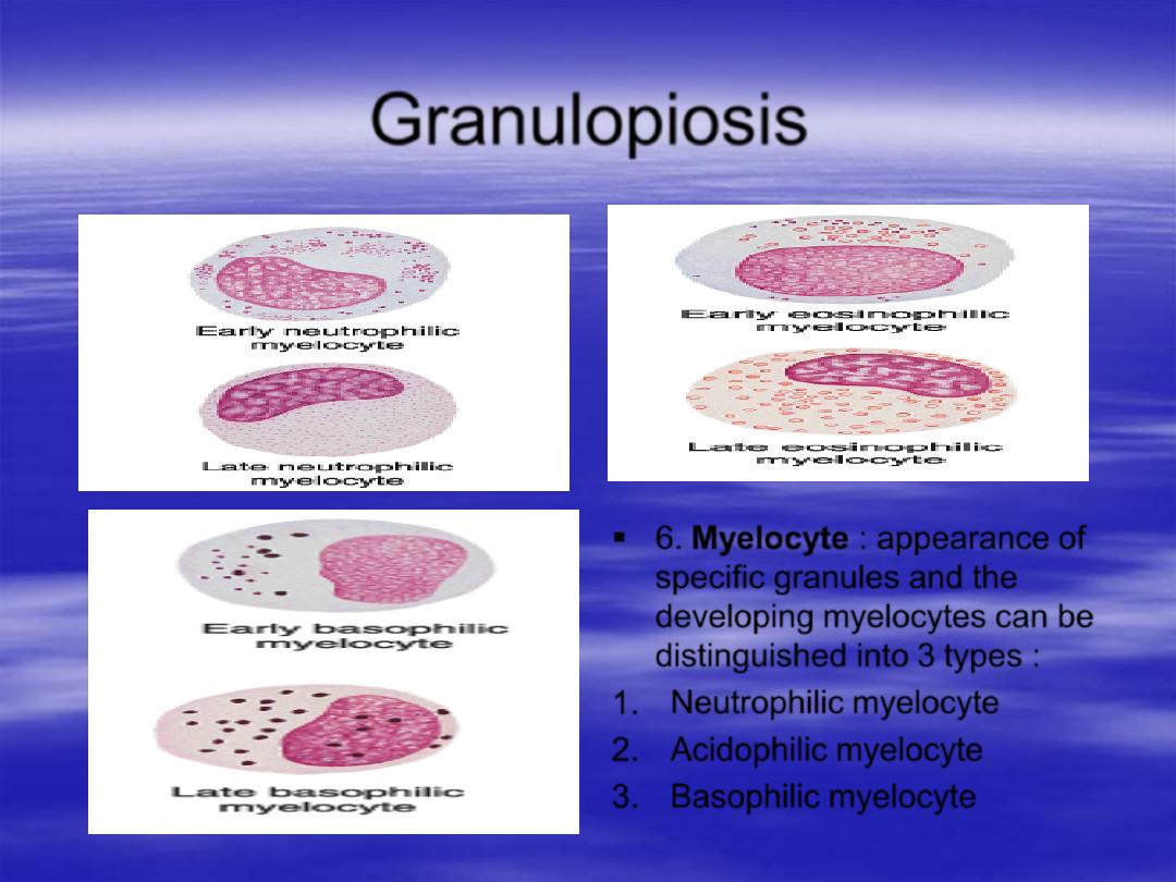
6.
Myelocyte
: appearance of
specific granules and the
developing myelocytes can be
distinguished into 3 types :
1.
Neutrophilic myelocyte
2.
Acidophilic myelocyte
3.
Basophilic myelocyte
Granulopiosis
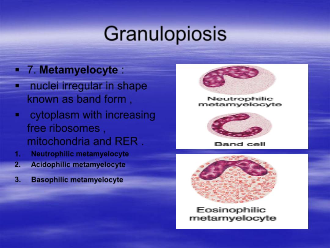
7.
Metamyelocyte
:
nuclei irregular in shape
known as band form ,
cytoplasm with increasing
free ribosomes ,
mitochondria and RER .
1.
Neutrophilic metamyelocyte
2.
Acidophilic metamyelocyte
3.
Basophilic metamyelocyte
Granulopiosis
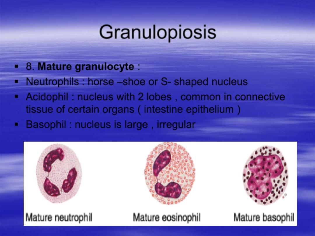
8.
Mature granulocyte
:
Neutrophils : horse
–shoe or S- shaped nucleus
Acidophil : nucleus with 2 lobes , common in connective
tissue of certain organs ( intestine epithelium )
Basophil : nucleus is large , irregular
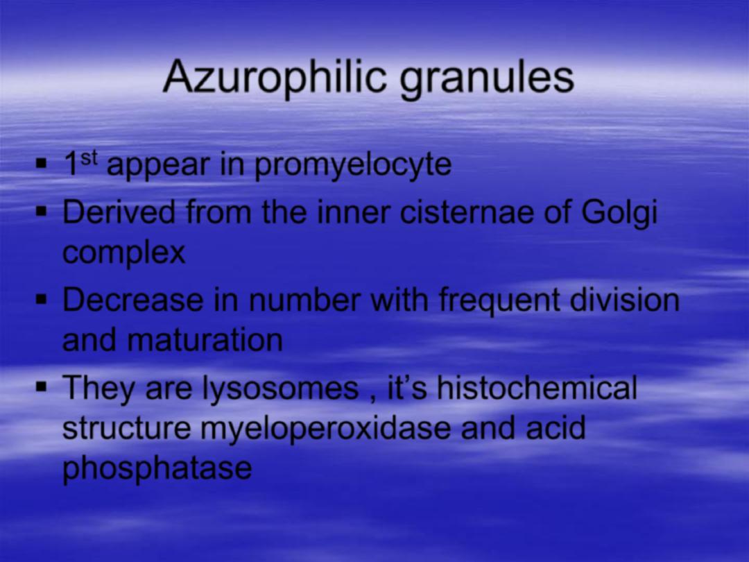
Azurophilic granules
1
st
appear in promyelocyte
Derived from the inner cisternae of Golgi
complex
Decrease in number with frequent division
and maturation
They are lysosomes
, it’s histochemical
structure myeloperoxidase and acid
phosphatase

Specific granules
Initial appearance in myelocyte
Derived from an outer cisternae of Golgi complex
Increase in number with maturation
neutrophil contain alkaline phosphatase and
antibacterial lysozyme
Acidophil contain sulphatase , peroxidase and
histaminase
Basophil contain heparin and histamine
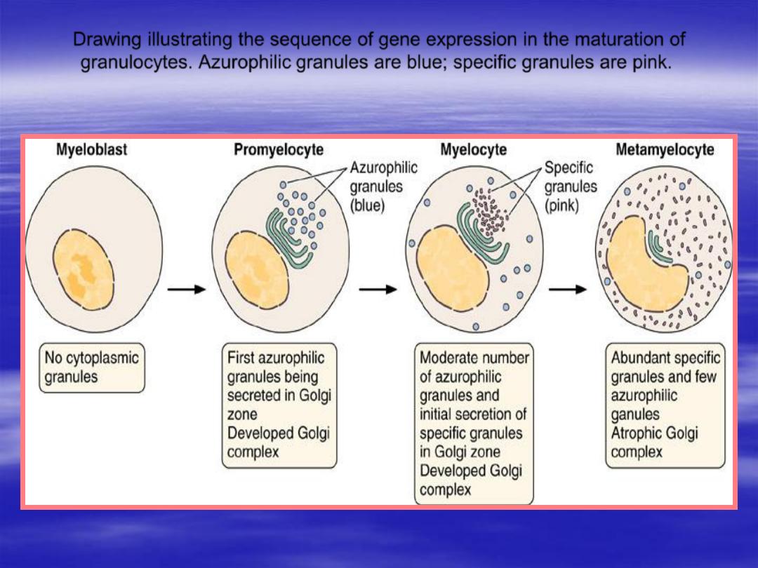
Drawing illustrating the sequence of gene expression in the maturation of
granulocytes. Azurophilic granules are blue; specific granules are pink.
MEDICAL APPLICATION
The appearance of large numbers of
immature neutrophils (band cells) in the
blood is called a shift to the left and is
clinically significant, usually indicating
bacterial infection.
Monopoiesis
Maturation of monocytes
Is regulated by GM-CSF
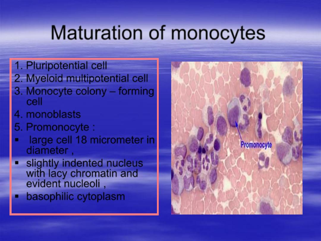
Maturation of monocytes
1. Pluripotential cell
2. Myeloid multipotential cell
3. Monocyte colony
– forming
cell
4.
monoblasts
5. Promonocyte :
large cell 18 micrometer in
diameter ,
slightly indented nucleus
with lacy chromatin and
evident nucleoli ,
basophilic cytoplasm
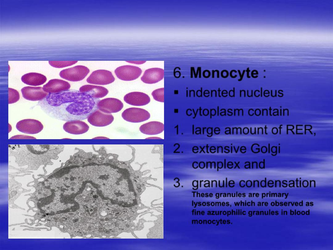
6.
Monocyte
:
indented nucleus
cytoplasm contain
1.
large amount of RER,
2.
extensive Golgi
complex and
3.
granule condensation
These granules are primary
lysosomes, which are observed as
fine azurophilic granules in blood
monocytes.
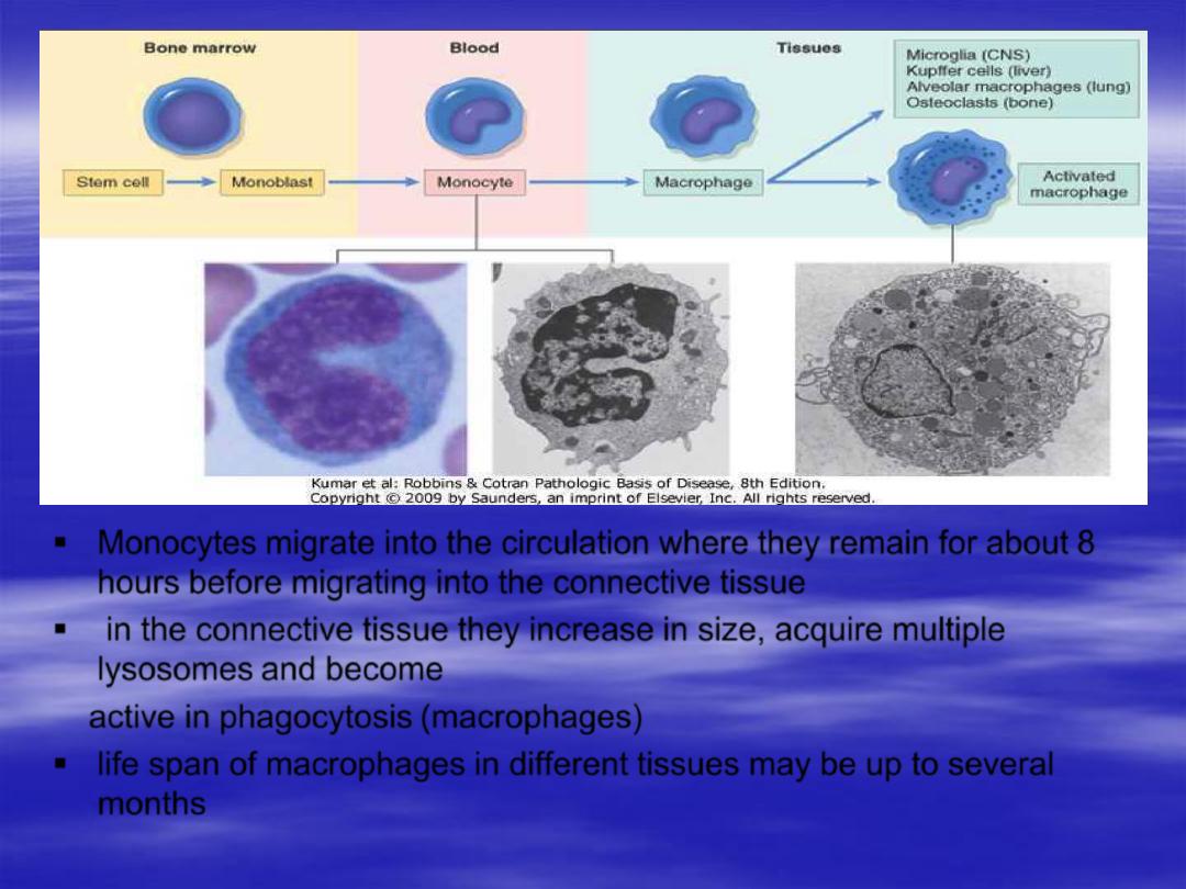
Monocytes migrate into the circulation where they remain for about 8
hours before migrating into the connective tissue
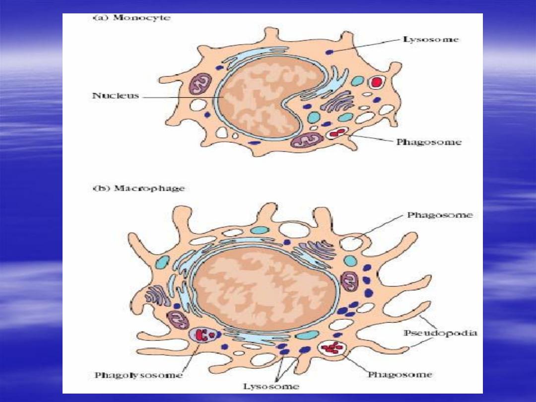
in the connective tissue they increase in size, acquire multiple
lysosomes and become
active in phagocytosis (macrophages)
life span of macrophages in different tissues may be up to several
months

Lymphopoiesis
Maturation of lymphocyte
Maturation of lymphocyte
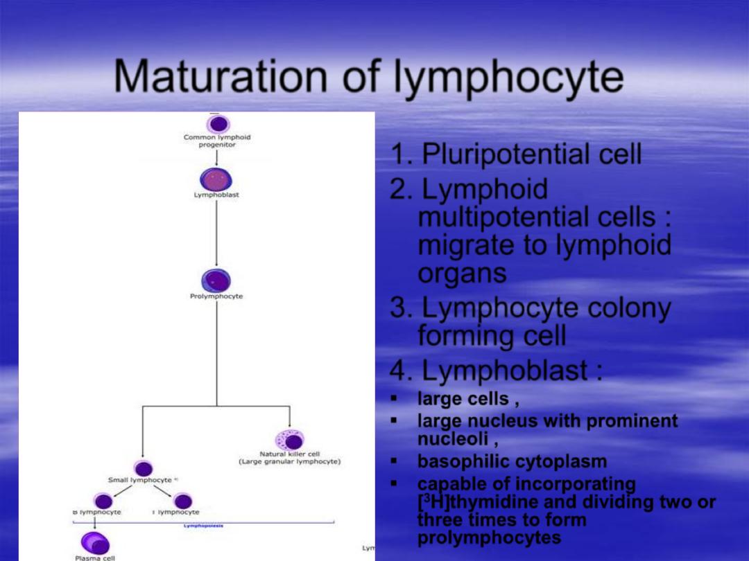
1.
Pluripotential cell
2.
Lymphoid
multipotential cells
:
migrate to lymphoid
organs
3.
Lymphocyte colony
forming cell
4.
Lymphoblast
:
large cells ,
large nucleus with prominent
nucleoli ,
basophilic cytoplasm
capable of incorporating
[
3
H]thymidine and dividing two or
three times to form
prolymphocytes
Maturation of lymphocyte
5.
Prolymphocyte
:
smaller with condensed chromatin ,
nucleoli less obvious ,
few azurophilic granules appear in the
cytoplasm ,
no cell surface receptor that mark them as T
or B lymphocyte
Maturation of lymphocyte
6. B and T lymphocytes
:
in the thymus or bone marrow , these cells
synthesize cell surface receptors but
they are not recognized as distinct cell
types using routine histological procedure .
They can be recognized by
immunohistochemistry
Medical application
Leukemias are malignant clones of leukocyte precursors.
They occur in
1.
lymphoid tissue (lymphocytic leukemias)
2.
bone marrow (myelogenous and monocytic leukemias).
In these diseases, there is usually a release of large
numbers of immature cells into the blood.
The symptoms of leukemias are a consequence of this
shift in cell proliferation, with a lack of some cell types and
excessive production of others (which are often abnormal
in function). The patient is usually anemic and prone to
infection.
Thrombopoiesis
Maturation of platelets
Regulated by thrombopoietin (TPO)
Mainly produced by the liver
Maturation of platelets
1.
Pluripotential cell
2.
Myeloid multipotential cell
3.
Megakaryocyte forming cell
4.
Megakaryoblast
:
large cell ( 15-50 micrometer ) ,
large ovoid or kidney shaped nucleus with
numerous nucleoli ( DNA 30X as much as a
normal cell ) ,
cytoplasm is homogenous and basophilic
Maturation of platelets
5.
Megakaryocyte
:
giant cell ( 35-150 micrometer),
irregular lobulated nucleus , coarse chromatin , no
visible nucleoli ,
Cytoplasm contain numerous mitochondria RER,
extensive Golgi complex , conspicuous granules
contain biologically active substances such as
platelet derived growth factor , fibroblast growth
factor
The demarkation membranes arise from
numerous invaginations of the plasma membrane
through out the cytoplasm
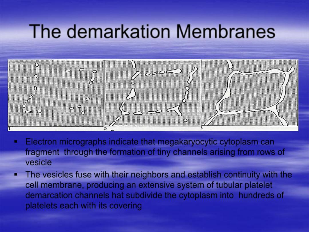
The demarkation Membranes
Electron micrographs indicate that megakaryocytic cytoplasm can
fragment through the formation of tiny channels arising from rows of
vesicle
The vesicles fuse with their neighbors and establish continuity with the
cell membrane, producing an extensive system of tubular platelet
demarcation channels hat subdivide the cytoplasm into hundreds of
platelets each with its covering
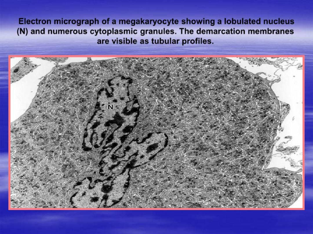
Electron micrograph of a megakaryocyte showing a lobulated nucleus
(N) and numerous cytoplasmic granules. The demarcation membranes
are visible as tubular profiles.
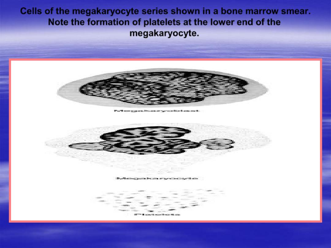
Cells of the megakaryocyte series shown in a bone marrow smear.
Note the formation of platelets at the lower end of the
megakaryocyte.
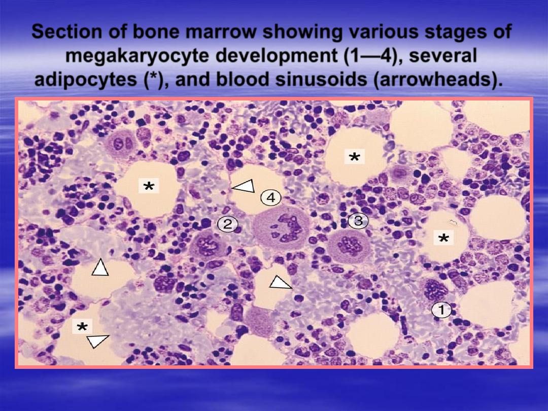
Section of bone marrow showing various stages of
megakaryocyte development (1
—4), several
adipocytes (*), and blood sinusoids (arrowheads).
MEDICAL APPLICATION
In certain forms of thrombocytopenic
purpura, a disease in which the number of
blood platelets is reduced, the platelets
appear to be bound to the cytoplasm of the
megakaryocytes, indicating a defect in the
liberation mechanism of these corpuscles.
The life span of platelets is approximately 10
days.

Thank you