
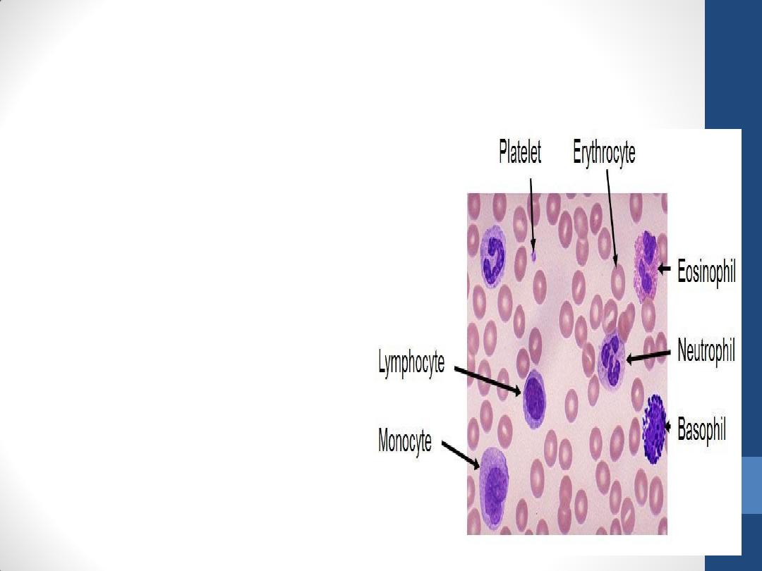
Blood
Prof. Dr. Malak A. Al-yawer

Objectives
At the end of this lecture, the medical
student will be able to
•
State the parts of blood
•
Name the dyes used for staining of blood
cells
•
identify the different types of blood cells
at the light and electron microscopic level
and describe their functions.

Blood
is made up of two parts:
•
formed elements
1.
erythrocytes (red blood cells),
2.
platelets,
3.
leukocytes (white blood cells).
•
plasma (Gr. plasma, thing formed), the liquid in
which the formed elements are suspended.
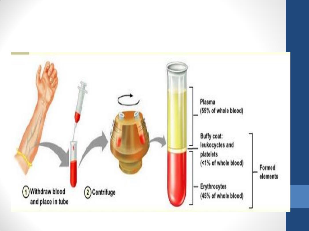
Components of Blood
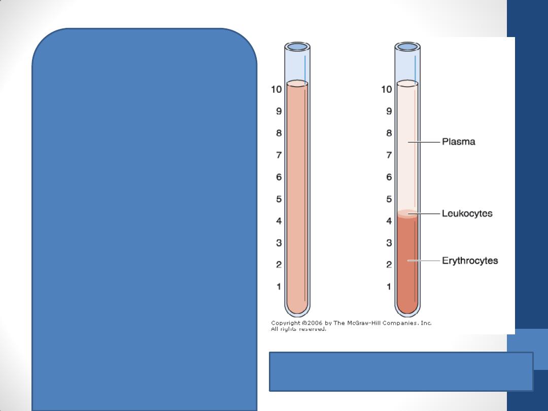
Hematocrit tubes with blood. Left: Before
centrifugation. Right: After centrifugation.
when whole blood is centrifuged
Plasma is the translucent,
yellowish, somewhat viscous
supernatant
The formed elements of the
blood separate into two easily
distinguishable layers.
1. The lower layer is red and is
made up of erythrocytes (42–
47% of the entire volume of
blood).
2. The layer immediately above
(1% of the blood volume),
which is white or grayish in
color, is called the buffy coat
and consists of leukocytes.
3. Covering the leukocytes is a
fine layer of platelets not
distinguishable by the naked
eye.
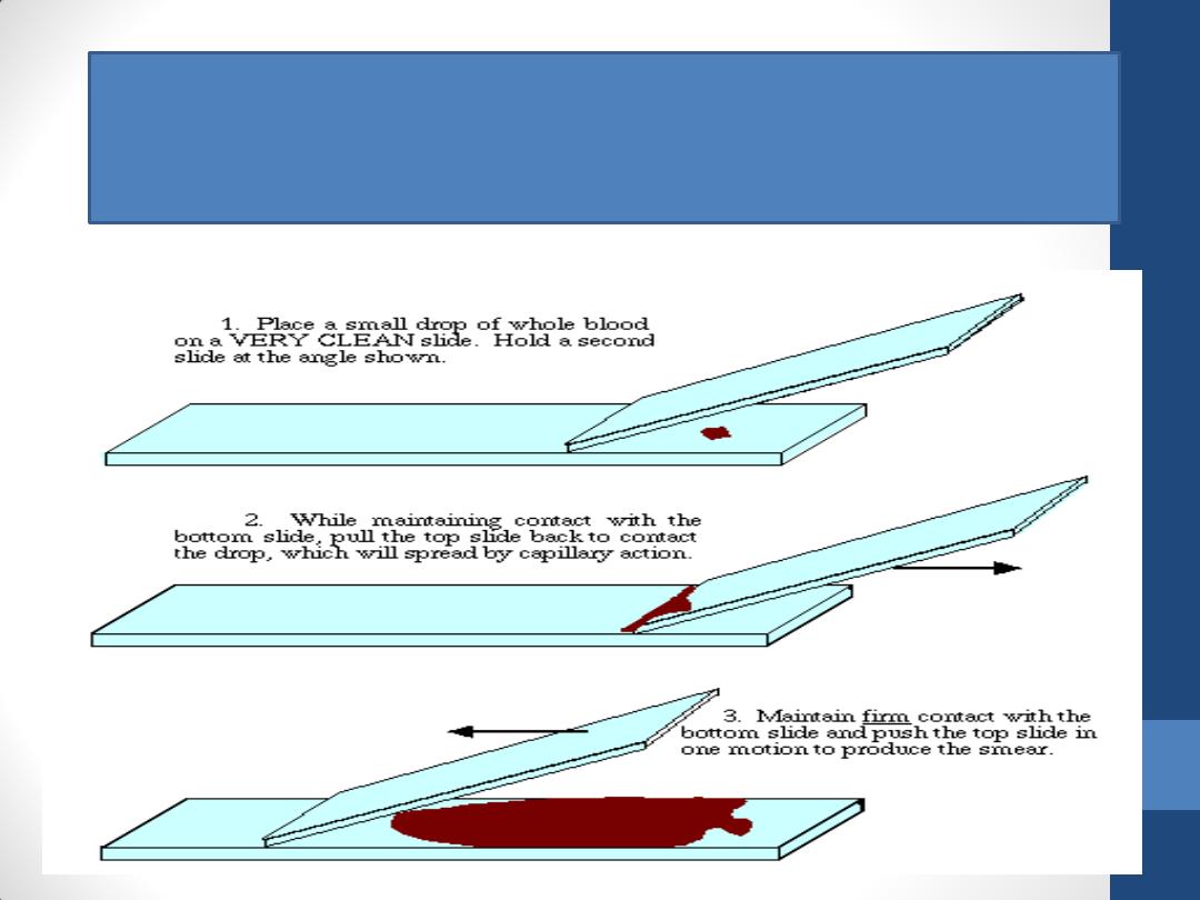
Blood cells are generally studied in smears or films prepared by spreading a drop
of blood in a thin layer on a microscope slide. The blood should be evenly
distributed over the slide and allowed to dry rapidly in air.

Staining of Blood Cells
•
Blood stains contain
1.
special mixtures of red (acidic) and blue (basic)
dyes.
2.
azures, dyes (useful in staining some structures
of blood cells known as azurophilics )
•
e.g. Giemsa, Wright's, Leishman's
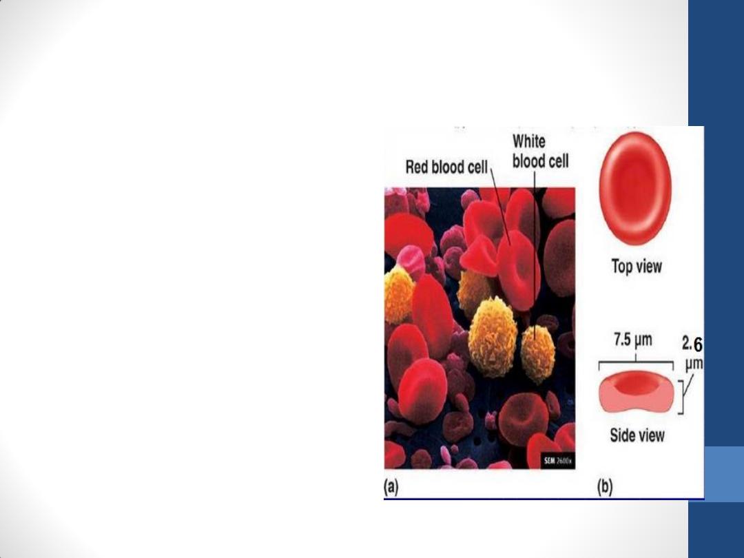
Erythrocytes (red blood cells)
•
are anucleate cells
•
Never leave the
circulatory system
under normal
conditions .
•
In an isotonic medium,
human erythrocytes
are
7.5 µm in diameter,
2.6 µm thick at the rim,
0.8 µm thick in the
center
.
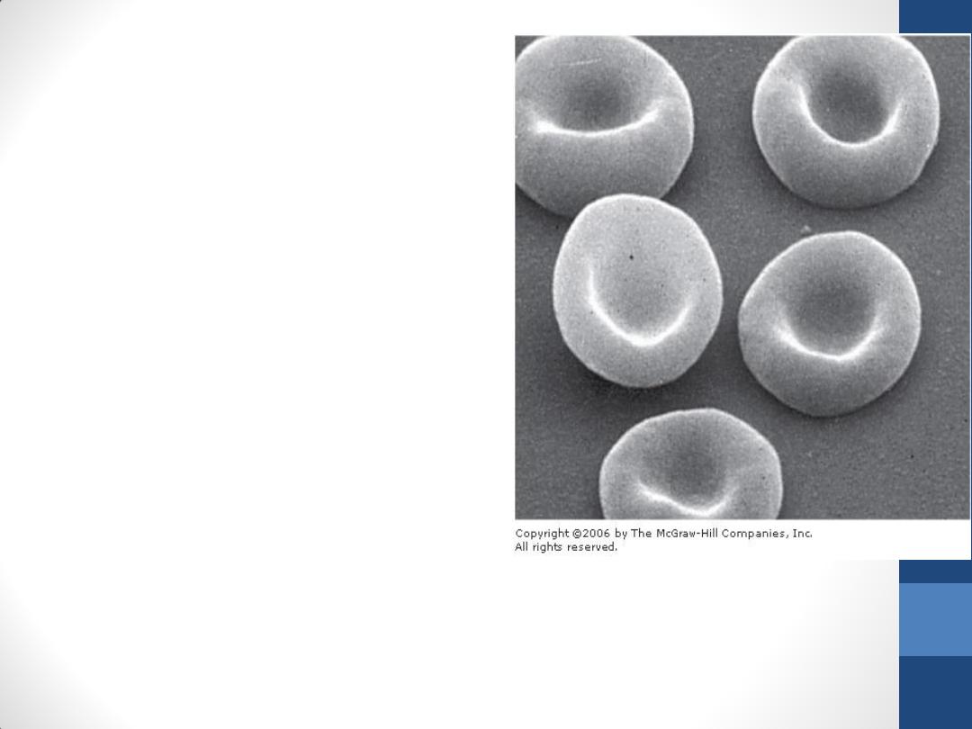
Scanning electron micrograph of normal
human erythrocytes. Note their biconcave
shape. x3300.
• The biconcave shape provides
erythrocytes with a large
surface-to-volume ratio, thus
facilitating gas exchange.
• Erythrocytes are surrounded
by a plasmalemma.
The normal concentration of
erythrocytes in blood is
3.9–5.5 million / microliter
in women
4.1–6 million /microliter in
men.
In their interiors, erythrocytes contain:
1. Hemoglobin ( O2-carrying protein) that accounts for
their acidophilia.
2. Enzymes of glucose metabolism

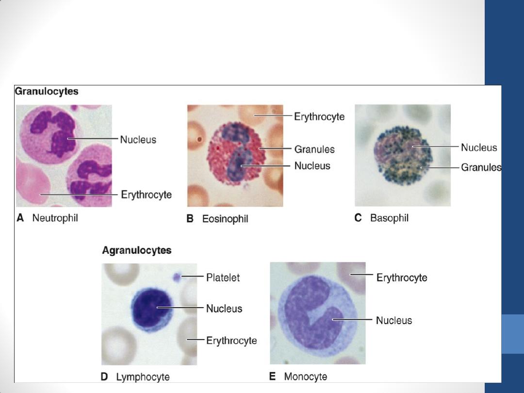
Leukocytes (white blood cells)
are divided into two groups according to the type
of granules in their cytoplasm and the shape of
their nuclei, :
1.
granulocytes (polymorphonuclear leukocytes)
2.
agranulocytes (mononuclear leukocytes).
•
They are spherical while suspended in blood
plasma, but some become ameboid after leaving
the blood vessels and invading the tissues.
Leukocytes (white blood cells)

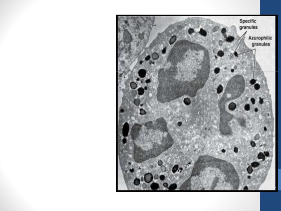
Granulocytes
•
possess two types of granules:
1.
Specific granules (neutral, basic, or acidic) and
have specific functions
2.
Azurophilic granules stain purple and are
lysosomes.
•
Have a life span of a few days, dying by apoptosis
(programmed cell death) in the connective
tissue.
Granulocytes
•
have poorly developed Golgi
complex and rough
endoplasmic reticulum (non-
dividing terminal cells, do not
synthesize much protein).
•
have few mitochondria (low
energy metabolism) and
depend more on glycolysis;
•
contain glycogen and can
function in regions scarce in
oxygen, such as inflamed
areas.
Agranulocytes
•
do not have specific
granules, but they do
contain azurophilic
granules (lysosomes)
that bind the azure
dyes of the stain.
•
The nucleus is round
or indented.
•
includes lymphocytes
and monocytes
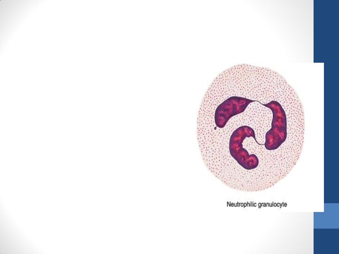
Neutrophils (Polymorphonuclear
Leukocytes)
•
constitute 60–70% of
circulating leukocytes.
•
are 12–15 µm in diameter
(in blood smears), with a
nucleus consisting of two
to five (usually three) lobes
linked by fine threads of
chromatin
•
In females, the inactive X
chromosome appears as a
drumstick-like appendage
on one of the lobes of the
nucleus(this characteristic
is not obvious in all
neutrophils in a blood
smear).
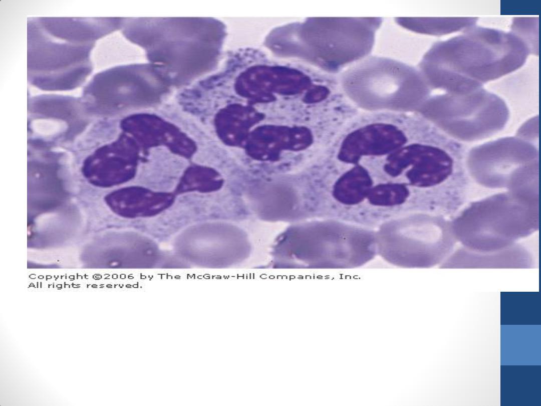
Photomicrograph of a blood smear showing three neutrophils and several
erythrocytes. Each neutrophil has only one nucleus, with a variable number of
lobes. Giemsa stain. High magnification.

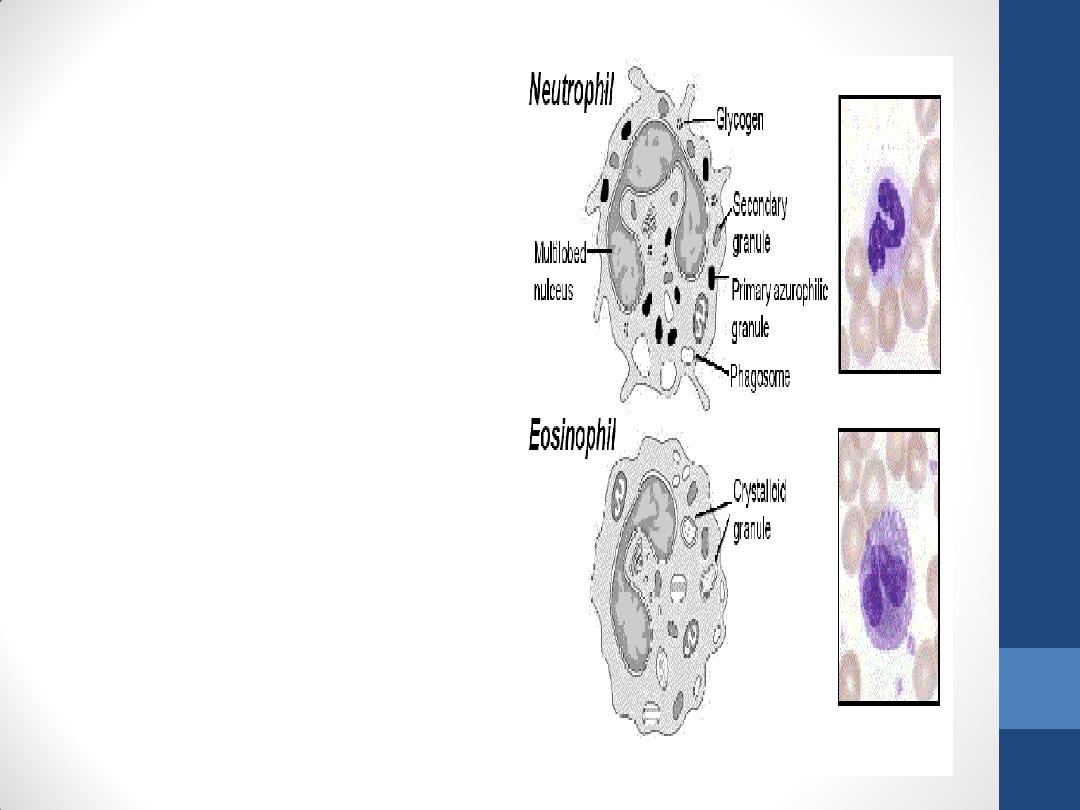
• The cytoplasm of the neutrophil contains two main types
of granules.
1. Specific granules more abundant granules , small( near
the limit of resolution of the light microscope).
2. Azurophilic granules, which are lysosomes 0.5μm in
diameter.
Neutrophils also contain glycogen in their cytoplasm
Glycogen is broken down into glucose to yield energy via the
glycolytic pathway of glucose oxidation. The citric acid cycle is
less important ( few mitochondria).
The ability of neutrophils to survive in an anaerobic
environment is highly advantageous, since they can kill
bacteria and help clean up debris in poorly oxygenated
regions, e.g., inflamed or necrotic tissue.
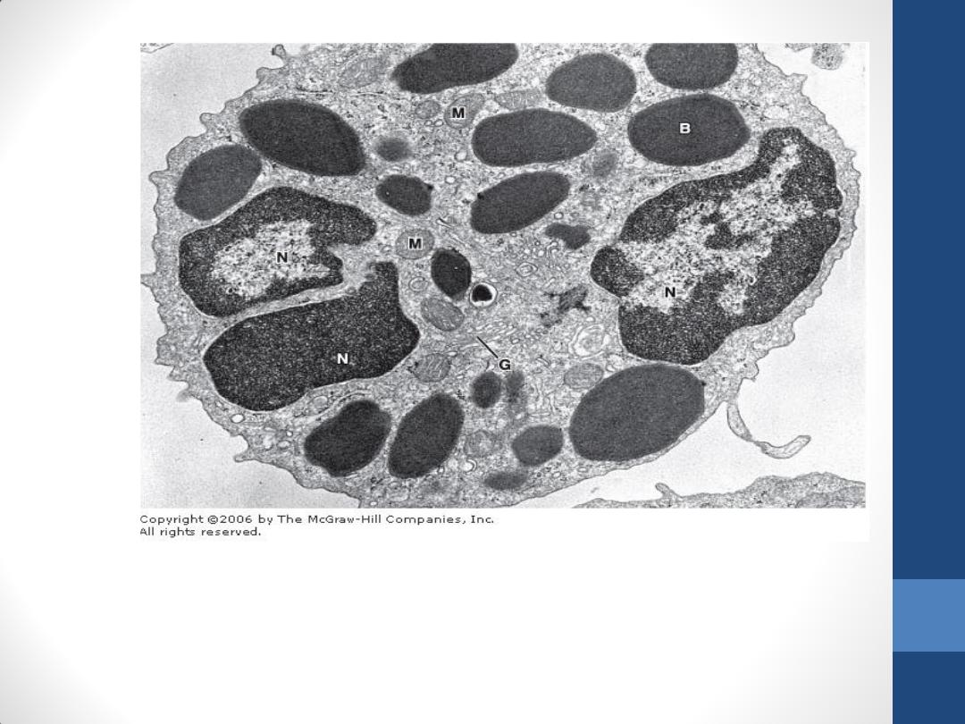
Electron micrograph of a human
neutrophil stained for peroxidase.
The cytoplasm contains
• two types of granules:
1. Specific granules the small,
pale, peroxidase-negative and
more abundant
2. Azurophilic granules the larger,
dense, peroxidase-positive.
•
Neutrophils also contain
glycogen in their cytoplasm
•
The nucleus is lobulated, and
the Golgi complex is small.
Rough endoplasmic reticulum
and mitochondria are not
abundant. x27,000.
Neutrophils
•
are short-lived cells with a
half-life of 6–7 h in blood
and a life span of 1–4 days
in connective tissues,
where they die by
apoptosis.
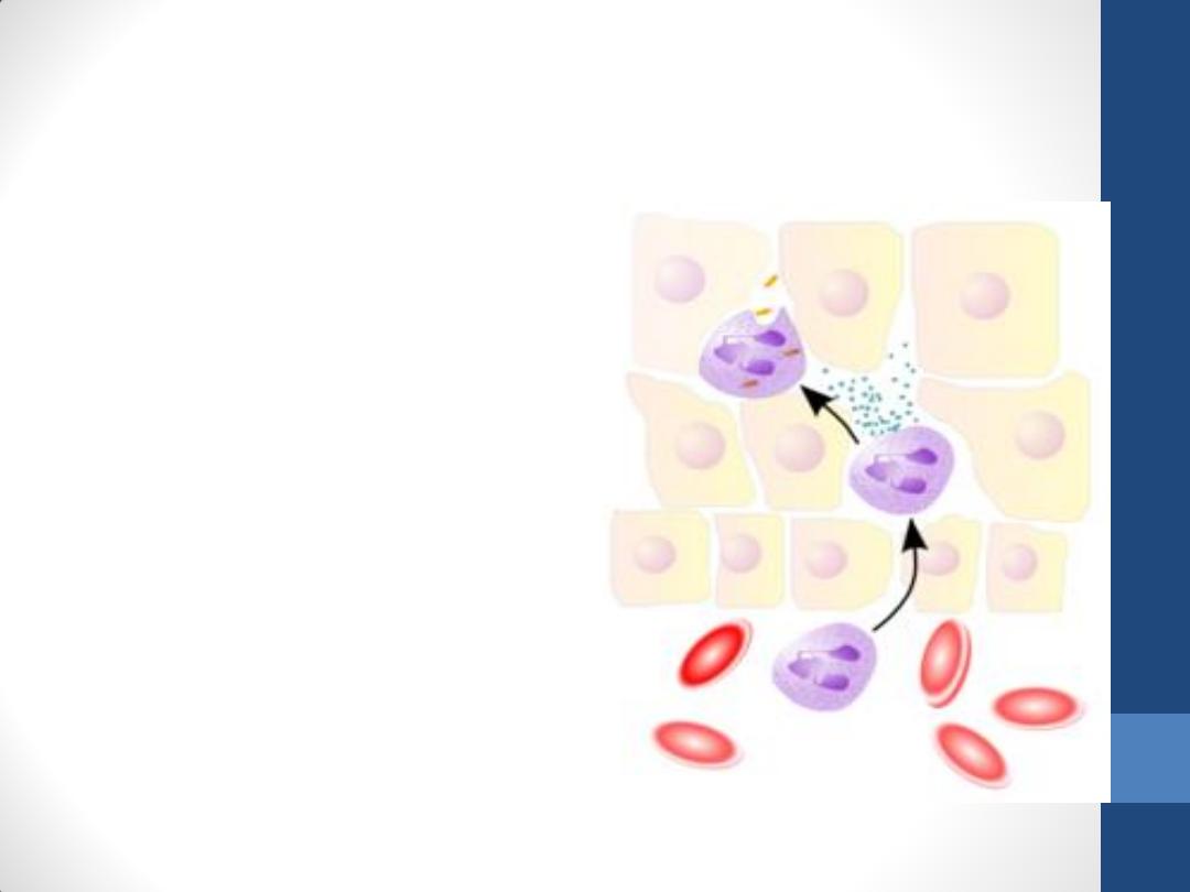
•
are active phagocytes of
bacteria and other small
particles. Neutrophils are
inactive and spherical while
circulating but show an
active ameboid movement
upon adhering to a solid
substrate, such as collagen
in the extracellular matrix.
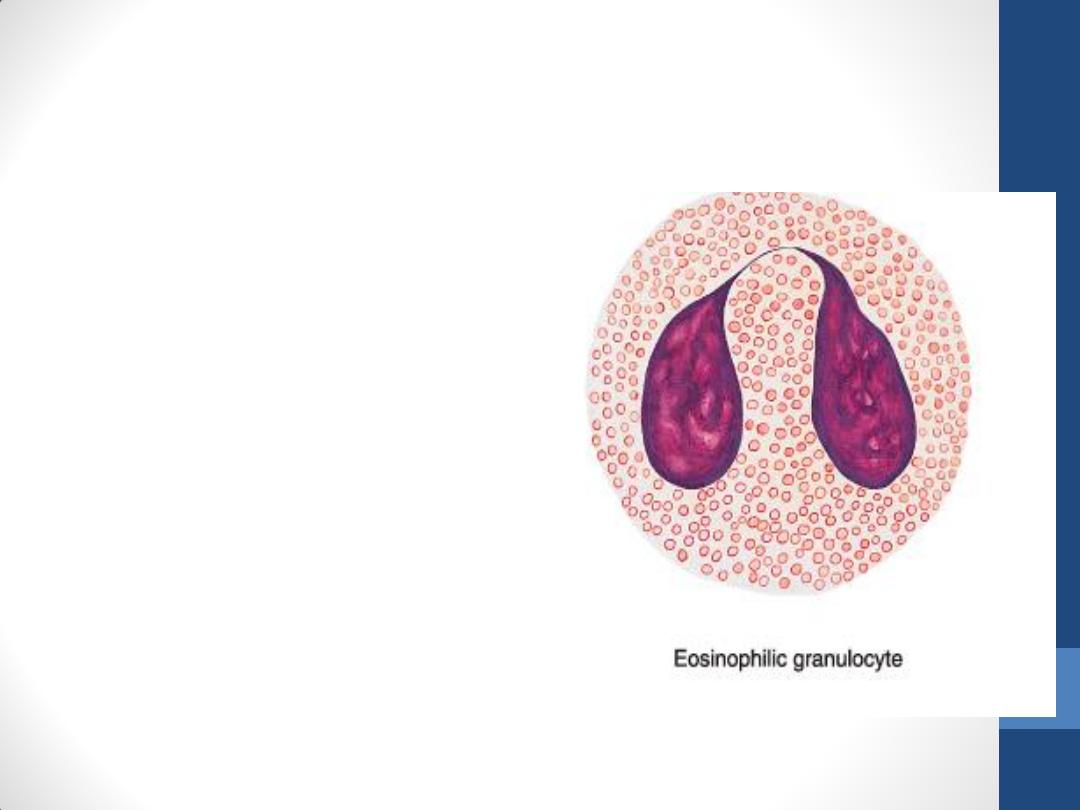
Eosinophils
•
constitute only 2–4% of
leukocytes in normal
blood.
•
In blood smears, this cell is
about the same size as a
neutrophil and contains a
characteristic bilobed
nucleus .
•
The main identifying
characteristic is the
presence of many large
and elongated refractile
specific granules (about
200 per cell) that are
stained by eosin
Photomicrograph of an eosinophil. Note its typical bilobed nucleus
and coarse cytoplasmic granules. Giemsa stain. High magnification.
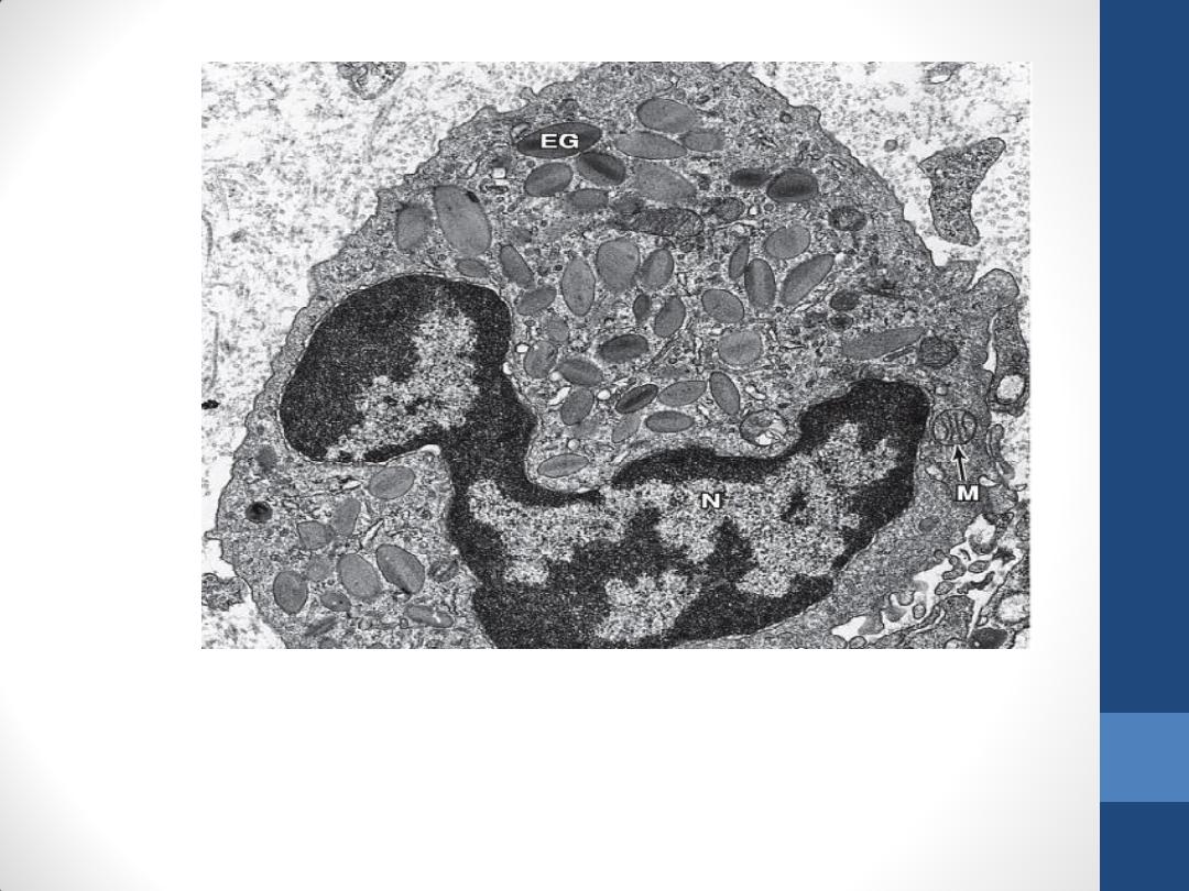
Electron micrograph of an eosinophil. Typical eosinophilic granules are clearly seen. Each
granule has a disk-shaped electron-dense crystalline core that appears surrounded by a
matrix enveloped by a unit membrane. EG, eosinophil granule; N, nucleus; M,
mitochondria. x20,000.
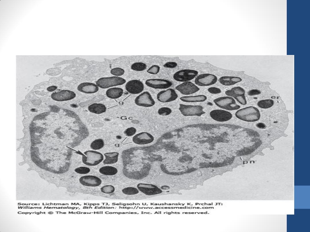
Human mature eosinophil incubated for peroxidase. Reaction product is
present only in granules (g). The rough endoplasmic reticulum (er), including
the perinuclear cisterna (pn) and the Golgi cisternae (Gc), does not contain
reaction product. Most of the granules (arrow) contain the distinctive
crystalline bar (x8000).
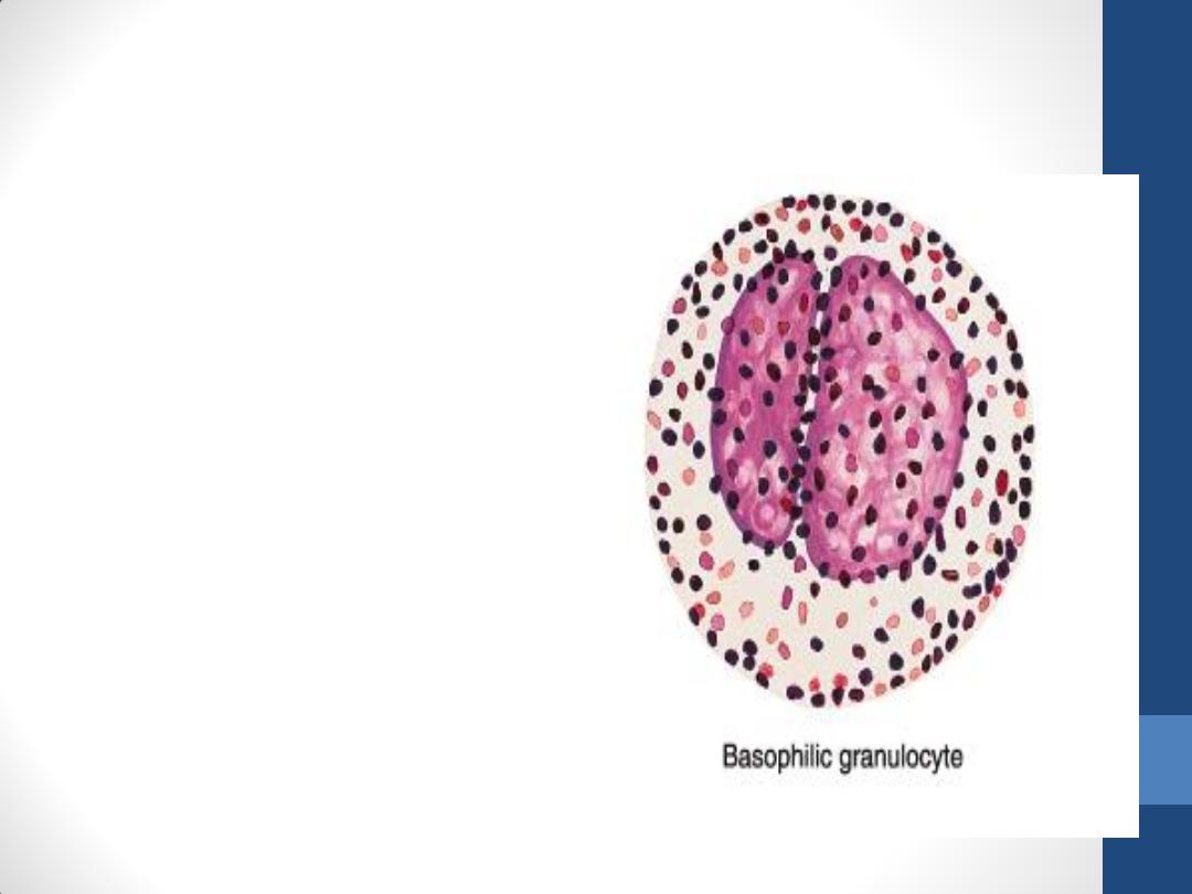
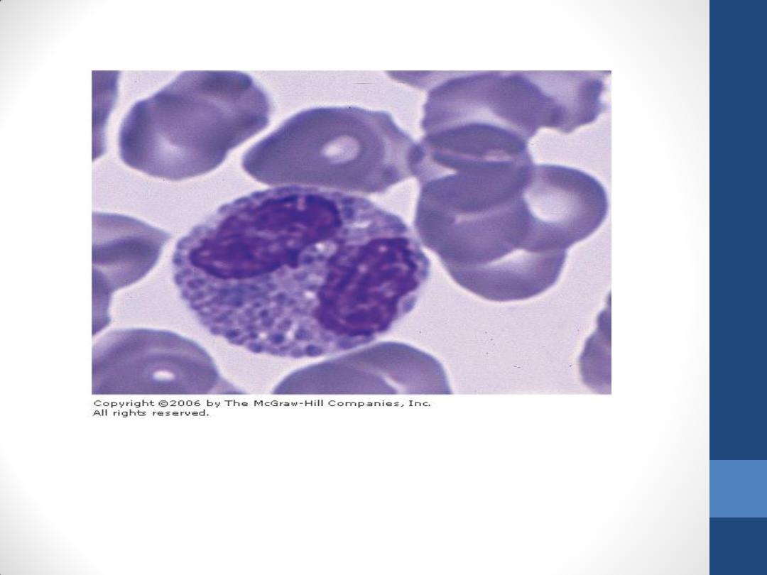
Basophils
•
make up less than 1% of
blood leukocytes and are
therefore difficult to find
in smears of normal
blood.
•
are about 12–15 µm in
diameter. The nucleus is
divided into irregular
lobes, but the overlying
specific granules usually
obscure the division.

Basophilic specific granules
•
They (0.5µm in diameter) stain metachromatically (change the
color of the stain used) with the basic dye of the usual blood
stains . This metachromasia is due to the presence of heparin.
•
They are fewer and more irregular in size and shape than the
granules of the other granulocytes.
•
They contain heparin and histamine.
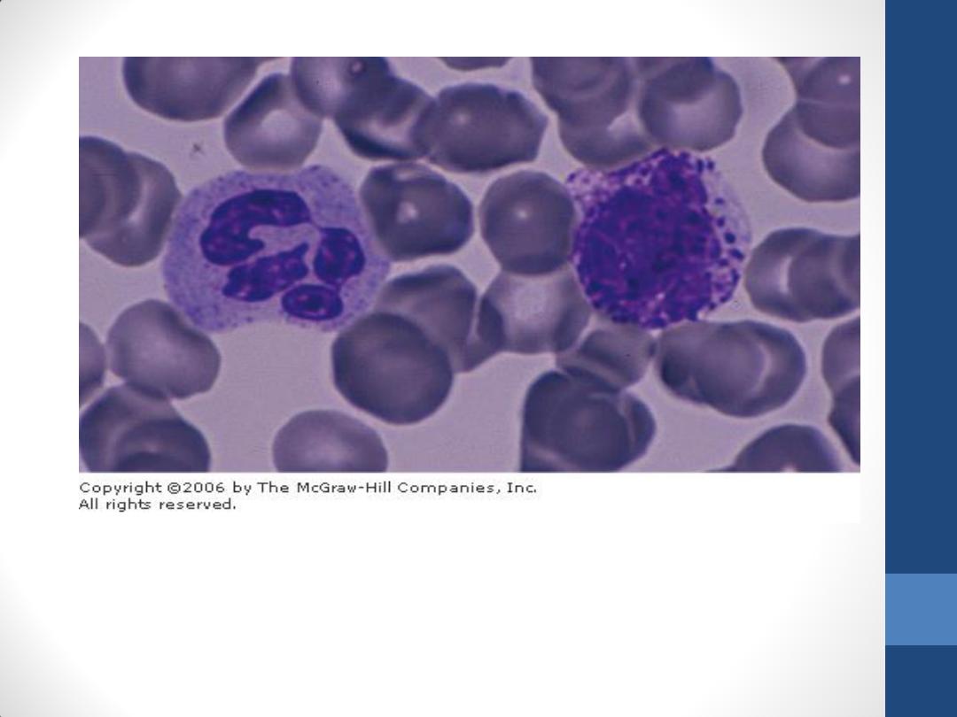
Two leukocytes and several erythrocytes. The cell on the right
is a basophil. The cell on the left is a neutrophil. In the basophil
there are many cytoplasmic granules over the nucleus. Giemsa
stain. High magnification.
Electron micrograph of a rabbit basophil. The lobulated nucleus (N)
appears as three separated portions. Note the basophilic granule (B),
mitochondria (M), and Golgi complex (G). x16,000.

Compare between basophils &
Mast cells
Basophil
Mast cell
Lymphocytes
•
They can be classified into several groups
according to distinctive surface molecules
(markers), which can be distinguished by
immunocytochemical methods.
•
They also have diverse functional roles, all related
to immune reactions in defending against invading
microorganisms, foreign macromolecules, and
cancer cells.
•
Lymphocytes vary in life span; some live only a
few days, and others survive in the circulating
blood for many years.
•
Lymphocytes are the only type of leukocytes that
return from the tissues back to the blood, after
diapedesis.
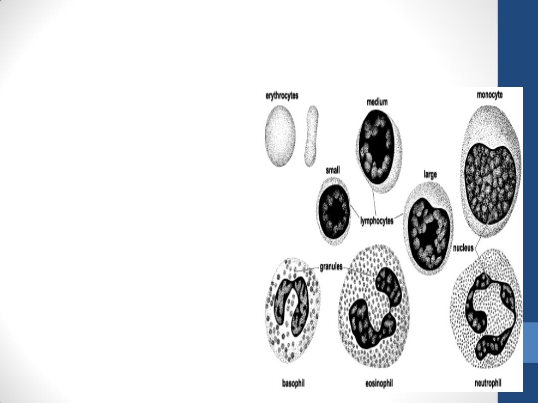
•
Lymphocytes with
diameters of 6–8 µm are
known as small
lymphocytes. A small
number of medium-sized
lymphocytes and large
lymphocytes with
diameters up to 18 µm are
present in the circulating
blood .
•
This difference has
functional significance in
that some larger
lymphocytes are believed
to be cells activated by
specific antigens.

Small lymphocyte
•
has a spherical nucleus, sometimes with an indentation. The
nucleus is intensely stained(condensed chromatin). Its
nucleolus is not visible
•
The cytoplasm is scanty, it appears as a thin rim around the
nucleus. It is slightly basophilic, assuming a light blue color in
stained smears. It may contain a few azurophilic granules. The
cytoplasm of the small lymphocyte has a few mitochondria
and a small Golgi complex; it contains free polyribosomes
Two small lymphocytes with their
round, dark-stained nuclei. Giemsa
stain. High magnification.
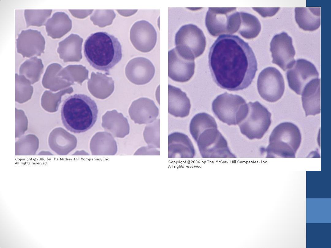
Photomicrograph of a large lymphocyte
and several erythrocytes. The nucleus of
this cell is round, and the cytoplasm is
devoid of specific granules. Giemsa stain.
High magnification
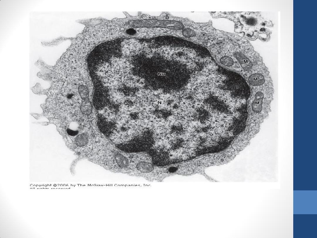
Electron micrograph of a human blood lymphocyte. This cell has little rough
endoplasmic reticulum but a moderate quantity of free polyribosomes.
Note the nucleus (N), the nucleolus (Nu), and the mitochondria (M).
Reduced from x22,000.
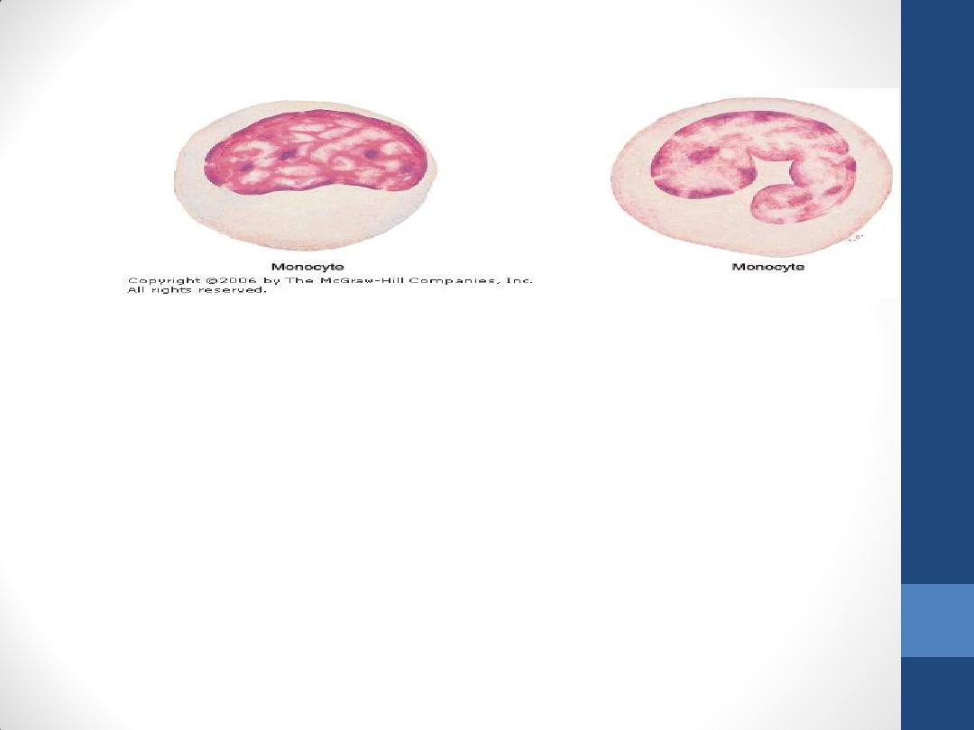
Monocytes
•
are bone marrow-derived agranulocytes with diameters varying from 12 to
20 μm.
•
The nucleus is oval, horseshoe, or kidney shaped and is generally eccentrically
placed.
•
The chromatin is less condensed than that in lymphocytes. Because of their
delicate chromatin distribution, the nuclei of monocytes stain lighter than do
those of large lymphocytes
•
The cytoplasm is basophilic and frequently contains very fine azurophilic
granules (lysosomes). These granules are distributed through the cytoplasm,
giving it a bluish-gray color in stained smears.
•
After crossing venule or capillary walls and entering connective tissues,
monocytes differentiate into macrophages.
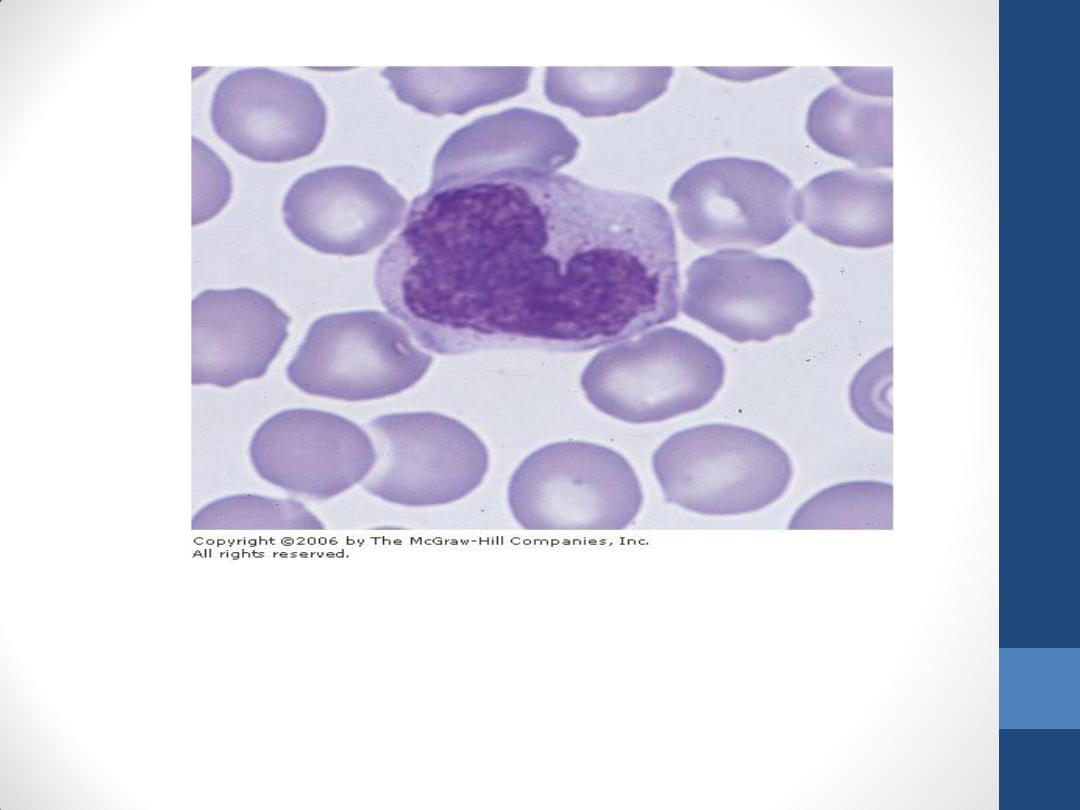
Photomicrograph of a monocyte. This cell type has a kidney-shaped
nucleus with delicately stained chromatin. The cytoplasm is slightly
basophilic. Giemsa stain. High magnification.
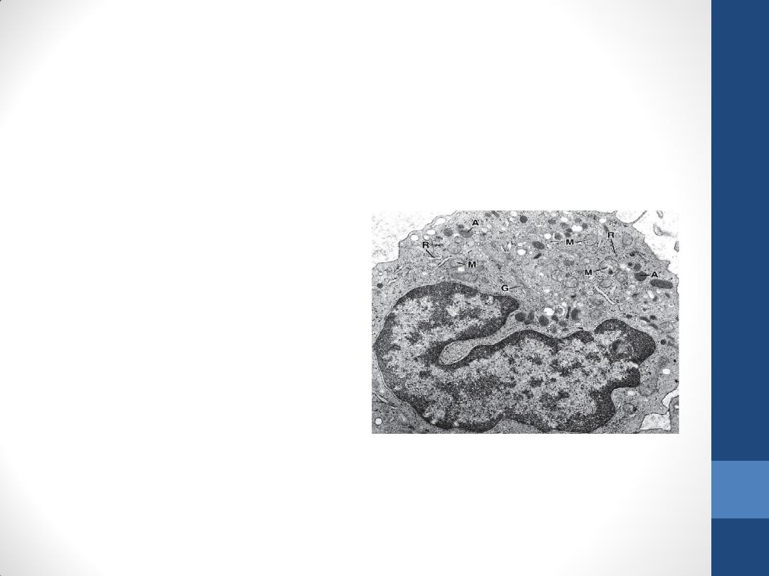
In the electron microscope,
one or two nucleoli are
seen in the nucleus
a small quantity of rough
endoplasmic reticulum,
polyribosomes
many small mitochondria
is observed.
A Golgi complex involved
in the formation of the
lysosomal granules is
present in the cytoplasm.
Many microvilli and
pinocytotic vesicles are
found at the cell surface

Platelets(thrombocytes)
•
are nonnucleated, disk-like cell fragments 2–4 µm in
diameter.
•
Platelets originate from the fragmentation of giant polyploid
megakaryocytes that reside in the bone marrow.
•
promote blood clotting and help repair gaps in the walls of
blood vessels, preventing loss of blood.
•
Normal platelet counts range from 200,000 to 400,000 per
microliter of blood.
•
have a life span of about 10 days.
•
often appear in clumps. Each platelet has a peripheral light
blue-stained transparent zone, the hyalomere, and a central
zone containing purple granules, called the granulomere.
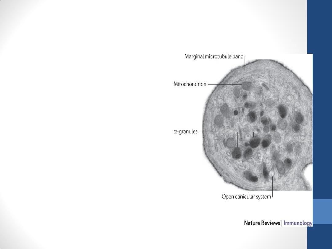
Platelets contain
•
the open canalicular system
(a system of channels), that
connects to invaginations of
the platelet plasma
membrane. This arrangement
is probably of functional
significance in facilitating the
liberation of active molecules
stored in platelets.
•
a marginal bundle of
microtubules around the
periphery of the platelet; this
bundle helps to maintain the
platelet's ovoid shape.
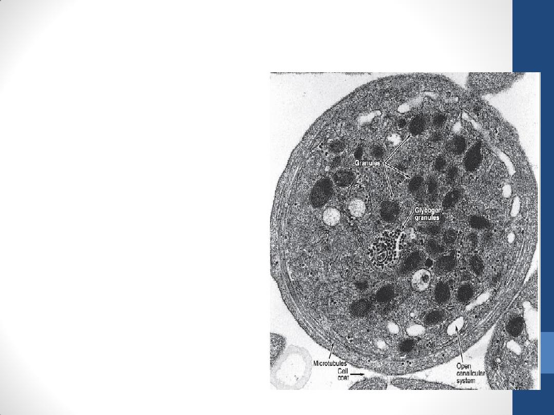
In the hyalomere
•
there are also a number of
electron-dense irregular
tubes known as the dense
tubular system.
•
Actin and myosin molecules
in the hyalomere can
assemble to form a
contractile system that
functions in platelet
movement and aggregation.
•
A cell coat rich in
glycosaminoglycans and
glycoproteins lies outside the
plasmalemma and is involved
in platelet adhesion.

The central granulomere
possesses a variety of membrane-bound granules and a sparse
population of mitochondria and glycogen particles.
•
Dense bodies (δ-granules) contain calcium ions, adenosine
diphosphate (ADP), adenosine triphosphate (ATP) and
serotonin .
•
α-granules contain fibrinogen, platelet-derived growth factor,
and several other platelet-specific proteins.
•
λ –granules(lambda granule) have been shown to contain
only lysosomal enzymes
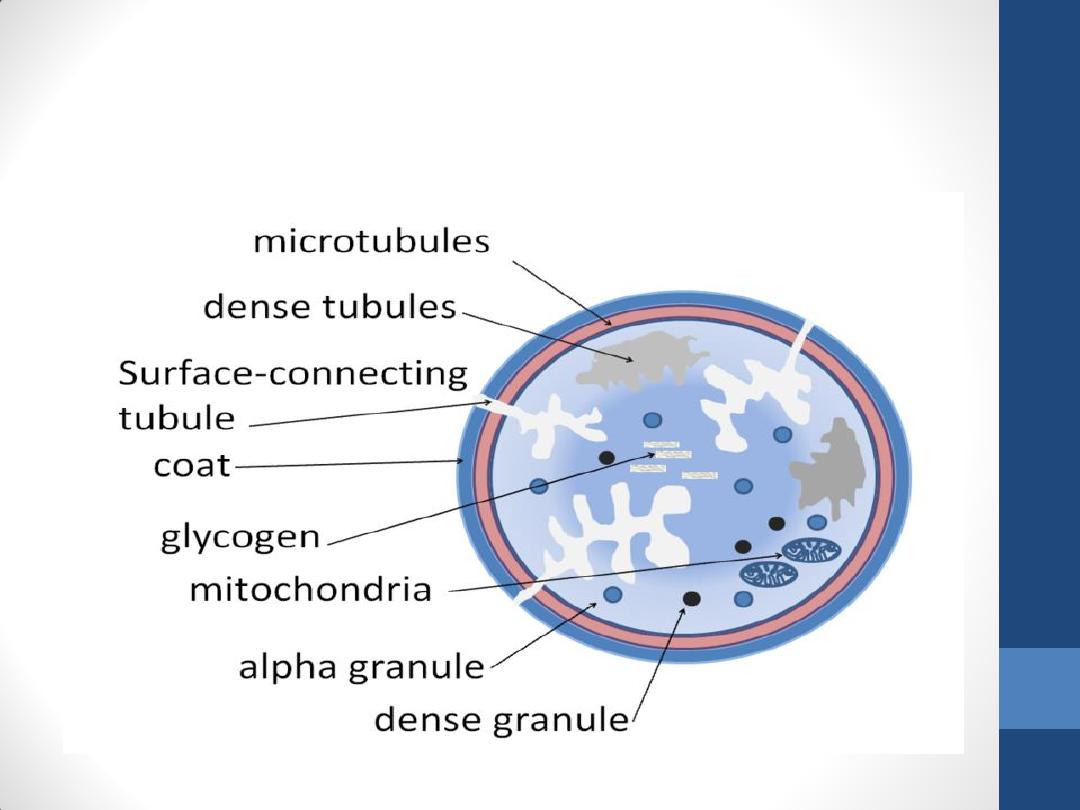
Diagram of the internal structure of
a platelet

Thank you
Next Lecture: Bone Marrow