
Dr.Maan Alkhalisy Embryology lec.11
1
Respiratory system
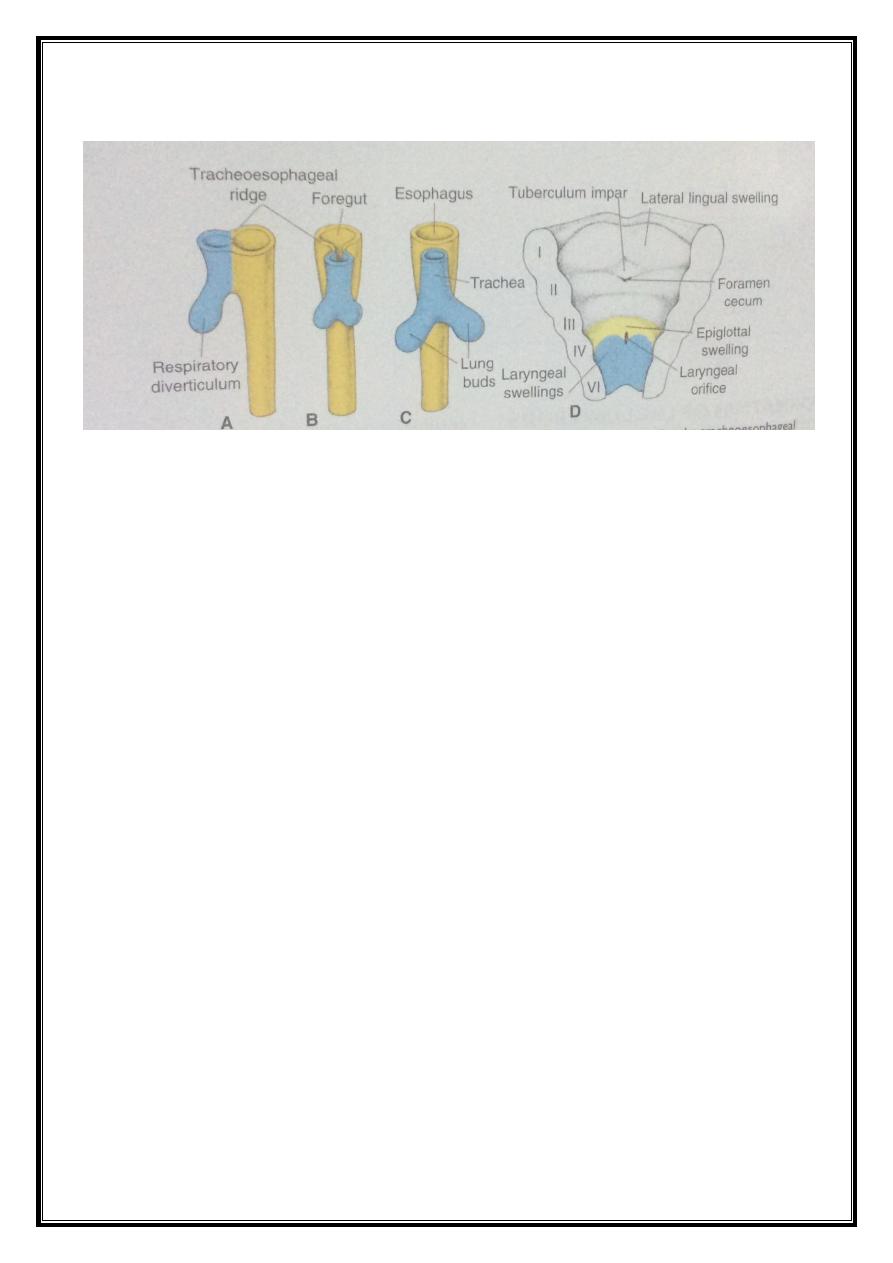
When the embryo is 4 weeks old, an outgrowth from the ventral wall
of the foregut will appear. This outgrowth is called the respiratory
diverticulum (lung bud).
The appearance and location of the lung bud are dependent upon
the increase in retinoic acid which is produced from the adjacent
mesoderm.
The epithelial lining of the larynx, trachea, bronchi, as well as the
lungs, is entirely of an endodermal origin. While the cartilaginous,
muscular, and connective tissue components of the trachea and lungs are
derived from the splanchnic mesoderm surrounding the foregut.
Initially, the lung bud is in open communication with the foregut.
As the respiratory diverticulum expands caudally, two longitudinal
ridges will form, the tracheo-oesophageal ridges separating it from the
foregut into a dorsal portion, the oesophagus, and a ventral portion called
the trachea and lung bud.
The respiratory primordium maintains its communication with the
pharynx through the laryngeal orifice.
Any defect in these processes could lead to oesophageal atresia or
tracheo-oesophageal fistula.
Dr.Maan Alkhalisy Embryology lec.11
2
Larynx :
The internal lining of the larynx originates from the endoderm, while
the cartilages and muscles originate from mesenchyme of the 4
th
and 6
th
pharyngeal arches
As a result of the rapid proliferation of this mesenchyme, the
laryngeal orifice changes in appearance from a sagittal slit to a T-shaped
opening. Subsequently, the mesenchyme of the two arches transforms
into thyroid, cricoid, and arytenoid cartilages. The characteristic adult
shape of the laryngeal opening can be recognized.
At the time of cartilages formation, the epithelial lining of the larynx
will proliferate rapidly in a way that causes the closure of the lumen.
Later on, recanalization of the lumen occurs, and becomes patent.
Besides, two lateral recesses, laryngeal ventricles, will occur.
These recesses are bounded by folding tissues forming the false and true
vocal cords.
Dr.Maan Alkhalisy Embryology lec.11
3
Since the muscles of the larynx are derived from the mesenchyme of
the 4
th
and 6
th
pharyngeal arches; therefore, the nerve supply will be by
the tenth cranial nerve (vagus n.).
The superior laryngeal nerve innervates the derivatives of the 4
th
pharyngeal arch, while the recurrent laryngeal nerve innervates the
derivatives of the 6
th
pharyngeal arch.
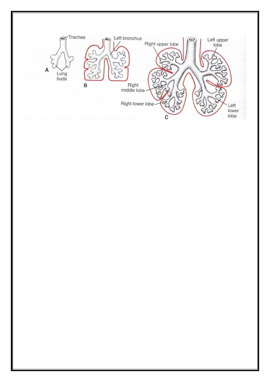
Trachea, bronchi, and lungs :
During the separation from the foregut, the lung bud forms the
trachea, and two bronchial buds.
At the beginning of the 5
th
week, each of these buds enlarges
forming the right and left main bronchi. The right then forms three
secondary bronchi, which later on form three the lobes of the right lung,
while the left one forms two secondary bronchi, which later on form the
two lobes of the left lung.
Dr.Maan Alkhalisy Embryology lec.11
4
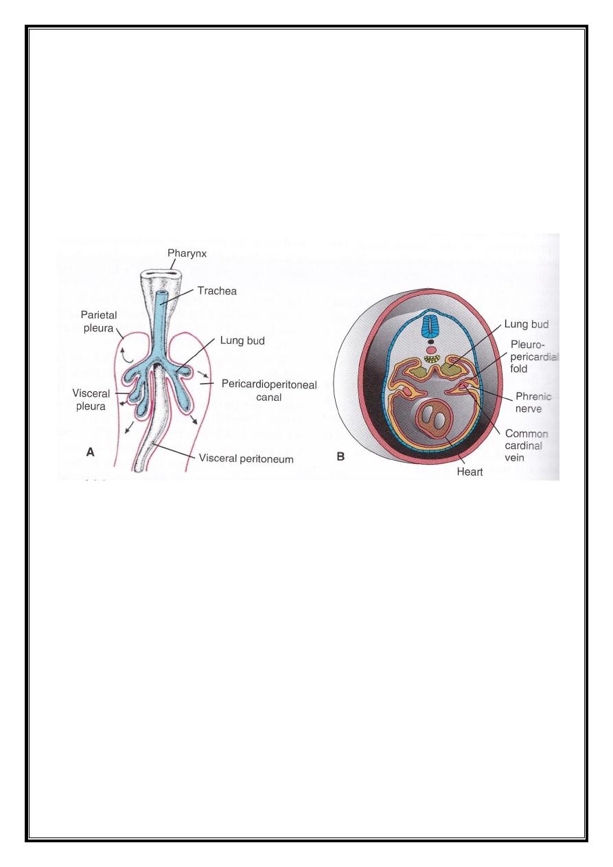
The pericardio-peritoneal canals, which lie on each side of the
foregut, become filled with the expanding lungs; therefore, the spaces
become narrow
Later on, the pleuro-peritoneal & pleuro-pericardial folds separate
the pericardio-peritoneal canals from the peritoneal and pericardial
cavities respectively, and the remaining spaces from the primitive pleural
cavity.
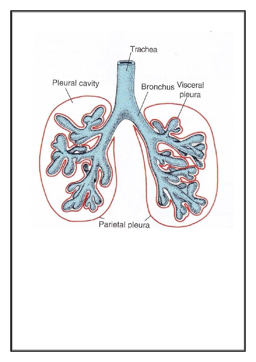
The mesoderm which covers the outside of the lungs develops into
the visceral pleura. The somatic mesoderm covering the body wall from
the inside becomes the parietal pleura, and the space between these two
pleurae is called the pleural cavity.
Dr.Maan Alkhalisy Embryology lec.11
5
Further development of the lungs leads to the division of the
secondary bronchi repeatedly, leading to the formation of 10 tertiary
(segmental) bronchi on the right side, and 8 ones on the left side. These
will create the broncho-pulmonary segments of the adult’s lung.
By the end of the sixth month, about 17 generation of subdivisions
have been formed. Before the bronchial tree reaches its final shape, an
additional 6 divisions is formed during the postnatal life.

Dr.Maan Alkhalisy Embryology lec.11
6
Maturation of the lung :
Up to the seventh prenatal months, the bronchioles are divided
continuously into more many smaller canals, and the vascular supply
increases steadily.
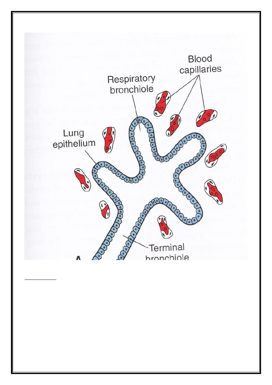
The terminal bronchioles divide into respiratory bronchioles, and
the respiratory bronchioles will divide into 3-6 alveolar ducts. The ducts
end in the terminal sacs (primitive alveoli) that are surrounded by flat
alveolar cells in close contact with the neighbouring capillaries.
By the end of the seventh month, sufficient amount of alveoli and
capillaries are present, which are sufficient to keep the infant survived.
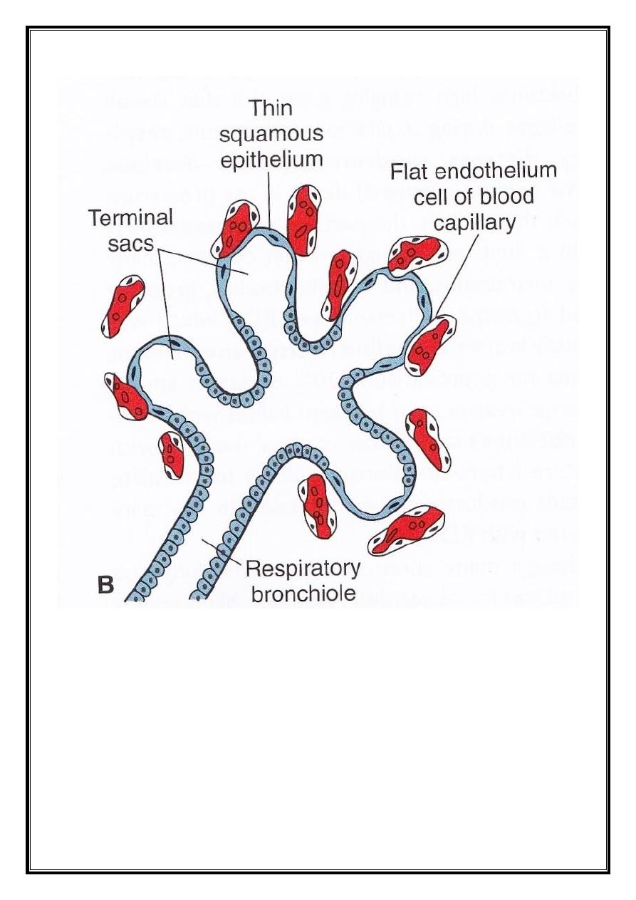
During the last 2 months prenatally, there are cells lining the sacs,
called type I alveolar epithelium, which becomes thinner, so that the
surrounding capillaries protrude into the alveolar sacs. This intimate
contact between the epithelial and endothelial cells makes the blood-air
barrier. Besides, at the end of the sixth month, type II alveolar epithelium
appears, and begins to produce the surfactant.
Dr.Maan Alkhalisy Embryology lec.11
7
rich fluid capable of lowering the surface
-
is a phospholipid
:
Surfactant
tension at the air-alveolar interface.
Before birth, the lungs are full of fluid containing high chloride
concentration, little protein, and some mucus from the bronchial glands.
As the concentration of the surfactant increases during the 34
th
week
of gestation, some of this surfactant enters the amniotic fluid, and acts on

Dr.Maan Alkhalisy Embryology lec.11
8
the macrophages of the amniotic fluid. Once this is activated,
macrophages migrate into the chorion to the uterus. It begins to produce
immune system’s proteins, including interleukin-1 beta (1L-1β).
The production of the interleukins will stiumulate prostaglandin
secretion, and stimulates the uterus to contract (beginning of labour).
Foetal breathing movements begin before birth, causing the
aspiration of the amniotic fluid. This movement is very important for
stimulating lungs development, and conditioning of the respiratory
muscles.
Due to this movement, the amniotic fluid will fill the lungs.
During birth, most of the fluid is reabsorbed, while the remaining
fluid in the trachea and bronchi will be expelled to the outside.
Here (during birth), the surfactant is very important for respiration of
the newborn baby, and prevents the collapse of the alveoli by lowering the
surface tension.
Dr.Maan Alkhalisy Embryology lec.11
9
THE END