Dr.Maan Alkhalisy Embryology lec.7
1
Cardio-vascular system
The cardio-vascular system starts to develop when the
embryo can’t satisfy its nutritional requirements by diffusion. This
occurs in the middle of the 3
rd
week of gestation.
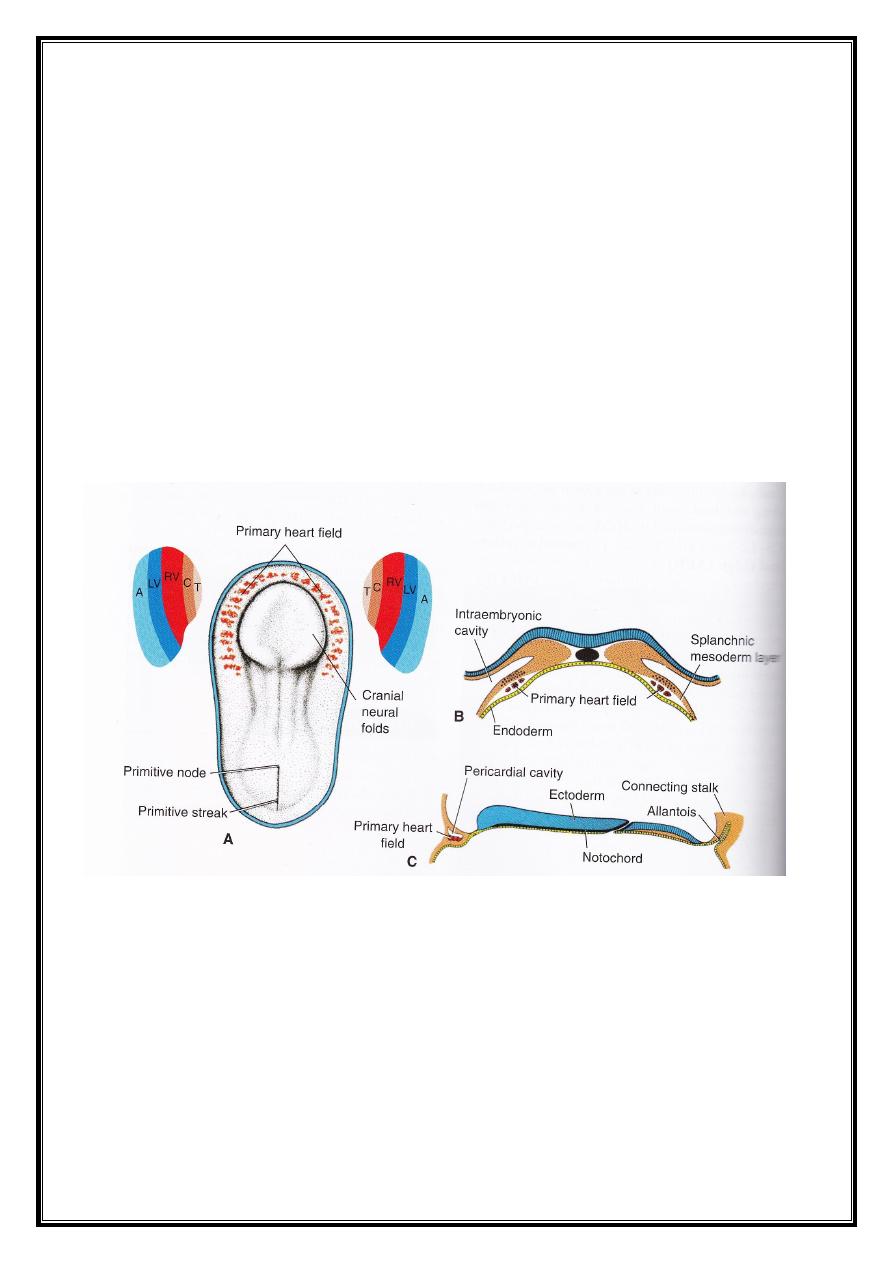
Progenitor heart cells (which lie in the epiblast) will migrate
and form the primary heart field (PHF). This is located at the
cranial end of the primitive streak, forming a horse shoe-shaped
cluster.
During days 16-18 the PHF will form- from medial to lateral-
the atria, the left ventricle, and most of the right ventricle. The
remainder of the heart, including part of the right ventricle and
the outflow tract (conus cordis and truncus arteriosus), will be
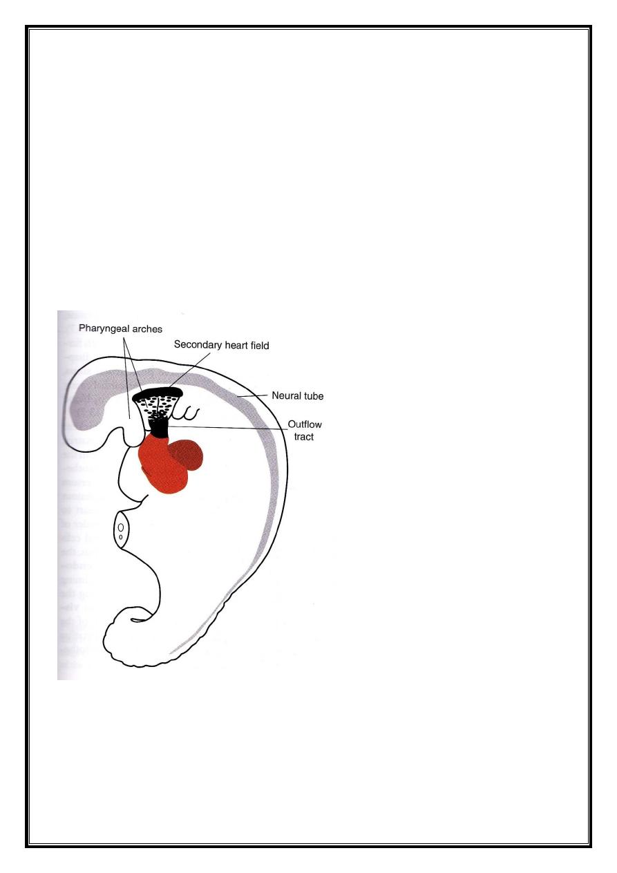
defined as the secondary heart field (SHF). This field appears
between days 20-21 of development, it is located ventral to the
Dr.Maan Alkhalisy Embryology lec.7
2
posterior pharynx, and is responsible for lengthening of the
outflow tract.
Once the cells establish PHF, they are induced by the
underlying pharyngeal endoderm to form cardiac myoblast and
blood islands that will form blood and blood vessels in a process
called vasculogenesis. With time, the islands unite and form a
horseshoe-shaped endothelial-lined tube covered by myoblast.
This region will be called the cardiogenic region which lies in a
primitive cavity called pericardial cavity.
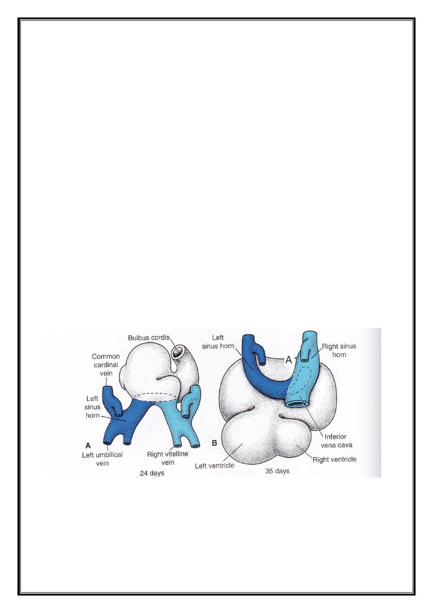
In the same time, on both side of mid line, bilaterally there is
blood islands appear informed of parallel longitudinal vessels
called dorsal aortae .
Dr.Maan Alkhalisy Embryology lec.7
3
Formation and position of the heart tube :
At the beginning, the cardiogenic area is located anterior to
the oropharyngeal membrane and the neural tube. With the
closure of the neural tube and formation of the brain, the central
nervous system grows cranially over the cardiogenic area, and the
oropharyngeal tube is pulled forward (due to cephalic folding of
the cranial cavity), and this leads the heart tube and the
pericardial cavity move downward to the cervical region and
finally to the thorax.
As the embryo grows and bends cephalo-caudally, is also
folds laterally. As a result the caudal regions of the paired cardiac
tube merge except at their cardial most end. Simultaneously, the

Dr.Maan Alkhalisy Embryology lec.7
4
central part of the horseshoe-shaped tube expands to form the
future outflow tract and ventricular regions. Thus, the heart
becomes a continuous expanded tube consisting of an inner
endothelial lining and an outer myocardial layer. It receives
venous drainage at its caudal pole and begins to pump blood out
of the first aortic arch into the dorsal aorta at its cranial pole.
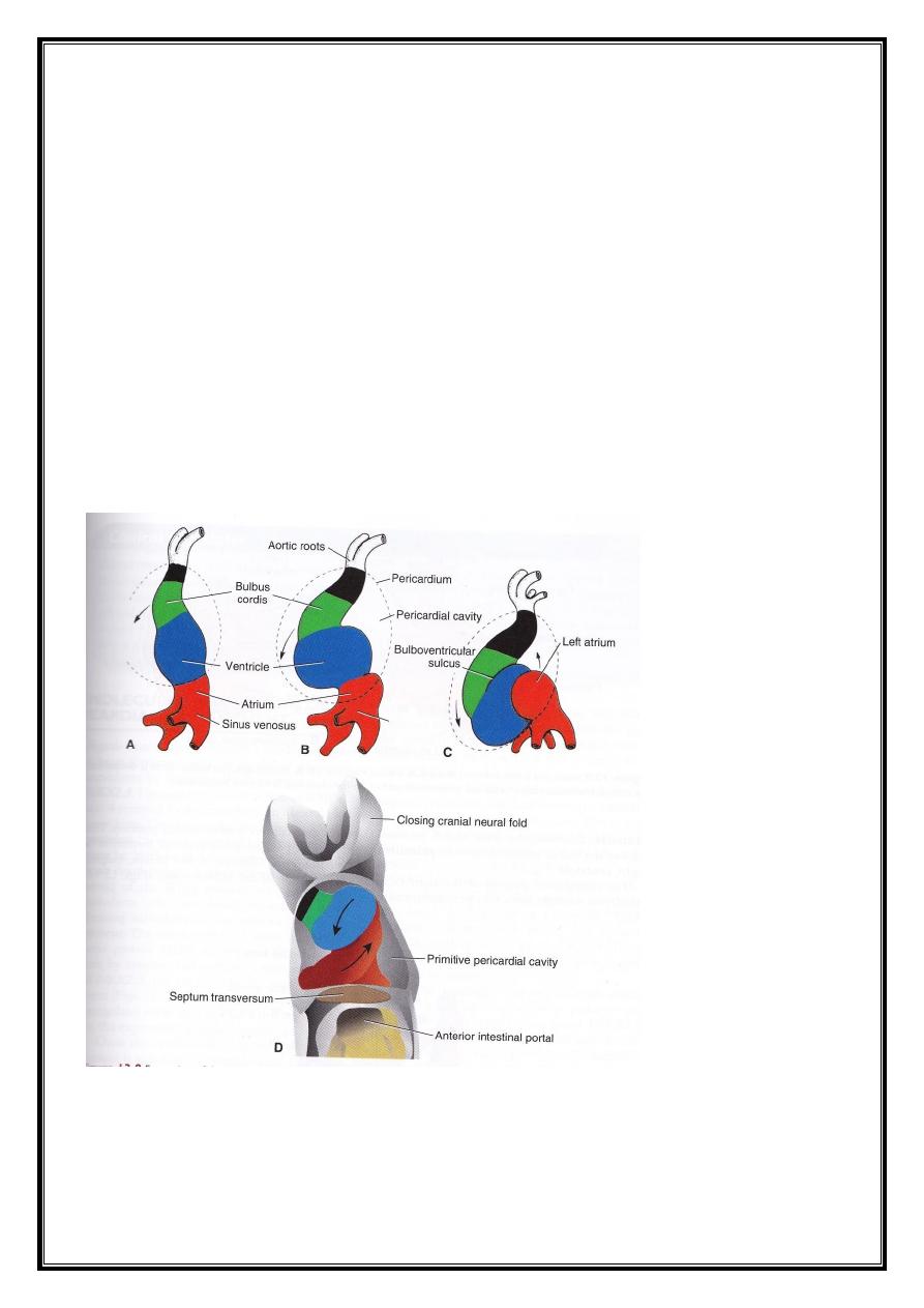
The developing heart tube bulges more and more into the
pericardial cavity. First, it attaches dorsally to the pericardial
cavity by the dorsal mesocardium. Yet, no ventral mesocardium is
present. The dorsal mesocardium will form the transverse
pericardial sinus. The heart is now suspended in the cavity by the
blood vessels at its cranial and caudal poles.
During this period, the myocardium secrets an extracellular
layer forming the proepicardium which will travel caudally
forming most of the epicardium, and the remaining part of the
epicardium will be formed by the mesothelial cells. Thus, the
heart tube cosists of 3 layers :
1- The endocardium : forming the endothelial lining of the
heart.
2- The myocardium : forming the muscular layer of the heart.
3- The epicardium (or visceral pericardium) : covering out of the
outside of the heart, forming the coronary arteries, including their
endothelial lining and smooth muscle.
Dr.Maan Alkhalisy Embryology lec.7
5
Formation of the cardiac loop :
As the heart tube continues to enlarge, this will leads to
lengthening of the SHF which is essential for the formation of part
of the right ventricle, conus cordis, and tunica arteriosus.
As the outflow tract lengthens, the cardiac tube begins to
bend on day 23. The cephalic portion of the tube bends ventro-
caudally and to the right, while the caudal (atrial) portion bends
dorso-cranially and to the left. This bending will create the cardiac
loop, which is completed on day 28. The atrial portion, initially a
paired structure outside the pericardial cavity, forms a common
atrium and is incorporated into the pericardial cavity. The atrio-
ventricular junction remains narrow forming the atrio-ventricular
canal which connects the common atrium and ventricle.
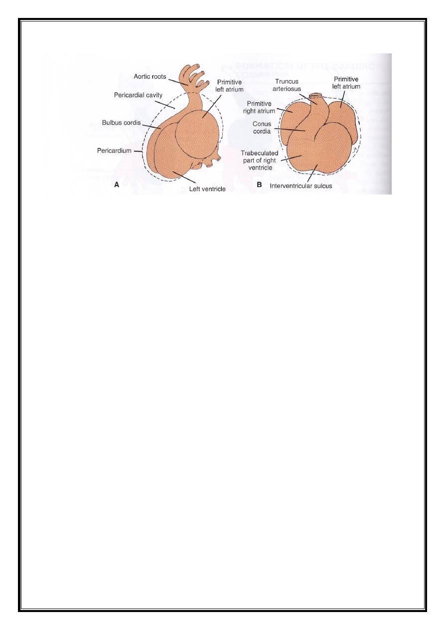
- The bulbus cordis is narrow except for its proximal 1/3 which
will form the trabeculated part of the right ventricle.
Dr.Maan Alkhalisy Embryology lec.7
6
- The mid-portion, conus cordis will form he outflow tract of
both ventricles (aorta and pulmonary vessels).
- The distal part of the bulbus (truncus arteriosus) will form the
root and proximal portion of the aorta and pulmonary artery.
The ventricular portion remains narrow in the middle leaving
a primary interventricular foramen. Therefore, the cardiac tube is
organized along its cranio-caudal axis from the cono-truncus to
the right ventricle to the left ventricle to the atrial region,
respectively.
At the end of the loop formation, the trabeculation of the
ventricles starts to appear. This trabeculation appears is the
primitive right ventricle and the primitive left ventricle.
Two chambers are formed, one on each side of the bulbus
cordis; therefore, the heart tube is shif ted from the right side to
the medial side.