Lecture 1
Carbohydrate (CHO)Metabolism
1- Glycolysis
Objectives:
a- Describe the role of ATP as an energy currency.
b- Explain what CHO are involved in by having an
overview to their role in human metabolism.
c- Explain glycolysis ( aerobic & anaerobic ) and
identify their importance.
d- Explain its regulation and the link with other
pathways through intermediate compounds.
By Prof.Dr. Munaf Salih Daoud

Source of Energy ( E ):
The ultimate source of E for all living matters is the
sunlight which converts CO
2
+ H
2
O into CHO( starch) in
plants. Starch converts into glucose (Glc or G ) in the
body which give E on oxidation.
Plants(CO
2
+H
2
O)-sunlight photosynthesis→ Glc
→ starch-body→Glc-oxidation with ADP+Pi→
ATP ( E is conserved in ATP )
ATP
is a nucleotide ( Adenine+Ribose+ 3
phosphates, it has 2 high-energy phosphate
bonds ( ~P ). It acts as a donor of a ~P to form
compounds of less free E of hydrolysis ( ∆G) like
Glc6-P , Fructose(Frc)6-
P …etc.
MSD
ADP
is another nucleotide ( with one
~P
) can
accept
~P
from compounds of higher ∆G
like phosphoenolpyruvate(PEP) or creatine
–P found in muscle to form ATP in the
ATP/ADP cycle which shows the link
between processes that generate
~P
&
those that utilize it.
Three major sources of ~P take part in E
conservation (E capture).
1
-Glycolysis
2
- TCA cycle
3
- ETC&Ox.Phosph.
MSD

Phosphagen
- act as storage forms of
~P
e.g.
Creatine~P found in skeletal
muscles,heart,spermatozoa,brain).In rapid ATP
utilization as a source of E for muscle
contraction,phosphagens act as a donor of ~P to
maintain its concentration & when ATP/ADP ratio
is high,then phosphagens increase acting as a
store.
Biological E are either
:
Exergonic-
∆G is negative, so reactions
proceeds spontaneously with loss of free E ( E
liberating) reactions. This occurs in Catabolic
Reactions( breakdown of molecules)
MSD

Glycogen→Glc→CO
2
+ H
2
O (Glycogenolysis &
Glycolysis)
Endergonic
-
∆G is positive so reactions proceeds
only if freeE can be gained (E is needed ). This
occurs in Anaerobic Reactions ( Synthesis of
molecules).
Glc
→ Glycogen ( glycogenesis)
Besides ATP,other nucleotides of high ~P are
GTP,CTP,& UTP used to supply E in protein,lipid,
& polysaccharide synthesis,respectively.Each is
formed by combination of ATP with GDP,CDP,&
UDP in order by a
kinase enzyme
.
MSD

CHO Metabolism
1- carried out in every cell in the body.
2- Found in
cytoplasm ( Cytosol).
Glycolysis,Glycogenesis,& Glycogenolysis.
in
mitochondria
( Membranes& Matrix), TCA
cycle, ETC& OX Phosph..
in
both ,
Gluconeogenesis.
3- Alternative pathways are HMP shunt , Uronic acid
pathway.
1- Glycolysis
. Used by all tissues for oxidation ( breakdown ) of
Glc in 9 reactions to give E ( ATP )& intermediates for
other metabolic pathways ( Link ).
MSD
Aerobic
( in presence of O
2
) Glc→ 2 pyruvates
or pyruvic acids.
Anaerobic
( in absence of O
2
) → 2 lactates
or lactic acids.
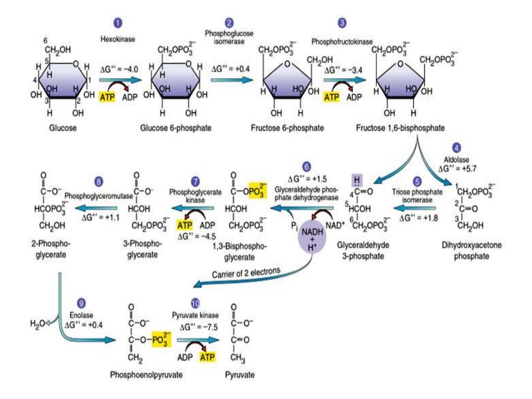
Reactions:
Glc(6-C ) with 1
ATP→ Glc6-P by
hexokinase(HK)
or
glucokinase(GK)
then
→Frc6-P by
isomerase
then with 1
ATP →
Frc 1,6 bisP ( 6-C) by
PFK-1
then cleaved
into 2 (3-C )
by Aldolase
A ( cleavage
enzyme) → Glyceraldehyde 3-P (3-C)+
dihydroxyacetone phosphate DHAP(3-C)
MSD

This step is interconvertible & 2( Glycerald. 3-P) is
formed by
isomerase
then with 2NAD by
its
dehydrogenase
→ 2NADH + 1,3
bisphosphoglycerate (1,3bisPG) which combine
with 2ADP by
kinase
→ 2(3PG) + 2ATP then →
2(2PG)by
mutase
then → PEP by
enolase
then
with ADP → 2ATP +
2 pyruvates
by
pyruvate
kinase(PK)
(
Aerobically
)….The 2 pyruvates with
2NADH
by lactate dehydrogenase ( LDH)
→ 2NAD
+
2 lactates
(
Anaerobically
)
[ N.B. 1,3 bisPG by
mutase
→2,3 bisPG then →by
phosphatase
3PG ,this occurs in RBCs].
MSD

The 2,3biPG of high concentration (4mM) equal to
Hemoglobin ( Hb) binds to it & act as regulator of O
2
transport by decreasing affinity of Hb to O
2
thus
allowing O
2
release in tissue capillaries .
▪ E production :
Aerobic glycolysis produces 2ATP+2NADH(
give 4 ATP if moves through Glycerol 3-Pshuttle
or 6 ATP if through malate shuttle) i.e. 6-8 ATP
per 1 Glc oxidized to 2 pyruvates.
Anaerobic glycolysis produces 2 ATP(
produced by substrate level phosphorylation.In
its pathway which occurs in exercised muscle (
due to lack of O
2
or Hypoxia) or in RBCs ( lack of
mitochondria),the NADH cannot be oxidized

through ETC but used by pyruvate to form lactate by
LDH
.This enzyme have Clinical significance & have 5
isoenzymes
.The increased level of blood lactate above
normal limit is known as
Lactic Acidosis
. (a pathological
condition of many causes).
▪Anaerobic Glycolysis occurs in exercised
muscle,RBCs,Cancer cells in
CancerCachexia
. During
extended muscle exercise, ↑[lactate] move in blood to
liver to be reconverted to pyruvate which form Glc
by Gluconeogenesis ( Cori ̛s cycle).
Aerobic Glycolysis occurs in most tissues ( organs )
when O
2
is available but it is low in Cardiac muscles
msd

Ischemic heart
diseases.The brain is highly dependent
on Glc for its E supply & needs continuous supplement .
Regulation :
The key enzymes are
HK,PFK-1 & PK
.
1-
PFK-1
is inhibited by ATP & citrate and activated by
cAMP,Frc6-P,Frc 2,6 bisP ( in liver).
2- Allosteric activation or inhibition of
HK,PFK-1 &PK
by phosphorylation & dephosphorylation ( short- term
influences , minutes-hours)
3- Hormonal influence on the amount of enzyme
synthesized ( long- term increase of activity by 10-20
folds , hours-days).
MSD

4- Well-fed ( after a meal of CHO) or high insulin
→high enzyme activity.
5-
Starvation or Diabetes →low enzyme
activity.
6-
PK
, activated by Frc1,6 bisP & inhibited
by ATP, glucagon & epinephrine( adrenaline).
☻ Genetic defect , Inherited deficiences of
HK&PK
cause Hemolytic Anemia due to
↓[ATP] important in maintaining the biconcave
shape of RBCs membranes and ↓[ 2,3 bisP]
important in O
2
release in tissue capillaries .
☻glycolysis is inhibited by
iodoacetate,arsenate& fluoride. msd

Other CHO that enters glycolysis:
1- Glycogen through formation of Glc6-P (
muscle).
2- Fructose through formation of Frc1-P by
fructokinase
( liver,kidnney,intestine,testis) i.e.
Fructolysis.Frc1-P is cleaved by
Aldolase B
(
predominantly found in liver) & bypass the main
regulatory steps catalyzed by
PFK-1
resulting in the
formation of more pyruvate ( and Acetyl CoA) than
is required for ATP formation.
MSD