ANATOMY
HEAD & NECK
Dr. Nawfal K. Al-Hadithi
Lec . 15
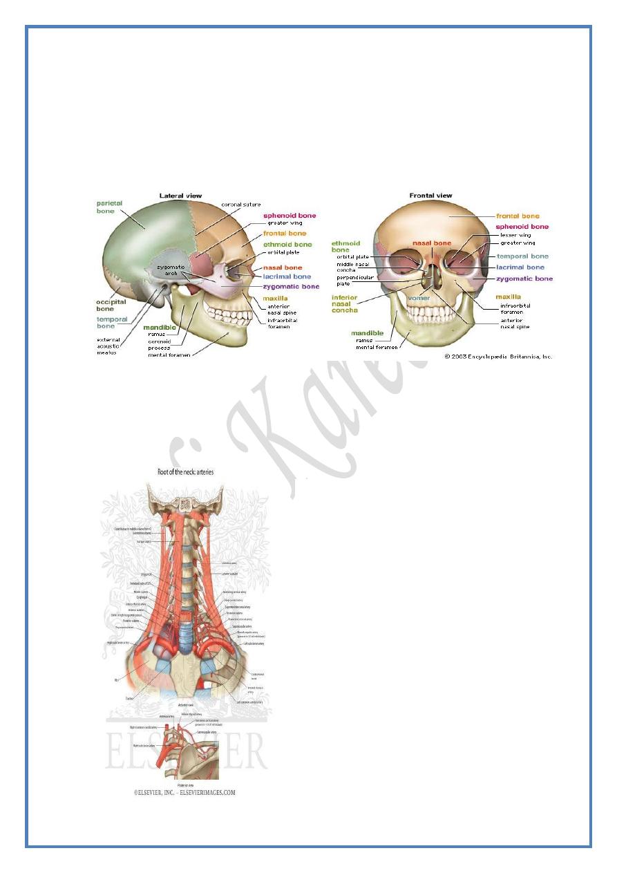
The Cranial Cavity
By : Ali Kareem
مكتب اشور لالس تنساخ
2013 – 2014

Head & Neck Dr. Nawfal K. Al-Hadithi
Anatomy
2
Lec. 15 The Cranial Cavity
The cranial cavity
The Cranial Meninges :
The brain & spinal cord are enveloped by three layers of meninges
variable in consistency & arrangement, they are from within outward:
1) Pia mater.
2) Arachnoid mater.
3) Dura mater, which is divided into:
A; Inner fibrous layer, the dura proper or fibrous dura.
B; Outer membranous layer, the endosteal (periosteal) dura.
Pia mater :
• This is a delicate, intimate areolar investment which faithfully
follows the contour of the brain & cannot be dissected from it.
• It is invaginated into the nervous tissue with blood vessels
surrounded by a tubular prolongation of CSF-filled subarachnoid
space named the perivascular space.
• It is evaginated with cranial (& spinal) nerves which leave the
nervous tissue.
• It is meshed with blood vessels so it exhibits the name (vascular
membrane).
Arachnoid mater :
• This is a delicate, transparent membrane made mainly of
collagenous & elastic fibers & lined by squamous mesenchymal
epithelium.
• It is separated from the pia by the CSF-filled subarachnoid space.
• It is loosely applied to the brain & don’t strictly follows its contour,
so it approaches (& may come in touch with) the pia over brain
gyri but diverge from it over the sulci.
• The only invagination of the arachnoid on the brain is into the
longitudinal fissure of the brain.
The subarachnoid space :
• This CSF-filled space is crossed by trabeculae derived from the
two bounding meninges.
• In regions where brain contour change markedly, the arachnoid
mater bridges over wide intervals of pia-covered brain tissue
resulting in large CSF lakes called subaracnoid cisterns.
• These cisterns are mainly located under the brain & they are
traversed by cranial nerves & blood vessels in the region of the
cistern. The main cisterns are four:

Head & Neck Dr. Nawfal K. Al-Hadithi
Anatomy
3
Lec. 15 The Cranial Cavity
a) Cisterna cerebellomedullaris; between the undersurface of the
cerebellum above & the roof of the fourth ventricle below.
b) Cisterna interpeduncularis; lies in front of the midbrain between
the two cerebral peduncles.
c) Cisterna pontis; lies in front of the basilar part of the pons between
it & the basilar part of the occipital bone.
d) Cisterna chiasmatica; lies just below the optic chiasma
• Arachnoid villi; finger-like projections of arachnoid through the
fibrous dura into the superior sagittal sinus where CSF diffuses into
the venous blood
• Arachnoid granulations; are the folded round ends of the villi
which have special histological structure to permit CSF diffusion
Dura mater :
Endosteal layer :
- This is the periosteum of the undersurface of the cranial bones lies
external to the fibrous layer.
- Because of its more intimate relation & fusion with the fibrous
dura than cranial bones from which it can easily be pealed off, this
layer is regarded as an external layer of cranial dura mater
- Venous sinuses are located in separations of the two layers of dura
or between folds of the fibrous layer of dura mater
Fibrous layer “dura proper” :
- This is a tough, dense, fibrous membrane which encloses &
protects brain tissue
- Folds of this layer between various parts of brain tissue are
responsible for preventing the effects of movements of the head on
the floating brain
- Dural folds are four in number :
1) Falx cerebri
2) Tentorium cerebelli
3) Falx cerebelli
4) Diaphragma sellae
Falx cerebri :
- This is a sickle-shape dural fold which occupies the median
longitudinal fissure of the brain
- It is attached anteriorly to crista galli & broadens as it goes
backward to end by becoming continuous with the dorsal surface
of the tentorium cerebelli
Head & Neck Dr. Nawfal K. Al-Hadithi
Anatomy
4
Lec. 15 The Cranial Cavity
- Its upper convex border is attached to the midline of cranial bones
& sagittal suture enclosing the superior sagittal sinus, it ends
posteriorly at the internal occipital protuberance
- The inferior concave margin arches over corpus callosum of the
brain to end posteriorly in the straight sinus
Tentorium cerebelli :
- This fold separates the cerebellum from the undersurface of
occipital lobes of the cerebrum
- It is elevated in the midline like a tent where it meets the posterior
end of falx cerebri
- Its attached posterior margin encloses the lateral (transverse)
sinuses which diverge from the internal occipital protuberance on
the occipital bone
- Its attached anterior margin encloses the superior petrosal sinuses
along the top of the petrous bone
- The anterior concave free margin arches around the brainstem & is
attached anteriorly to the anterior clinoid processes
- The straight sinus lies in the line of junction of the tentorium with
the falx cerebri
Falx cerebelli :
- This shallow fold separates the two cerebellar hemispheres
- It is attached to the internal occipital crest
- It contains the occipital venous sinuses
Diaphragma sellae :
- This is a horizontal fold of dura projecting from the margins of
sella turcica
- It roofs the sella leaving a midline opening for the pituitary stalk
Dural venous sinuses :
• These are endothelial-lined spaces between the two dural layers or
between folds of fibrous dura
• They are the collecting channels of cerebral, meningeal & diploic
veins
• They communicate with external veins by emissary veins
• They end in the IJV either directly or indirectly
• They are classified by many classifications, one of them is :
1- Midline unpaired sinuses
2- Paired sinuses
Head & Neck Dr. Nawfal K. Al-Hadithi
Anatomy
5
Lec. 15 The Cranial Cavity
Unpaired sinuses :
1- Superior sagittal sinus :
- Begins at foramen caecum where it receives a vein from the roof of
the nose
- Passes back in the root of the falx cerebri to the internal occipital
protuberance being triangular in coronal section
- As it goes backward it becomes larger as it receives more blood
- The lateral blood lakes (lacunae) lie on each side of it at parietal
level, into these lakes the arachnoid granulations bulge for
diffusion of CSF
- It receives cerebral, diploic & emissary veins
- At the internal occipital protuberance it ends in the sinus
confluence or bifurcates into right & left lateral sinuses or
continues to one side “usually the right” as the lateral sinus
2- Inferior sagittal sinus :
- Begins one third the distance behind the attachment of the falx
cerebri
- It occupies the inferior margin of the falx
- Ends posteriorly in the straight sinus
- It receives tributaries from the medial surface of the hemispheres &
from the falx
3- Straight sinus “sinus rectus” :
- It is the continuation of the inferior sagittal sinus in the tentorium
cerebelli
- It lies in the line of junction of the falx cerebri & the tentorium
- It receives the great cerebral & superior cerebellar veins
- Ends as the superior sagittal sinus at the internal occipital
protuberance buts it has the tendency to continue as the left
transverse sinus
Paired sinuses :
1- Cavernous sinus :
- They lie in the MCF on either side of sella turcica extending from
the superior orbital fissure anteriorly to the apex of petrous bone
posteriorly
- They are trabeculated as to be given the name “cavernous”
- They begin anteriorly by union of the superiorophthalmic vein &
sphenoparietal sinus
- They end posteriorly by division into the superior & inferior
petrosal sinuses
Head & Neck Dr. Nawfal K. Al-Hadithi
Anatomy
6
Lec. 15 The Cranial Cavity
- They communicate with the angular vein & pterygoid plexus
a) Contents of the lateral wall :
- Oculomotor nerve
- Trochlear nerve
- Ophthalmic & maxillary divisions of trigeminal nerve
b) Contents of the lumen :
- Abducent nerve
- ICA enters the sinus from below posteriorly at foramen lacerum &
passes in its floor grooving the bone to pierce its roof anteriorly
medial to the anterior clinoid process to the circle of Willis
2- Intercavernous sinuses :
- Connect the two cavernous sinuses
- Pass anterior & posterior to the stalk of the hypophysis
3- Sphenoparietal sinuses :
- Lie in the dural fold at the lesser wing of sphenoid
- Receive radicles from the midle meningeal veins
- End in the cavernous sinus
4- Superior petrosal sinus :
- Lie in the attached anterior margin of the tentorium at the top of the
petrous bone
- They connect the cavernous with sigmoid sinuses
- They receive the cerebellar, inferior cerebral & tympanic veins
5- Inferior petrosal sinus :
- Arise from the postero-inferior part of cavernous sinus
- Groove the petro-occipital fissue
- Pass through the anterior compartment of jugular foramen
- End in the IJV
- Receive pontine, labyrinthine, medullary & cerebellar veins
6- Basilar plexus :
- Lie on the basilar part of the occipital bone
- Connect the two inferior petrosal sinuses
- Connected to the anterior vertebral venous plexus
7- Occipital sinuses :
- Arise from union of small veins near foramen magnum
- Pass through the root of falx cerebelli
Head & Neck Dr. Nawfal K. Al-Hadithi
Anatomy
7
Lec. 15 The Cranial Cavity
- Ends at the internal occipital protuberance at the confluence of
sinuses
8- Sphenoparietal sinuses :
- Arises by receiving a radical from the middle meningeal vein deep
to the pterion
- Lies in dura on the undersurface of the lesser wing of sphenoid
- Ends in the anterior part of the cavernous sinus
9- Transverse (lateral) sinuses :
- Begin at the internal occipital protuberance
- They lie in the posterior attached margin of the tentorium cerebelli
- They extend laterally to reach the base of the petrous temporal
where they receive the superior petrosal sinus & descend in the
posterior cranial fossa the sigmoid sinuses
- They receive the inferior cerebral & superior cerebellar veins
10- Sigmoid sinuses :
- Are large S-shape sinuses formed at the root of the petrous bone by
confluence of the lateral & superior petrosal sinuses
- They groove the deep surface of the mastoid process of temporal
bones
- They continue on the occipital bone to reach the jugular foramen
- At the jugular foramen they continue as the IJV
- They receive the mastoid emissary veins & inferior cerebellar veins
Blood supply of cranial dura :
Arteries :
The endosyeal layer of dura is very richly supplied by blood, unlike the
fibrous layer which needs little supply.
1- Supratentorial part is supplied by the middle meningeal artery, a
branch of maxillary artery which enters the cranium through foramen
spinosum & divides into anterior & posterior divisions.
The anterior division courses up passing over the precentral gyrus,
haematoma in this region causes contralateral paralysis.
The posterior one passes back courses over the superior temporal
gyrus, haematoma from this artery causes contralateral deafness.
2- Cranial fossae :
A) Anterior CF: ophthalmic & anterior ethmoidal arteries.
Head & Neck Dr. Nawfal K. Al-Hadithi
Anatomy
8
Lec. 15 The Cranial Cavity
B) Middle CF: middle & accessory meningeal arteries.
C) Posterior CF: vertebral arteries.
Veins :
Blood is collected into two main sources :
1- Middle meningeal veins, accompany the arteries & leave the cranium
through foramen spinosum to enter the pterygoid plexus.
2- Diploic veins, which drain either to the exterior or to dural venous
sinuses especially the superior sagittal.
Nerves :
1- Supratentorial dura : tentorial branches of Va.
2- Cranial fossae :
A) ACF: Anterior & posterior ethmoidal n.
B) MCF: Vb & Vc.
C) PCF: meningeal branches of IX & X cranial nerves
D) The area around foramen magnum: the upper three cervical spinal
nerves.
:P
لحد يسألني ع الصور الن
م
ـل
ﮔ
يت
Edited & Published by :
Ali Kareem
