
ANATOMY
Dr.Nawfal
Lec.9
The Orbit
-It is pierced by nerve to mylohyoid & mylohoid artery
The orbit:
The bony orbit:
•
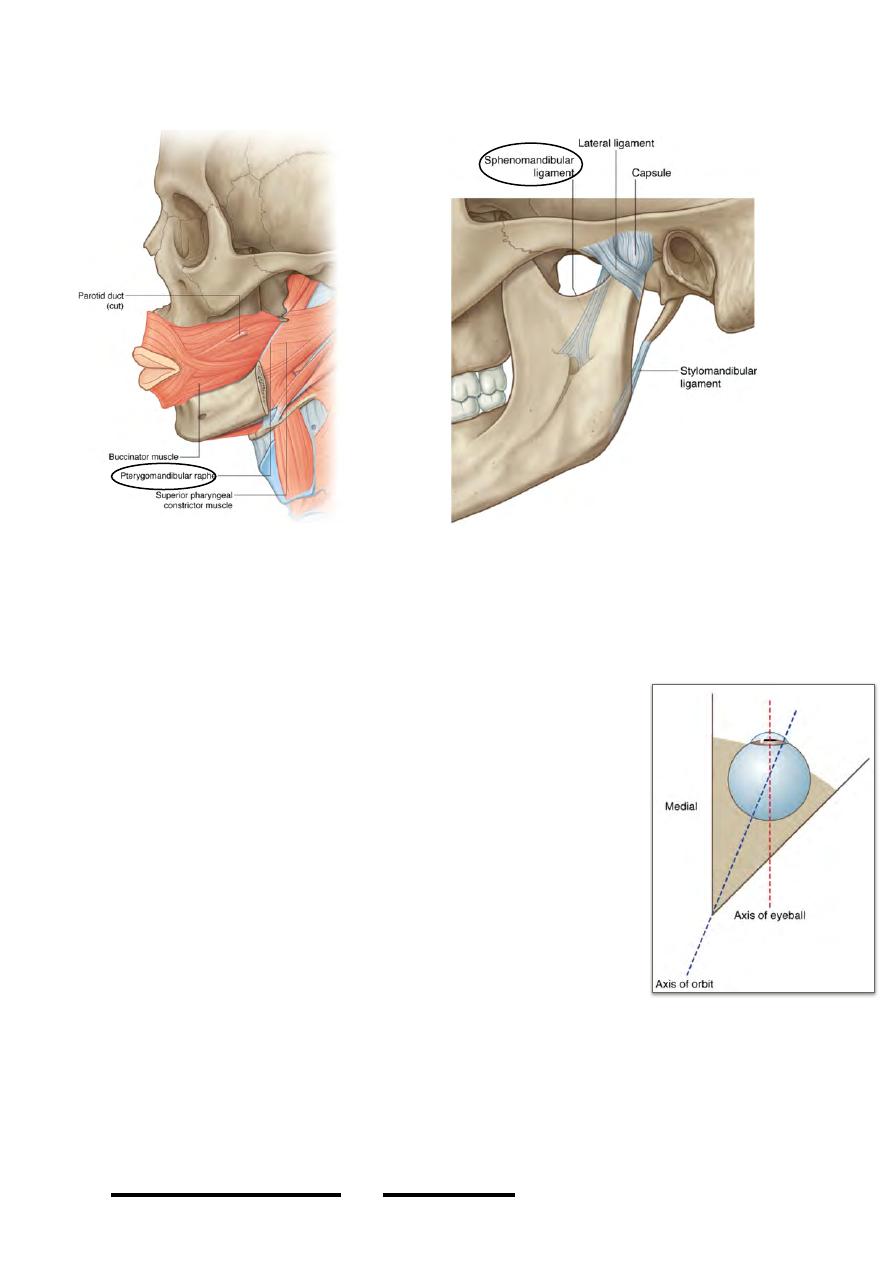
The orbit is a four-sided pyramidal shape space whose base lies anterior & its
apex posterior
•
The base is almost 3.5 X 4 cm & the depth is about 5 cm
•
Medial walls are parallel to each other with a 2 cm
distance separating them
•
Lateral walls diverge laterally at 45O from medial walls
thus the lateral walls are 90O at each other
•
Orbital axis lies along the center of the orbit & both will
also be perpendicular on each other
Orbital margins:
The margins of the orbit are strong bones, they are even stronger
than its four walls
•
Superior: supraorbital arch of the frontal bone
•
Lateral: frontal process of zygomatic bone & zygomatic
process of frontal bone
•
Inferior: zygomatic bone & maxilla
•
Medial: frontal process of maxilla & maxillary process of frontal bone
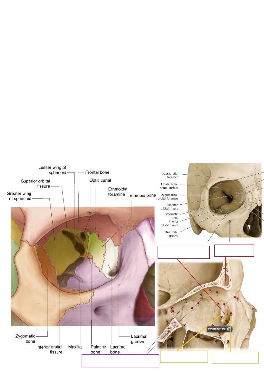
Roof:
-Formed by orbital process of the frontal bone completed posteriorly by the lesser
wing of sphenoid
!
67
Head & Neck
Dr. Nawfal K. Al-Hadithi
45ْ
-It is concave especially laterally where the lacrimal fossa which accomodates the
lacrimal gland lies
Floor:
-Formed by the the orbital surface of the maxilla supplemented laterally by the
zygomatic
-It slopes upward in the direction of the medial wall
-It contains the infraorbital groove which connects the inferior orbital fissure to the
infraorbital canal
Lateral wall:
-Formed by the zygomatic bone in front & greater wing of sphenoid behind
Medial wall:
-Formed from in front backwards by: frontal process of maxilla, lacrimal bone,
orbital lamina of ethmoid & near the apex by the body of sphenoid
-It is very thin & lies almost vertical
-It separates the orbit from the ethmoidal & spheboidal air cells
-It shows the site of the lacrimal sac which is bounded by anterior & posterior
lacrimal crests
-It contains anterior & posterior ethmoidal foramina at its junction with the roof
!
68
Head & Neck Dr. Nawfal K. Al-Hadithi
Fossa for lacrimal sac
Frontal air sinus
Post. Lacrimal crest
Ant. Lacrimal crest
Ant. & post. Ethmoidal foramina
Relations:
The orbit is bounded :
•
Above: anterior cranial fossa & frequently the frontal air sinus
•
Medially: sphenoidal & ethmoidal air cells
•
Inferiorly: maxillay air sinus
•
Laterally: temporal fossa
Anatomy of the eyelids:
The eyelid is composed of five layers:
1-Skin:
-very thin & moist
2-Subcutaneous tissue:
-lax, scanty & rarely contains any fat
-contains the roots of the eyelashes with the
accompanying sebaceous glands “of Zeis”
& modified sweat glands “of Moll”.
-contains vessels & nerves of the lid
3-Muscular layer :
-consists of the palpebral & lacrimal parts of
O. oculi
-palpebral part “discussed”
-lacrimal part connects the lacrimal sac &
posterior lacrimal crest to the tarsus
- i t s p o s t e r o m e d i a l d i r e c t i o n o f
contraction provides better contact
between the eyeball & eyelid &
consequently better distribution of tear
film, also it dilates the lacrimal sac
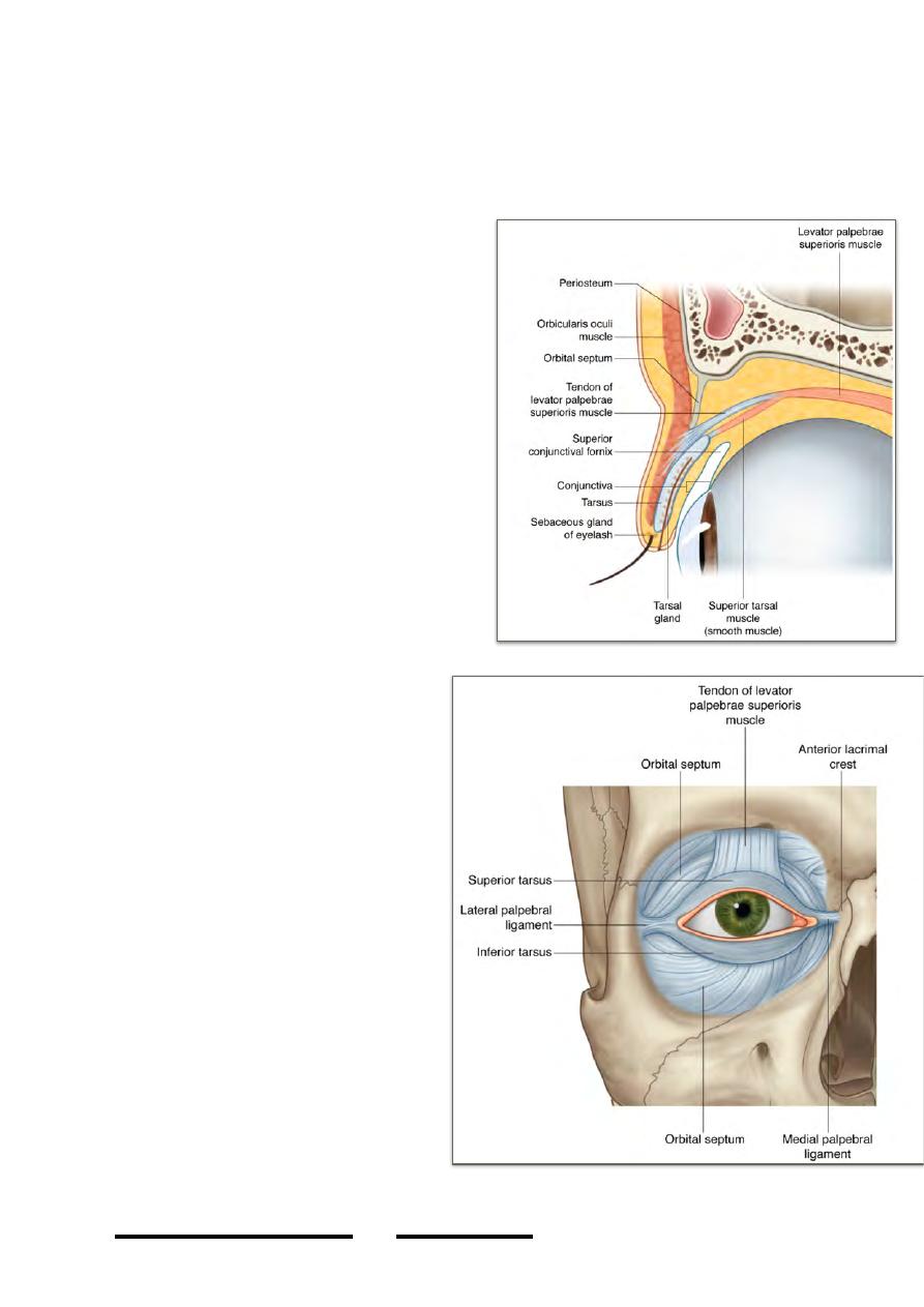
4-Tarso-fascial layer:
-is the skeleton of the eyelid
-formed of two layers, the tarsal plate
“tarsus” & orbital septum:
*Tarsal plate:
-tough fibrous layer extends between
the medial & lateral palpebral
ligaments
-2.5 X 1 cm in dimensions
-semilunar in shape with the straight
edge at the lid margin
*Orbital septum:
-thin membrane which is continuous
with the periosteum of the superior &
inferior orbital margins
-the superior one is perforated by the
levator palpebrae superioris
-away from this muscle, the tarso-fascial
!
69
Head & Neck Dr. Nawfal K. Al-Hadithi
layer forms a complete septum between the superficial compartment of the eyelid
which is continuous with the face & deep compartment which is continuous with the
orbit
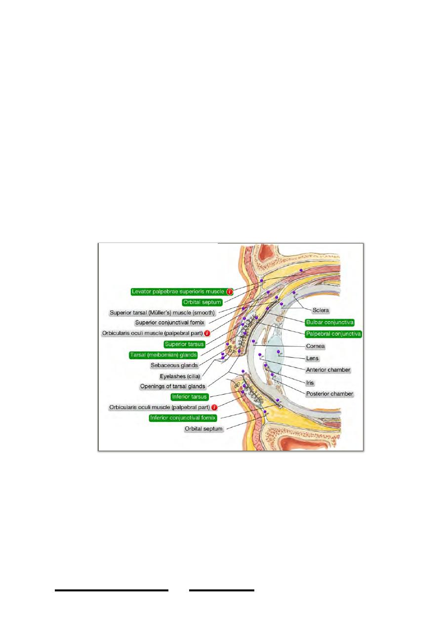
Tarsal glands:
Are modified sebaceous glands on the deep surface of the tarsus secrete an oily layer
to prevent tear overflow at the lid margins
5- Conjunctiva:
-the transparent membrane which lines the lids (palpebral c.) & onto the eyeball
(bulbar c.)
-the site of reflection is called the fornix, so we have superior & inferior fornices
-palpebral c. differs from the bulbar in being thicker, opaque & more vascular
-modifications in the conjunctiva:
1-lacrimal lake: a shallow bay on the medial angle of the eye bounded laterally by the
semilunar fold, it acts as reservoir for lacrimal fluid.
2-semilunar fold: a rudimentary fold in the conjunctiva
3-lacrimal caruncle: a rounded elevation in the lacrimal lake formed of moist skin
with fine hairs, sebaceous & sweat glands.
Contents of the orbit:
1-Eyeball.
2-Muscles: - LPS
- four recti
- two oblique
3-Nerves: - motor (III, IV & VI)
- sensory (Va)
4-Vessels: - ophthalmic artery
!
70
Head & Neck Dr. Nawfal K. Al-Hadithi
- ophthalmic veins
5-Fascial modifications: - periorbita
- muscular fasciae
- check & suspensory ligaments
- retrobulbar fat
6-Lacrimal apparatus: - lacrimal gland
- lacrimal sac
- nasolacrimal canal
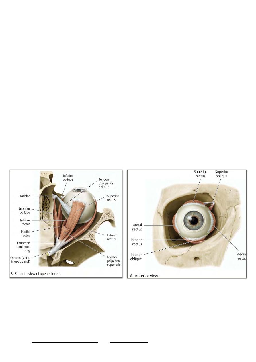
Muscles o the orbit:
The recti are 4 in number:
-
Superior rectus
-
Medial rectus
-
Inferior rectus
-
Lateral rectus
Origin:
All the 4 recti arise from a tendinous ring surrounding the medial end of the SOF
Insertion:
The muscles, narrow at their origin broaden as they come forward to be inserted into
the sclera anterior to the coronal equator forming a muscular cone around the eyeball
3- Oblique muscles:
a) Superior oblique:
Origin: from the bone just above the optic canal
Insertion:
-
the muscle passes forward in the junction between the roof & medial wall of
the orbit to reach the anterior part of the orbit as a thin tendon which hooks
!
71
Head & Neck Dr. Nawfal K. Al-Hadithi
around the trochlea “pulley” which is attached in the roof of the orbit above the
lacrimal crest.
-
From this pulley the tendon returns postero-laterally to be inserted into the
sclera deep to SR tendon behind the equator of the globe
b) Inferior oblique:
Origin: from the orbital surface of the maxilla lateral to the lacrimal groove
Insertion: the muscle is located below the eyeball, passes postero-laterally below IR
to be inserted in the sclera beneath LR
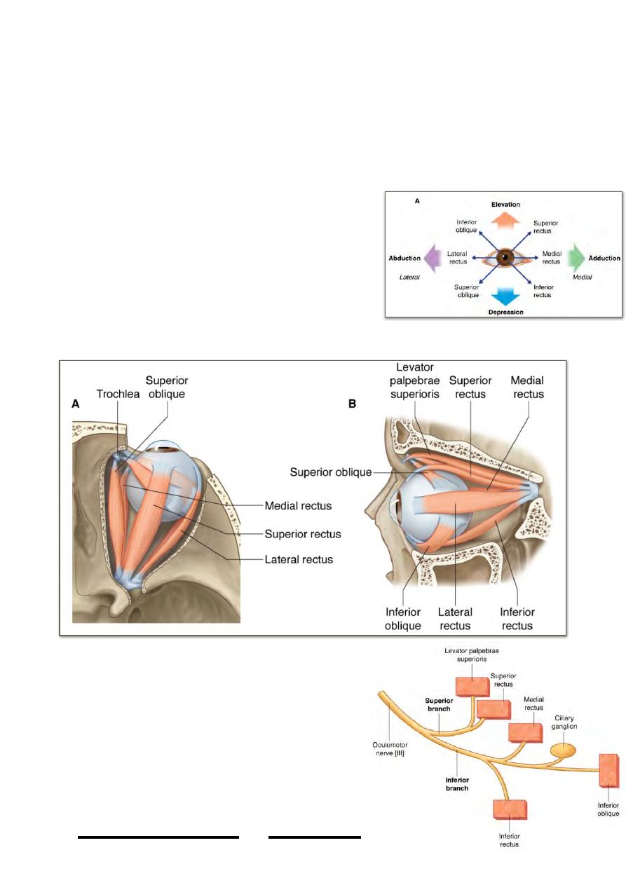
* The recti will move the globe:
- SR superiorly + nasally (elevation + adduction)
- IR inferiorly + nasally (depression + adduction)
- MR nasally (adduction)
- LR temporally (abduction)
* The oblique muscles move the globe:
- SO inferiorly + temporally (depression +
abduction)
- IO superiorly + temporally (elevation +
abduction)
Nerve supply of ocular muscles:
LR 6 SO 4 Others 3
Motor nerves of the orbit:
1- Oculomotor n.:
-enters the orbit through the SOF as superior &
inferior branches
-the superior branch crosses over the optic n.
under SR supplying it & passes medial to it to
!
72
Head & Neck Dr. Nawfal K. Al-Hadithi
terminate in the undersurface of LPS
-the inferior branch crosses below the
optic n. to supply
GSE to MR, IR, & IO
GVE to sphincter pupillae & ciliary
muscle “parasympathetic” with a relay
in the ciliary ganglion, this component
reaches the globe via branch to IO
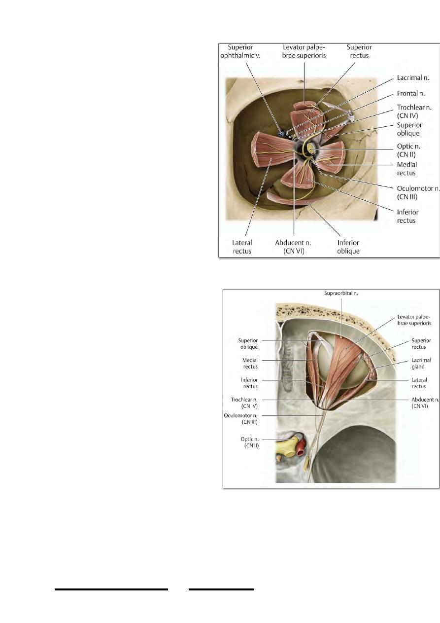
2- Trochlear n.:
-the smallest of all cranial nerves, enters
the orbit through the SOF being the
highest of all nerves entering the orbit
-lies in the roof of the orbit medial to
the frontal n.
-supplies SO at its posterior 1/3
3- Abducent n.:
-enters the orbit through the SOF
inferior to all nerves
-enters the ocular surface of LR
supplying it
N.B:
The above three motor nerves have a
communication with Va in the cavernous
sinus which make them able to carry the
proprioceptive sensation from the
muscles they supply.
Sensory nerves of the orbit:
-the ophthalmic division of trigeminal
nerve “Va” is the smallest of the three
divisions of V nerve, it is entirely
sensory
-from the semilunar ganglion, Va leaves
forward in the lateral wall of the
cavernous sinus together with motor
nerves of the orbit with which it forms
some communication
-it divides into its three terminal division
short of the way to the SOF after it gives
the tentorial branch to the tentorial dura
-the three divisions of Va, namely the lacrimal, frontal & nasociliary nerves enter the
orbit through the SOF to supply its contents
-in addition to the orbit & its contents, Va supplies:
*some skin of the face & scalp
*some mucous membranes of the nasal cavity & paranasal sinuses
!
73
Head & Neck Dr. Nawfal K. Al-Hadithi
-ALL STRUCTURES WHICH ENTER THROUGH THE S.O.F DO WITHIN THE
CONE OF MUSCLES “THROUGH THE TENDINOUS RING” EXCEPT
{LACRIMAL N., FRONTAL N., TROCHLEAR N. & THE OPHTHALMIC
VEINS}
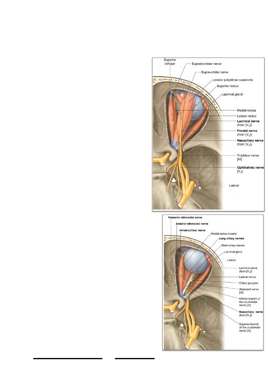
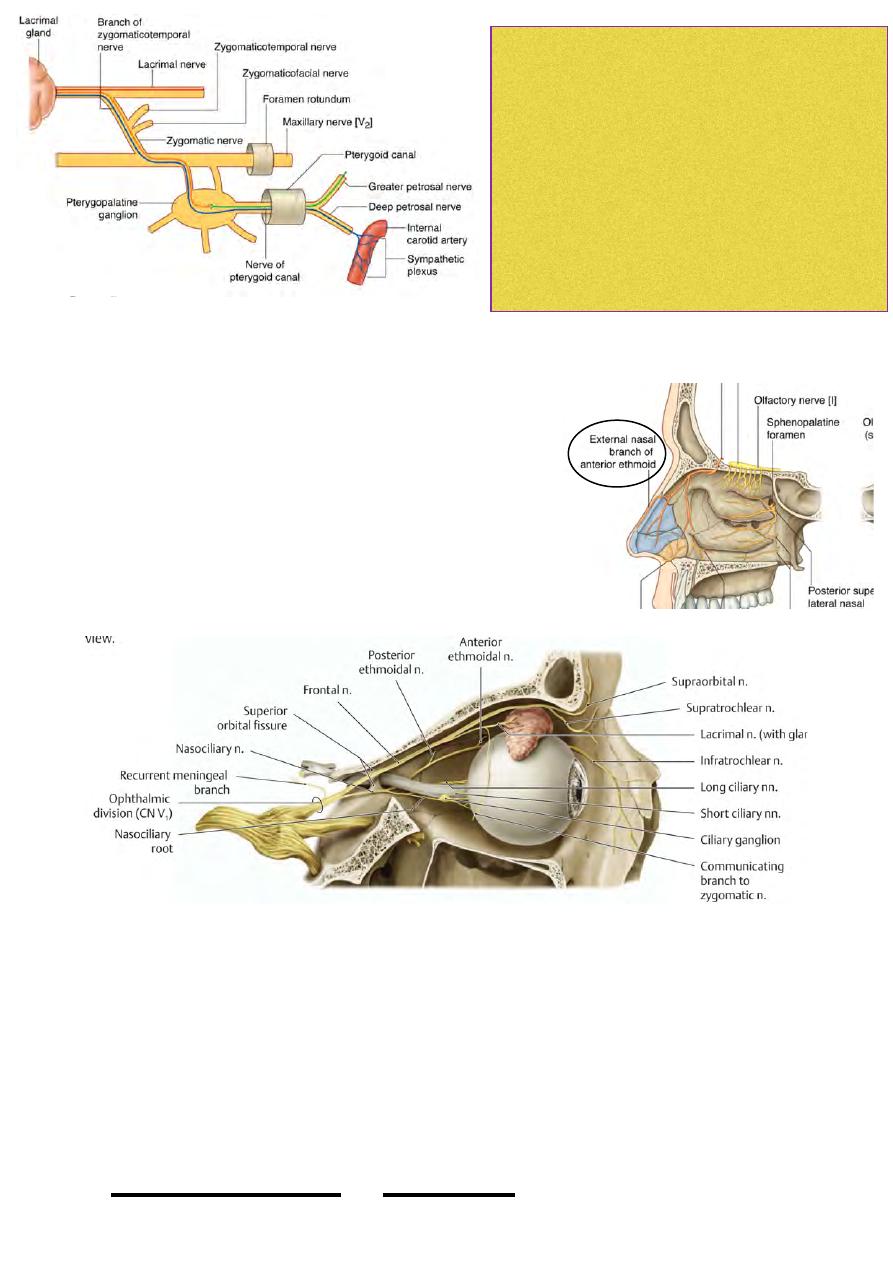
1-Lacrimal nerve:
-the smallest of Va branches, passes over the
LR muscle
-half its way in the orbit it receives contribution
from the zygomaticotemporal branch of Vb
supplying it with parasympathetic component
from the pterygopalatine ganglion to the
lacrimal gland
-it supplies the gland with sensory &
parasympathetic supply, together with the
lateral ½ of the upper lid & its conjunctiva
2-Frontal nerve:
-the largest of Va branches, passes between LPS
& the roof of the orbit
-in the middle of the orbit it divides into its
terminal branches:
*the supraorbital n.; leaves the supraorbital
notch (or foramen), supplies the lateral part of
the skin of the forehead & the anterior ½ of the
scalp up to the vertex
*the supratrochlear n.; lies medial to the
former, it leaves the orbit above the trochlea of
SO to supply skin of the middle of the forehead
below the hairline
3-Nasociliary nerve:
-enters through the muscle cone & crosses the optic
nerve from lateral to medial
-passes under the SR & LPS, the nerve is directed
to the medial wall of the orbit where it divides into
its principal branches; the posterior ethmoidal,
anterior ethmoidal & infratrochlear nerves
Branches:
1-sensory root of ciliary ganglion; runs on the
lateral aspect of the optic nerve to enter the
ganglion
2-long ciliary nerves; pierce the sclaera to supply
the eyeball with sensation
3 - p o s t e r i o r e t h m o i d a l n e r v e ; e n t e r s t h e
corresponding foramen to supply sensation to the
posterior ethmoidal & sphenoidal air cells.
4-infratrochlear nerve; leaves the orbit below the
!
74
Head & Neck Dr. Nawfal K. Al-Hadithi
trochlea of SO to supply the medial ½ of the
upper lid & its conjunctiva together with the skin of the bridge of the nose
5-anterior ethmoidal nerve;
-leaves the orbit through the anterior ethmoidal
foramen
-supplies the anterior & middle ethmoidal air sinuses
-enters the floor of ACF & courses over the cribriform
plate
-enters the nasal cavity through the nasal slit on each
side of crista galli
-supplies mucous membranes of the anterosuperior ¼
of the lateral wall of nasal cavity & upper part of nasal
septum
-leaves the nasal cavity between the nasal bone & cartilage as the external nasal nerve
which supplies the middle of the skin of external nose below the bridge
The optic nerve:
-is the 2nd cranial nerve
-wholly sensory
-enters the back of the eyeball just medial to its posterior pole
-its medial fibers transmit image from nasal side of the retina (temporal field)
-its lateral fibers transmit image from temporal side of the retina (nasal field)
!
75
Head & Neck Dr. Nawfal K. Al-Hadithi
Lacrimal gland needs sympathetic ,sensory and
parasympathetic innervation
- sensory from lacrimal nerve branch of ophthalmic
nerve
- sympathetic to the head carried around internal
carotid artery as nerve fibers --> deep petrosal nerve
--> pterygopalatine ganglion --> maxillary nerve -->
zygomatic nerve --> zygomaticotemporal branch -->
lacrimal nerve
Parasympathatic from facial nerve
-decussation of nasal fibers occur in optic chiasma so each eye will see the opposite
½ of visual field
-the nerve is crossed inside the orbit by many structures like the ophthalmic artery,
nasociliary nerve, some motor nerves …
-ciliary ganglion lies on its lateral side
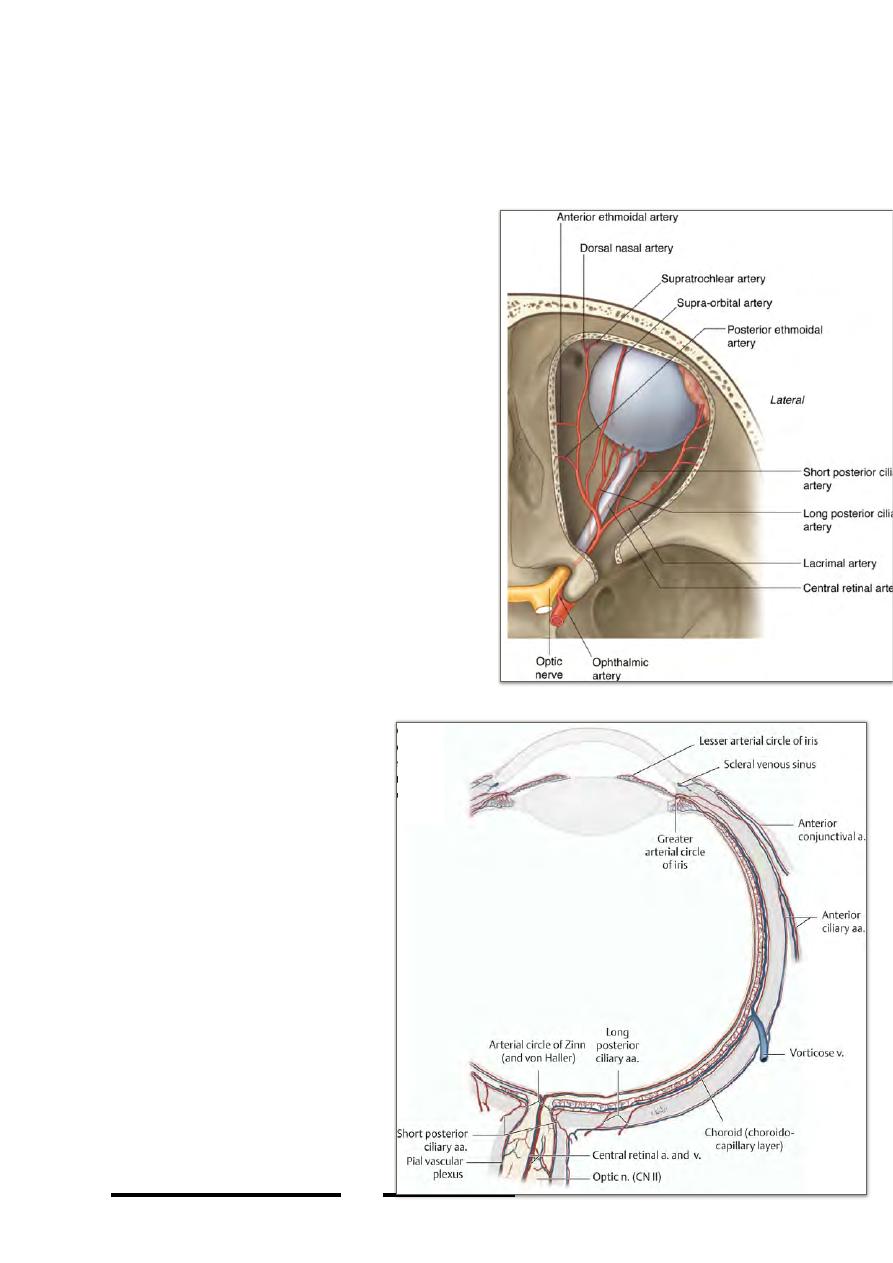
Arterial supply of the orbit:
-the ophthalmic artery, branch of ICA just after
it leaves the cavernous sinus, enters the orbit
through the optic canal
-it is directed in the orbit from lateral to medial
across the optic nerve
Branches:
1-Branches to the eyeball:
- central artery of the retina
- long & short posterior ciliary branches
- anterior ciliary branches
2-Branch with each of the sensory nerves of
the orbit; taking its course & destination
3-Muscular branches; with the motor nerves of
the orbit supplying ocular muscles & give the
anterior ciliary arteries
*The central artery of retina:
-pierces the optic n. near the middle of its
intraorbital course
-supplies the distal 1/3 of the optic nerve & the
whole retina
- i t s d a m a g e l e a d s t o t o t a l
blindness of that eye with optic
atrophy
*The short posterior ciliary
arteries:
-pierce the back of sclera near the
optic nerve
-supply the choroid
*The long posterior ciliary
arteries:
-pierce the back of sclera near the
optic nerve
-pass between the sclera &
choroid to the iris
*Anterior ciliary arteries:
-branches of muscular arteries
-pierce the sclera near the cornea
-end in the greater arterial circle of
the iris
!
76
Head & Neck Dr. Nawfal K. Al-Hadithi
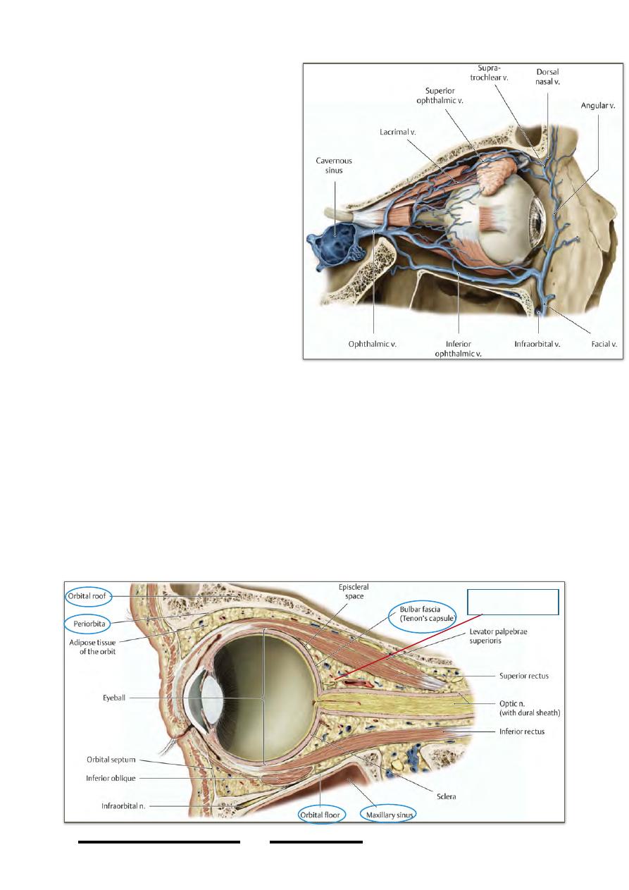
Venous drainage of the orbit:
1-Superior ophthalmic vein:
-formed at the supraorbital foramen by
u n i o n o f t h e s u p r a o r b i t a l &
supratrochlear veins
-has the same course & branches of
the ophthalmic artey
-joined by the inferior ophthalmic vein
at the medial end of SOF
-enters the cavernous sinus after
leaving the orbit
2-Inferior ophthalmic vein:
-formed in the floor of the orbit by
union of muscular veins
-communicates with pterygoid venous
plexus through the inferior orbital
fissure
-empties in the SOV at the medial end
of SOF
-sometimes empties directly in the cavernous sinus
Fasciae of the orbit:
1-Periorbita:
-the double-layered dura mater of the cranial cavity enters the orbit with the optic
nerve
-the fibrous coat remains with the nerve & the endosteal layer leave the fibrous layer
to form the periosteal layer of the orbit (periorbita)
-unlike in the cranial cavity, periorbita could be easily stripped from bones of the
orbit
-the site where the two dural layers diverge in the orbit represents the site of complete
separation of the orbital from cranial cavities
!
77
Head & Neck Dr. Nawfal K. Al-Hadithi
Retrobulbar fat
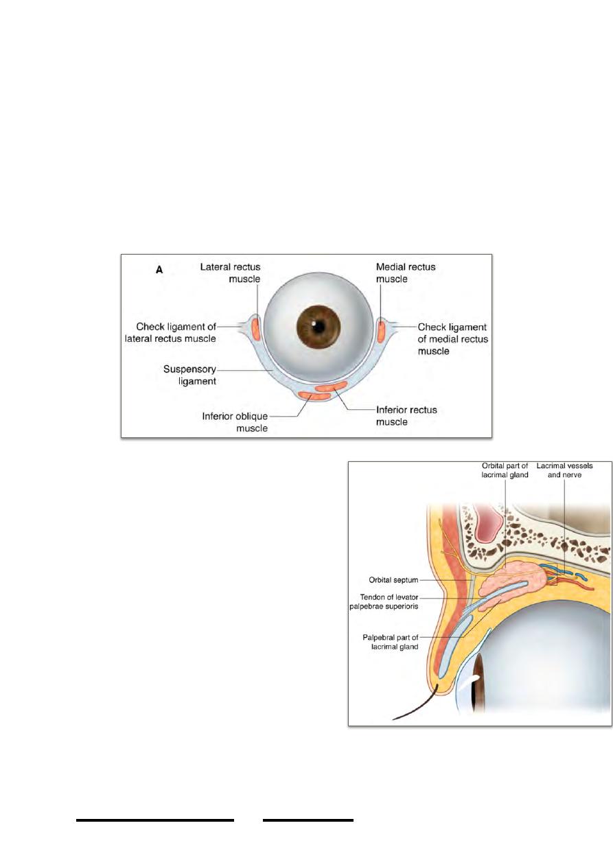
2-Muscular fasciae:
-fascia covering ocular muscles
-muscular fascia of MR thickened at certain site to be attached to the posterior
lacrimal crest forming the “medial check ligament”
-the same thing occur in LR fascia & attaches it to the zygomatic bone forming the
“lateral check ligament”
-these two thickenings fuse with fasciae of IO & IR to form the hammock-like sling
on which the eyeball rests “suspensory ligament of the eyeball”
3- Retrobulbar (orbital) fat:
A fixed-sized cushion of fatty tissue on which the globe rests with a fixed position of
its center.
The lacrimal apparatus:
1- Lacrimal gland:
-an oval gland occupies the superolateral part
of the orbit “lacrimal fossa”
- i t i s p i e r c e d b y L P S m u s c l e w h i c h
incompletely divides it into orbital part which
remains in the roof of the orbit partially
invested by fascia of SR & LR muscles, &
palpebral part which projects inside the upper
eyelid with its deep surface in relation to the
conjunctiva
-ducts of the gland are 6-10 in number, all
empty in the superior fornix of conjunctiva
-supplied by lacrimal branch of ophthalmic
artery
-drained by lacrimal v. which empties in the
superior ophthalmic v.
-supplied by lacrimal nerve which carries
autonomic component derived from the zygomaticotemporal branch of Vb
2- Lacrimal canaliculi:
!
78
Head & Neck Dr. Nawfal K. Al-Hadithi
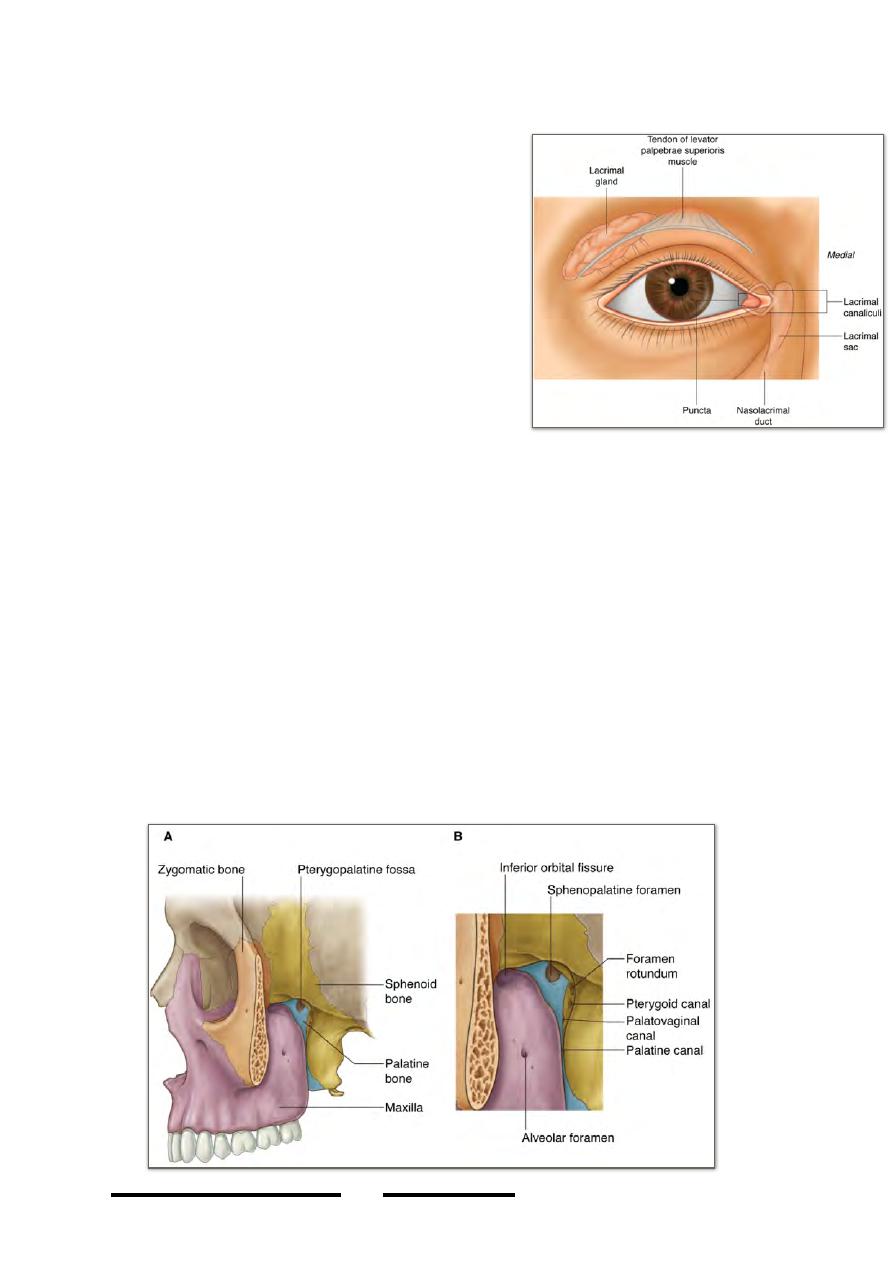
-open in the eyelids as the lacrimal puncta whose openings are directed toward the
lacrimal lake
-course over the corresponding eyelids, the puncta
open in the lacrimal sac
-they collect tears from the lake to the sac
3- Lacrimal sac:
-it is the upper dilated end of nasolacimal duct
-measures 0.5 X 1 cm
-receives the lacrimal canaliculi separately
-lies in front of the lacrimal part of orbicularis
oculi & behind the medial palpebral ligament
-contraction of the lacrimal part of O. oculi dilates
the sac making negative pressure which sucks
tears from the lake by the canaliculi
4- Nasolacrimal duct:
-extends from the lacrimal sac downward, backward
& laterally towards the inferior nasal meatus
-transmits tears from the sac to the nasal cavity
-is about 2 cm long
The pterygo-palatine fossa:
•
Apyramidal space located in the interval between the root of the pterygoid
process posteriorly & the back of maxilla anteriorly.
•
Boundaries:
-Anterior: back of maxilla
-Posterior: root of pterygoid process & body of sphenoid
-Lateral: ITF
-Medial: nasal cavity
-Superior: apex of the orbit
-Inferior: maxillary sinus
!
79
Head & Neck Dr. Nawfal K. Al-Hadithi