ANATOMY
HEAD & NECK
Dr. Nawfal K. Al-Hadithi
Lec . 5
The Suprahyoid, Prevertebral,
Suboccipital Regions
By : Ali Kareem
مكتب اشور لالس تنساخ
2013 – 2014
Head & Neck Dr. Nawfal K. Al-Hadithi
Anatomy
2
Lec. 5 The Suprahyoid, Prevertebral, Suboccipital Regions
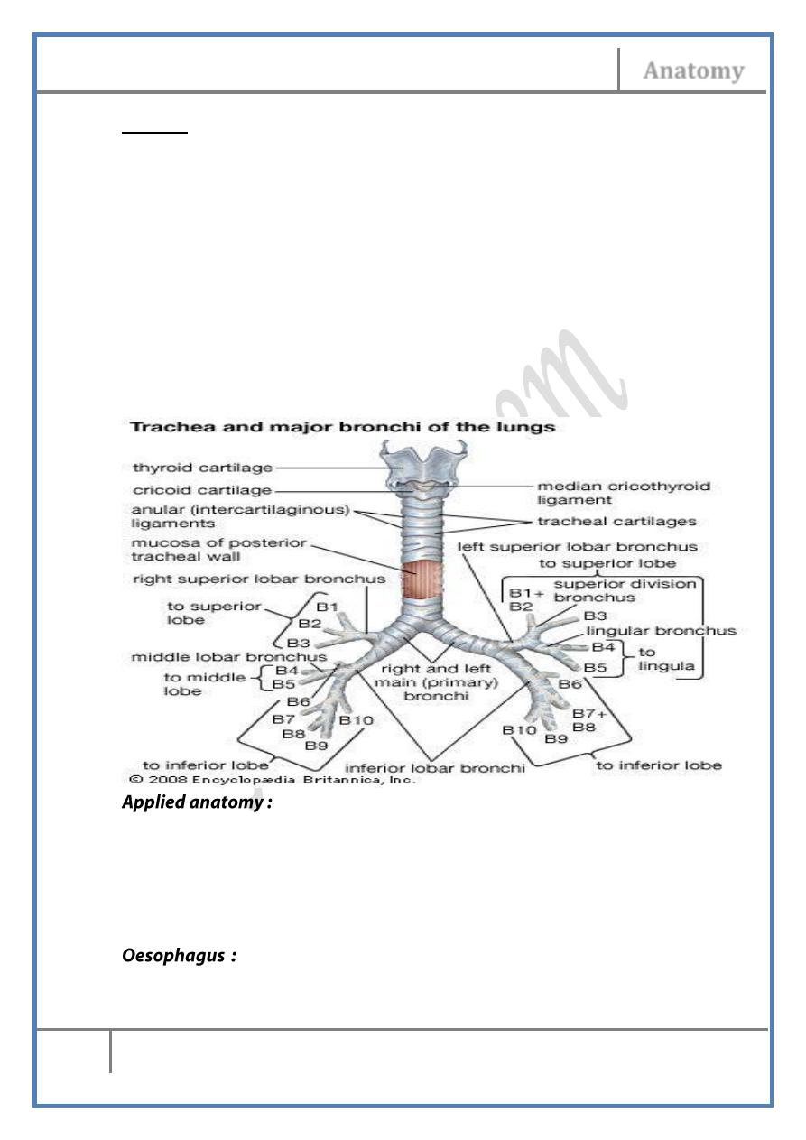
Trachea :
• Begins at the cricoid cartilage (C6) & ends at the carinaT4 level, a
12 cm long tube ½ of which lies in the neck
• The CCA lie on each side of the trachea being separated from it
above by the lateral lobes of the thyroid.
• The oesophagus lies behind it & the intervening groove is occupied
by the recurrent laryngeal nerves
• Thyroid isthmus lies anterior to the rings 2-4
• Trachea is formed of C-shape cartilages opened behind & the
opening is closed by trachealis muscle.
• Cervical trachea is supplied by the inferior thyroid a., drained by
inferior thyroid v. , its nerve supply is similar to the thyroid gland
& its lymph goes to the postero-inferior group of deep cervical
nodes.
• Tracheostomy is a life-saving operation to open the airway in acute
& chronic airway obstruction.
• The incision is done below the cricoid cartilage & the trachea is
exposed after dividing the successive layers in front of it
• A tube is inserted to keep the airway patent
• 25 cm tube begins at the level of C6 vertebra (cricopharyngeus) &
ends at the cardiac opening of the stomach
Head & Neck Dr. Nawfal K. Al-Hadithi
Anatomy
3
Lec. 5 The Suprahyoid, Prevertebral, Suboccipital Regions
• The carotid sheaths lie on each side being separated from it by the
posterior part of the lateral lobe of the thyroid.
• The trachea lies anterior to it & the intervening groove is occupied
by the recurrent laryngeal nerves
• The prevertebral muscles lie behind it with the sympathetic trunk
on each side.
• Cervical oesophagus is supplied by the inferior thyroid a., drained
by inferior thyroid v. , its nerve supply is similar to the thyroid
gland & its lymph goes to the postero-inferior group of deep
cervical nodes.
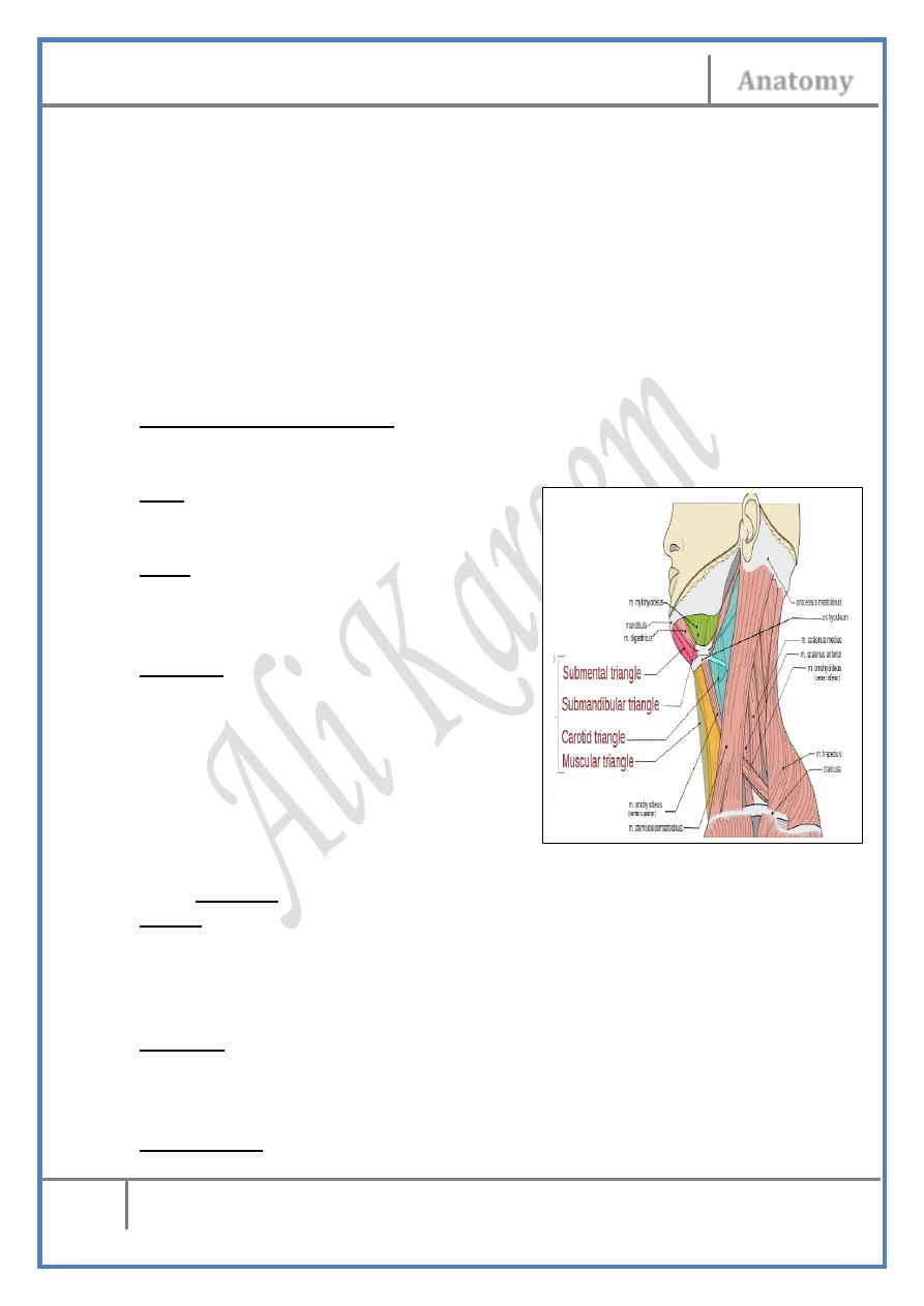
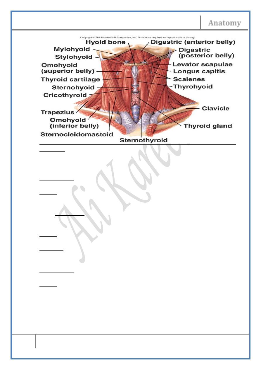
The submandibular triangle :
Is the space between the two bellies of digastric & the lower border of the
mandible.
Roof :
- Skin, subcutaneous connective tissue,
platysma and investing fascia.
Floor :
- Myelohyoid “anteriorly”
- Hyoglossus & part of the middle
- constrictor “posteriorly”
Contents :
1- Suprahyoid muscles.
2- Submandibular gland.
3- Facial artery.
4- Common facial vein.
5- Submandibular lymph nodes.
6- Cervical branch of facial nerve.
Suprahyoid Muscles :
1- Digastric :
Origin :
- Anterior belly: From the digastric fossa at the lower border of the
mandible on each side of symphysis menti
- Posterior belly: From the digastric notch on the medial aspect of
the mastoid process.
Insertion :
The 2 bellies are directed to the body of the hyoid bone where they are
united by an intermediate tendon which is held to the hyoid bone by a
fibrous sling.
Nerve supply :
Head & Neck Dr. Nawfal K. Al-Hadithi
Anatomy
4
Lec. 5 The Suprahyoid, Prevertebral, Suboccipital Regions
- Anterior belly : Nerve to myelohyoid (branch from the mandibular
n. Vc)
- Posterior belly : Facial nerve
Action :
- Elevate the hyoid bone, larynx & pharynx during swallowing
- Opens the mouth widely
2- Stylohyoid :
Origin :
- From the posterior aspect of the styloid process near its root.
Insertion :
The muscle descends superomedial & parallel to the posterior belly of
digastric to reach the intermediate tendon of digastric, here it divides into
2 slips which pass on each side of the intermediate tendon of digactric &
inserted on the hyoid bone near the greater horn.
Nerve supply :
Facial nerve
Action :
- Pulls the hyoid backward & upward.
3- Myelohyoid :
Origin :
From the whole length of myelohyoid line on the inner surface of the
mandible
Insertion :
- Fibers descend medially & backward
- Posterior part of it will reach the body of hyoid bone to which they
are inserted
- Anterior fibers meet each other in a midline raphe which extends
between the hyoid bone & the mandible
Nerve supply :
Nerve to myelohyoid (from the mandibular nerve Vc)
Action :
- Forms the floor of the mouth on which the tongue rests & move
- Plays a major role in swallowing.
4- Geniohyoid :
Origin :
Inferior genial tubercle.
Head & Neck Dr. Nawfal K. Al-Hadithi
Anatomy
5
Lec. 5 The Suprahyoid, Prevertebral, Suboccipital Regions
Insertion :
The two ribbon like muscles descend side by side between myelohyoid &
genioglossus to be inserted into the upper border of the body of hyoid
bone.
Nerve supply :
C1 fibers from the XII nerve.
Action :
Pulls the hyoid anterosuperiorly.
5- Hyoglossus :
- This is a tongue muscle & is an important key around which the
structures of the floor of the mouth are distributed
Origin :
The upper border of the greater cornu of the hyoid.
Insertion :
The rectangular muscle ascends up to be inserted into the side of the
tongue posteriorly
Nerve supply :
XII nerve.
Action :
- Pulls the hyoid up
- Depresses the side of the tongue
- Both retract the tongue
Submandibular gland :
Will be discussed later.
Head & Neck Dr. Nawfal K. Al-Hadithi
Anatomy
6
Lec. 5 The Suprahyoid, Prevertebral, Suboccipital Regions
Facial artery :
Discussed.
Common facial vein :
• Formed at the lower border of the mandible by union of the
anterior facial v. & anterior division of retromandibular v.
• Passes superficial to the submandibular gland in the direction of
the carotid triangle of the neck
• Terminates in the IJV either separately or together with the
superior thyroid & lingual veins
• It drains the structures in the submandibular triangle
Submandibular lymph nodes :
Will be discussed later.
Cervical branch of VII :
Descends vertically from behind the mastoid process to platysma.
The submental triangle :
Is the space between the anterior bellies of the 2 digastrics & the hyoid
bone.
Roof :
- Skin, subcutaneous connective tissue, platysma and investing
fascia.
Floor :
- Myelohyoid
Contents :
1- Submental branch of facial artery.
2- Nerve to myelohyoid.
3- beginning of AJV.
4- Submental lymph nodes.
Submental branch of facial artery :
Discussed.
AJV :
Discussed.
Submental lymph nodes :
Head & Neck Dr. Nawfal K. Al-Hadithi
Anatomy
7
Lec. 5 The Suprahyoid, Prevertebral, Suboccipital Regions
Will be discussed later.
Myelohyoid nerve :
• Given from the inferior alveolar nerve at the mandibular foramen.
• Pierces the sphenomandibular ligament & passes forward in the
floor of the submental triangle inferior to myelohyoid between it &
the anterior belly of digastric supplying both.
• It is accompanied by the submental artery.
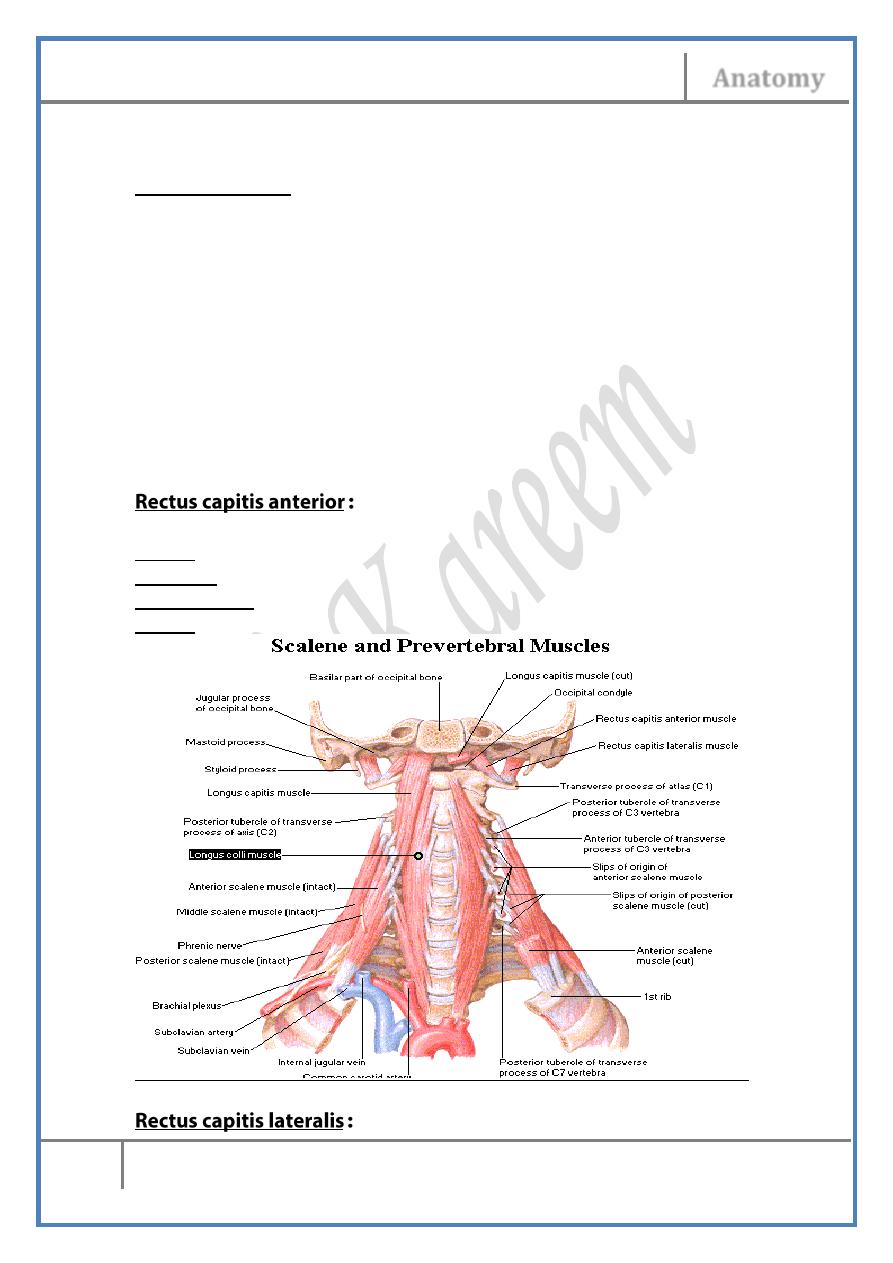
The prevertebral region :
Is the area which lies in front of the vertebral column & constitutes the
prevertebral & paravertebral muscles covered by the prevertebral fascia.
Prevertebral Muscles :
Origin; lateral mass of atlas
Insertion; basi-occiput
Nerve supply; C1
Action; flexes the head
Head & Neck Dr. Nawfal K. Al-Hadithi
Anatomy
8
Lec. 5 The Suprahyoid, Prevertebral, Suboccipital Regions
Origin; transverse process of atlas
Insertion; jugular process of occipital bone
Nerve supply; C1
Action; laterally flexes the head
Origin; anterior tubercles of T.P of the 4 typical cervical vertebrae end
to-end with the tendons of scalenus anterior
Insertion; basi-occiput anterior to foramen magnum
Nerve supply; C1-C4
Action; flexes the head & neck
This muscle extends on the anterior surface of the vertebral column from
C1 to C4
Origin;
- Vertical part : bodies of thoracic vertebrae
- Superior oblique part: T.P of upper cervical vertebrae
- Inferior oblique part : bodies of upper thoracic vertebrae
Insertion;
- Vertical part : bodies of cervical vertebrae
- Superior oblique part : anterior tubercle of C1
- Inferior oblique part : T.P of lower cervical vertebrae
Nerve supply; C1-T4
Action; flexes & rotates the neck
The Cervical Sympathetic Trunk :
• An upward extension of the thoracic S.T which is located behind
the carotid sheath on the prevertebral fascia
• In a cord-like form or multiple strands, the S.T develops three
ganglia in the neck; superior, middle & inferior
• Each one of the three has three sets of branches; somatic, vascular
& visceral.
• The body of the cervical S.T is formed mainly of preganglionic
fibers which traverse the white rami communicantes of the upper 5
thoracic segments
• Cervical S.T possesses no white rami communicantes attaching it
to the spinal nerves since there is no lateral gray column in the
cervical segments
Head & Neck Dr. Nawfal K. Al-Hadithi
Anatomy
9
Lec. 5 The Suprahyoid, Prevertebral, Suboccipital Regions
Vascular br.
Visceral br.
Somatic br.
Position
Ganglion
-Carotid
sympathetic
plexus
-Superior
cervical cardiac
nerves
-The left to the
superficial
cardiac plexus
-The right to the
deep C.P
-Gray
rami
communicants
to
C1-C4
spinal nerves
-Largest (2.5-
3.5) cm long
-C2 level
Superior
C.G
-Inferior
thyroid sym.
Plexus
-Middle
cervical cardiac
nerves to the
deep C.P
-Gray
rami
communicants
to C5 & C6
spinal nerves
-Smallest
(1.3 cm) long
-C6 level
Middle
C.G
-Vertebral
sym. plexus
-Inferior
cervical cardiac
nerves to the
deep C.P
-Gray
rami
communicants
to C7 & C8
spinal nerves
-In
82%
fused
with
the
T1
ganglion
to
form the the
stellate gan.
on the neck
of the 1st rib
Inferior
C.G
Root of the neck :
- The root of the neck is based on the supra-pleural membrane which
inclines downward & forward with the inclination of the first rib
- Behind & inferior to the membrane lies the lung apices with their
cervical pleura
- In front & superior to the membrane lies structures of the root.
- The neck root is studied according to structure relation to scalenus
anterior
The triangular space “of Chassaignac” :
- It is the space bounded laterally by the medial border of scalenus
anterior, medially by the sloping lateral border of longus colli &
inferiorly by the neck of the 1st rib
- Its apex is the carotid tubercle (C6 T.P) against which the CCA can
be compressed
Head & Neck Dr. Nawfal K. Al-Hadithi
Anatomy
10
Lec. 5 The Suprahyoid, Prevertebral, Suboccipital Regions
- It contains the 1st part of subclavian artery & the 6th cervical sym.
Ganglion
Anterior relations :
1- Phrenic nerve: passes vertically across the obliquity of the muscle
leaving its medial border inferiorly to pass to the thorax.
2- Vagus nerve: descends anterior & medial to the muscle to reach the
front of the subclavian artery & gives the recurrent laryngeal n.
around this vessel on the right side.
3- Ascending cervical artery: ascends from the inferior thyroid a. on
the muscle medial to the phrenic nerve.
4- Transverse cervical & suprascapular arteries: cross anterior to the
lower part of the muscle in their way to the posterior triangle.
5- IJV: lies anterior to the lower part of the muscle.
6- Subclavian vein: crosses the lower part of the muscle to meet the
IJV medial to it in the pyramidal space.
7- Deep cervical L.N: around the IJV, inferior nodes lie especially in
front of the muscle.
8- SCM: covers all the above structures.
Medial relations :
1- Vagus nerve.
2- Middle & inferior cervical sympathetic ganglia: lie in the
pyramidal space on the medial side of scalenus anterior connected
in front of the subclavian artery by the ansa subclavia.
3- First part of subclavian artery: with its three branches
4- Vertebral vein(s): leave the T.P of C7 & course forward to enter
the confluence of the subclavian v. & IJV.
5- Thoracic duct (on the left) & right lymph duct (on the right): arch
in the pyramidal space to enter the confluence of the subclavian v.
& IJV.
Posterior relations :
1- Roots of brachial plexus: as they emerge from the intervertebral
foramina to the posterior triangle, they lie superior to the
subclavian artery.
2- Second part of subclavian artery: behind the muscle on the 1st rib,
here it gives the costocervical axis.
Lateral relations :
1- Trunks of brachial plexus: in the posterior triangle.
Head & Neck Dr. Nawfal K. Al-Hadithi
Anatomy
11
Lec. 5 The Suprahyoid, Prevertebral, Suboccipital Regions
2- Third part of subclavian artery: behind the prevertebral fascia in
the floor of the posterior triangle.
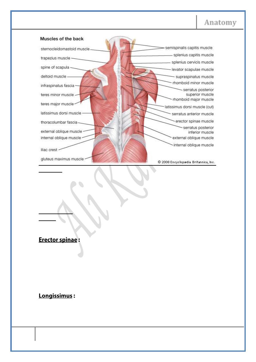
- The muscles of the back of the neck are enclosed by the
thoracolumbar fascia in the posterior compartment of which the
postvertebral muscles lie
- Externally the investing fascia wraps the whole neck
Back of the neck :
Ligamentum nuchae :
A strong, triangular sheet of fibrous tissue dividing the back of the neck
into two halves & provides an important origin for many muscles, it is
attached by its three borders to :
1- The external occipital crest, superiorly.
2- Tips of cervical spines & supraspinous ligaments, anteriorly.
3- Posterior free border to which the investing fascia comes from
either side of the neck to be attached.
Thoracolumbar fascia :
- A strong fascia attached to the spines & transverse processes of the
vertebral column enclosing within its two compartments the
muscles
- This arrangement persists in the the lumbar region, in the thoracic
& cervical regions the anterior lamella of the fascia disappears
leaving the middle & posterior lamellae only enclosing in between
the postvertebral muscles
Muscles of the back of the neck :
- Its name in latin means “bandage” revealing its shape & function
- The muscle lies deep to trapezius & covers the deep muscles of the
neck like a strap.
Origin: from ligamentum nuchae & spines of C6 & 7 with the
supraspinous ligaments.
Head & Neck Dr. Nawfal K. Al-Hadithi
Anatomy
12
Lec. 5 The Suprahyoid, Prevertebral, Suboccipital Regions
Insertion: The muscle is directed upward & laterally & divided into two
parts :
- Splenius capitis; inserted into the deep part of mastoid & the
lateral 1/3 of the superior nuchal line.
- Splenius cervicis; inserted into the posterior tubercles of T.P of the
upper 3 cervical vertebrae.
Nerve supply: posterior rami of C2-C6.
Action: pulls the head back & laterally in the direction of the active
muscle. Both extend the neck.
- This group of muscles extend from the sacrum to the skull in the
form of three longitudinal parallel columns & occupy the posterior
compartment of lumbar fascia
- Some of them are inserted into various regions (lumbar & thoracic)
& some muscles belong to the head & neck.
- Their nerve supply is segmental
Is the intermediate column of E.S
Has three parts (thoracis, cervicis & capitis).
Arise from T.P of lower vertebrae.
Head & Neck Dr. Nawfal K. Al-Hadithi
Anatomy
13
Lec. 5 The Suprahyoid, Prevertebral, Suboccipital Regions
Inserted into T.P. of higher vertebrae.
Capitis muscle is inserted into the back of the mastoid process deep
to splenius & SCM.
Occupy the medial column of the E.S
Its limited to the thoracic (thoracis), cervical (cervicis) & head
(capitis)regions
S. cervicis & thoracis arise from the T.P of all thoracic vertebrae
They are inserted into the spines of 4-6 vertebrae above. The highest
fibers of cervicis muscle are inserted into the undersurface of the spine of
axis
They extend the upper vertebral column & rotate it to the opposite side
S. capitis arises from T.P of C7-T6
It is the largest muscle in the back of the neck,being inserted into the
occipital bone between the 2 nuchal lines medially
It is the most powerful skull extensor
The suboccipital muscles :
Origin: spine of C2
Insertion: the area between inferior nuchal line & foramen
magnum, laterally
Origin: posterior tubercle of C1
Insertion: the area between inferior nuchal line & foramen
magnum, medially
Head & Neck Dr. Nawfal K. Al-Hadithi
Anatomy
14
Lec. 5 The Suprahyoid, Prevertebral, Suboccipital Regions
Origin: T.P of C1
Insertion: the area between the two nuchal lines, lateral to
semispinalis capitis
Origin: spine of C2
Insertion: T. P of C1
The suboccipital triangle :
Is the triangle embraced by the supoccipital muscles except rectus capitis
posterior minor which lies medial to it.
Floor: the posterio atlanto-occipital membrane & posterior arch of atlas
Contents :
1- Vertebral artery; in the floor.
2- Suboccipital nerve, C1.
3- Greater occipital nerve, C2: hooks below the inferior oblique &
ascends in the roof of the triangle.
4- Occipital artery: ascends medially in the roof of the triangle in its
way to the scalp together with the greater occipital nerve.
Edited & Published by :
Ali Kareem