ANATOMY
HEAD & NECK
Dr. Nawfal K. Al-Hadithi
Lec . 4
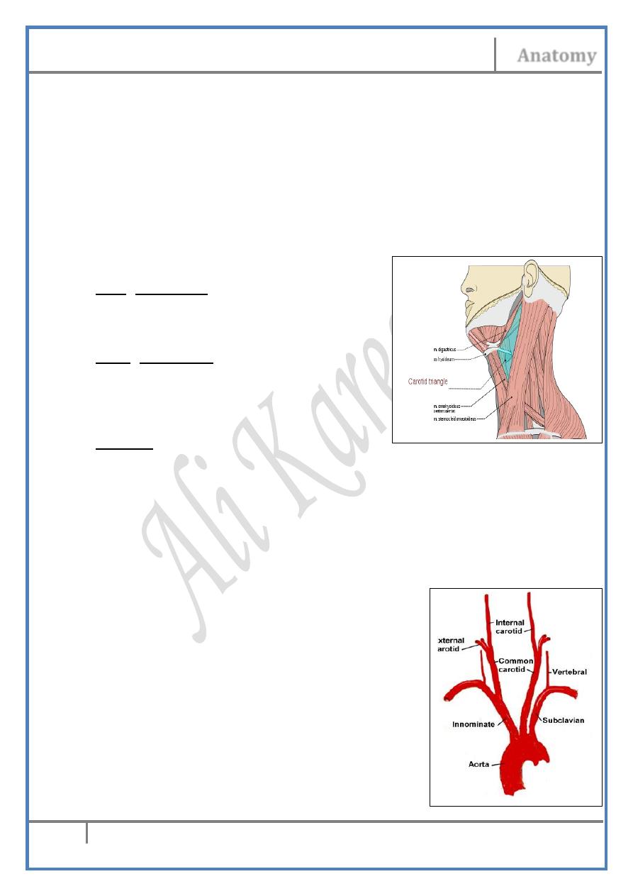
The Ant. Cervical Triangle
By : Ali Kareem
مكتب اشور لالس تنساخ
2013 – 2014
Head & Neck Dr. Nawfal K. Al-Hadithi
Anatomy
2
Lec. 4 The Ant. Cervical Triangle
The anterior cervical triangle :
• Is the area of the neck which lies between the anterior borders of
the two SCM muscles & the lower border of the mandible.
• The anterior midline divides this area into two anterior cervical
triangles.
• Digastric & the superior belly of omohyoid further divide each of
the two triangles into carotid, muscular & submandibular triangles.
• The remaining area between the anterior bellies of both digastrics
in the single submental triangle.
The carotid triangle :
Roof (lateral wall) :
Skin, subcutaneous tissue, platysma and
Investing fascia
Floor (medial wall) :
- Anteriorly : hyoglossus & thyrohyoid
- Posteriorly : middle and inferior constrictor
muscles
Contents :
1- Carotid sheath & its contents.
2- Nerves; ansa cervicalis, hypoglossal nerve.
3- Deep cervical lymph nodes.
Contents of the carotid sheath :
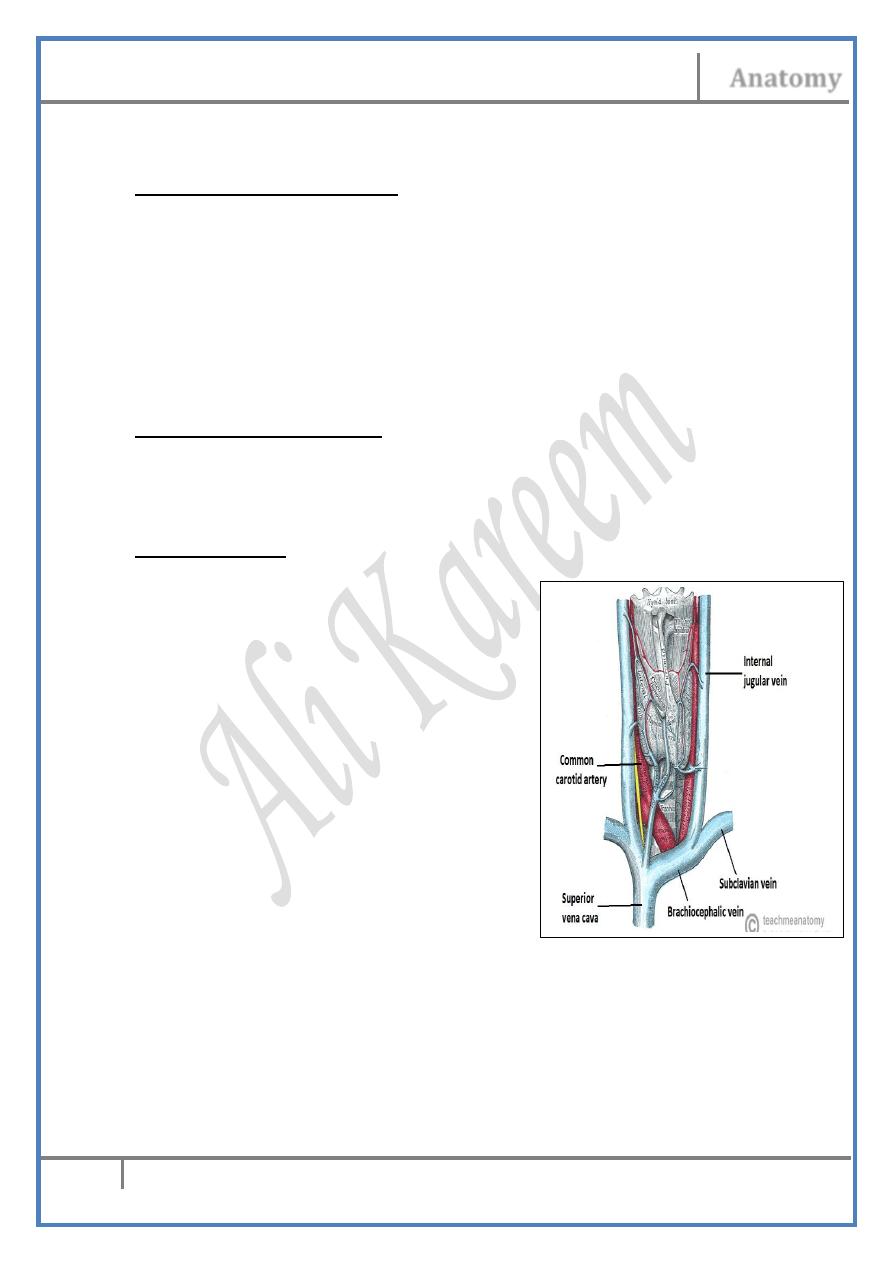
The Common Carotid Artery (CCA) :
- From behind the corresponding SCJ, the CCAs diverge as they
ascend on each side of the neck separated by the
larynx & thyroid gland
- Their course is in the carotid line
(a vertical line midway between the
angle of the mandible & the mastoid
process)
- At the upper border of thyroid cartilage
(C3-4) it divides into ECA & ICA
- The artery is crossed from below upward
by the IJV, SCM, superior belly of omohyoid,
superior laryngeal a. & thyroid veins
Head & Neck Dr. Nawfal K. Al-Hadithi
Anatomy
3
Lec. 4 The Ant. Cervical Triangle
The carotid sinus :
- It is a dilatation at the bifurcation of the CCA especially around the
origin of the ICA
- Its walls are especially elastic & contain baroreceptors responsible
for regulation of blood pressure
The carotid body :
- A mass of cells in the posterior wall of the sinus
- Contains chemoreceptors for regulation of blood osmolarity
• BOTH carotid sinus & body are supplied by the sinu-carotid
branch of IX nerve
• Carotid body tumor “Potato tumor”:
A tumor affecting the carotid body resulting in a pulsetile swelling in the
lateral aspect of the neck
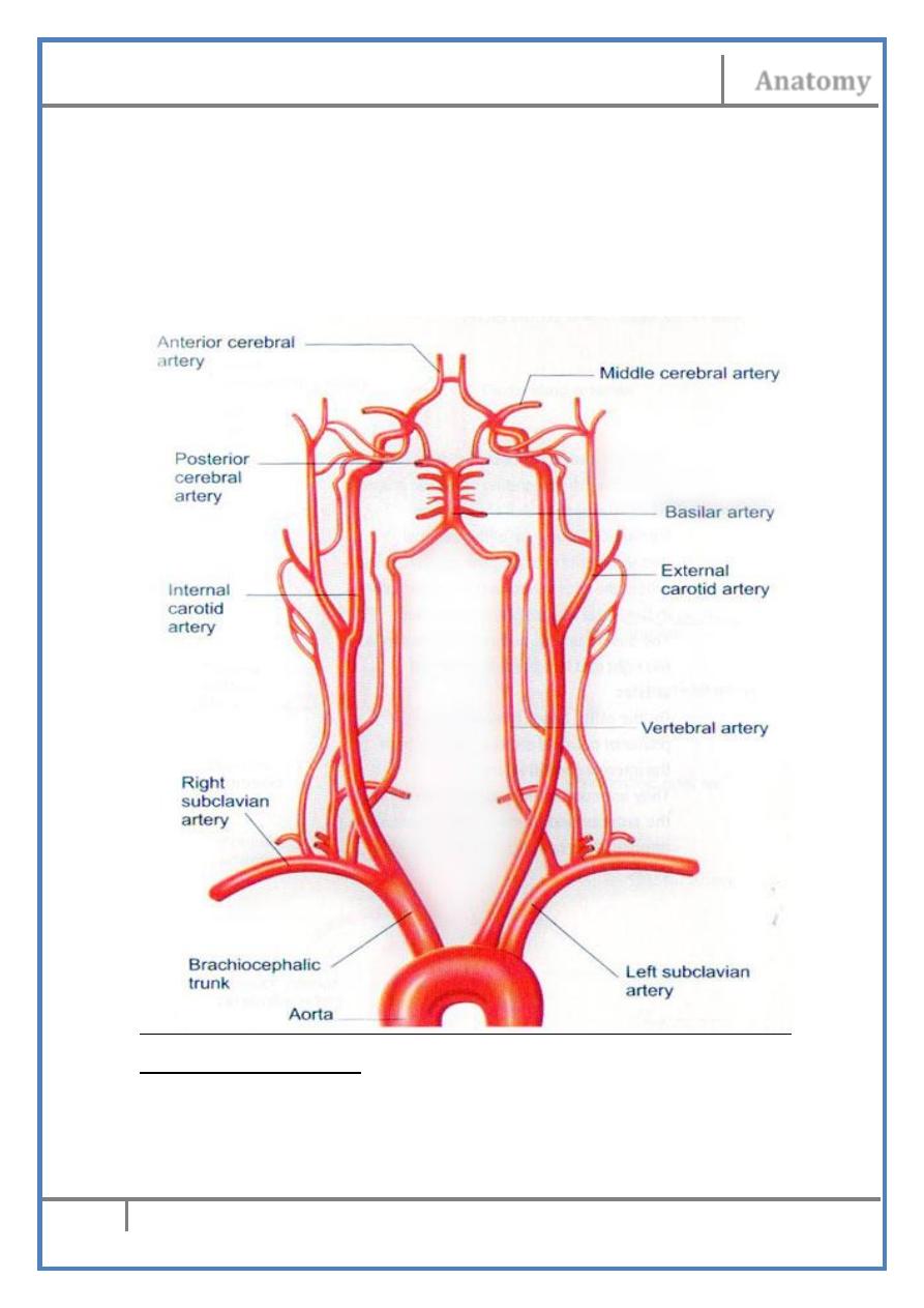
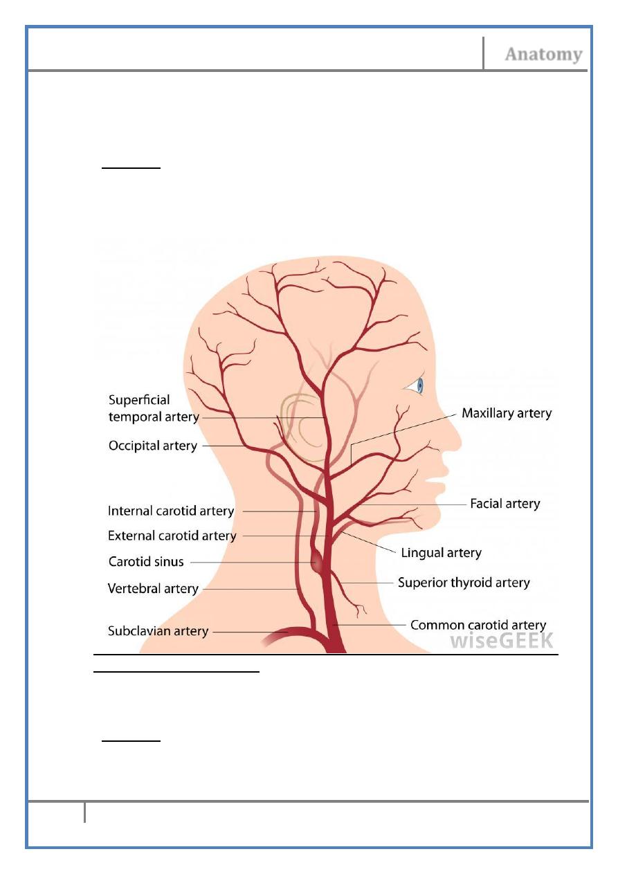
The Internal Carotid Artery (ICA) :
• At the bifurcation, the ICA is postero-lateral to the ECA, then it
takes a medial relation alongside the pharynx.
• It passes upward with the IJV to reach the base of the skull where it
enters the temporal bone through the carotid canal.
• It gives no branch in the neck.
• It supplies the brain & structures inside the orbit.
The External Carotid Artery (ECA) :
• About equal in caliber to the ICA, the ECA extends between the
upper border of thyroid cartilage & the neck of the mandible where
it ends by dividing into its two terminal branches
• In the carotid triangle, the ECA lies superficial being covered
directly by the investing fascia of the roof, it is crossed by the
facial, lingual & sometimes the superior thyroid veins
• Leaving this triangle, the artery passes deep to the posterior belly
of digastric & stylohyoid muscles separating them from
stylopharyngeus
• Here, the ECA lies in the deep lobe of the parotid gland together
with the retromandibular vein & facial nerve.
Branches of the ECA :
A) Anterior branches :
1- Superior thyroid a.
2- Lingual a.
3- Facial a.
B) Posterior branches :
Head & Neck Dr. Nawfal K. Al-Hadithi
Anatomy
4
Lec. 4 The Ant. Cervical Triangle
4- Occipital a.
5- Posterior auricular a.
C) Medial branch :
6- Ascending pharyngeal a.
D) Terminal branches :
7- Maxillary a.
8- Superficial temporal a.
Superior thyroid artery :
- Given at the beginning of the ECA just below the tip of greater horn of
the hyoid bone
- Descends downward towards the apex of the lateral lobe of the thyroid
gland accompanied by the external laryngeal nerve
Head & Neck Dr. Nawfal K. Al-Hadithi
Anatomy
5
Lec. 4 The Ant. Cervical Triangle
- Pierces the pretracheal fascia at the apex of the thyroid lobe & divides
into anterior & posterior branches which descend on the corresponding
surfaces of the gland & anastomose with the inferior thyroid branches
- Branches :
a) Superior laryngeal a. ; pierces the thyrohyoid membrane together with
the internal laryngeal n. & supplies the larynx above the level of vocal
folds
b) Sternomastoid a. ; to SCM
c)Muscular branches to the inferior constrictor muscle
Lingual artery :
- Arises opposite to the tip of the greater horn of hyoid bone.
- After an upward loop, the artery passes forward to disappear deep to
hyoglossus.
- It is crossed laterally by the hypoglossal nerve, posterior belly of
digastric & stylohyoid.
- Branches :
a) Dorsal lingual a.; given at the back of the tongue & supplies the tonsils,
soft palate & epiglottis.
b) Sublingual a.; to the sublingual gland
Facial artery :
- Given just above the tip of greater horn of hyoid bone.
- Ascends up deep to the posterior belly of digastric in the submandibular
triangle where it lies between the submandibular gland & myelohyoid.
- After an S-shape course in the triangle, the artery curves over the lower
border of the mandible at the anterior edge of masseter to ascend in the
face.
- Branches :
a) Ascending palatine a.; ascends between styloglossus &
stylopharyngeus to lie on the superior constrictor muscle. It supplies
the upper pharyngeal wall, soft palate, palatine tonsils & auditory tube
b) Tonsillar a.; to the palatine tonsils
c) Glandular branches to the submandibular gland
d) Submental a.; given before the facial a. curves on the mandible, it
passes back between the anterior belly of digastric & myelohyoid with
the nerve to myelohyoid in the submental triangle.
Occipital artery :
- Given from the back of the ECA at the level of the facial a.
- Ascends parallel to the lower border of the posterior belly of digastric to
hide deep to SCM.
Head & Neck Dr. Nawfal K. Al-Hadithi
Anatomy
6
Lec. 4 The Ant. Cervical Triangle
- It grooves the skull in the region of the occipito-mastoid suture to
appear in the apex of the posterior triangle
- Pierces the fascia of the neck & ascends in the back of the scalp
supplying it up to the vertex.
- Branches :
a) Muscular branches to digastric, stylohyoid & posterior cervical
muscles.
b) Sternomastoid a.; to SCM
c) Meningeal, auricular & mastoid branches
Posterior auricular artery :
- Ascends parallel to the upper border of the posterior belly of digastric.
- Passes in the region of the parotid gland supplying it
- Crosses superficial to the mastoid process behind the auricle.
- Branches :
a) Stylomastoid a.; enters the corresponding foramen & gives stapedial
arery to stapedius.
b) Auricular a.
Head & Neck Dr. Nawfal K. Al-Hadithi
Anatomy
7
Lec. 4 The Ant. Cervical Triangle
c) Occipital arteries to the scalp
Ascending pharyngeal artery :
- From the medial aspect of the ECA, this long & slender branch ascends
on the sidewall of the pharynx deep to the ICA.
- Branches:
a) Pharyngeal branches to the superior & middle constrictor muscles
b) Palatine a.; to the soft palate & palatine tonsils.
c) Inferior tympanic a.; to the middle ear
d) Meningeal branches to the dura mater after entering the hypoglossal
canal or the jugular foramen.
Superficial temporal artery :
- The smaller of the 2 terminal branches of the ECA
- Given deep to the neck of the mandible
- Ascends in the temporal fossa
Maxillary artery :
- The larger of the 2 terminal branches of the ECA
- Given deep to the neck of the mandible
- Passes forward into the infratemporal fossa
The Internal Jugular Vein (IJV) :
• It is the continuation of the dural
sigmoid sinus in the posterior
compartment of the jugular foramen.
• In the carotid sheath, the IJV is
posterolateral to the artery in the
upper part of the neck, then becomes
lateral & in the root of the neck it is
almost anterior
• Ansa cervicalis is usually formed
around it
• Deep cervical L. N. lie around it
• At its beginning it has a dilatation called superior bulb, a similar
(inferior) bulb lies at its termination
• Tributaries :
- Inferior petrosal sinus: just below the base of the skull.
- Thyro-glosso-facial confluence of v.: superior thyroid, lingual &
facial vv. usually open in common or separately in the IJV at the
level of the origin of lingual a. from the ECA.
Head & Neck Dr. Nawfal K. Al-Hadithi
Anatomy
8
Lec. 4 The Ant. Cervical Triangle
- Pharyngeal veins : from the pharyngeal plexus, terminate in the IJV
at the level of the angle of mandible.
- Middle thyroid v.
The Vagus Nerve :
• Leaves the skull with the IJV, IX & XI nerves through the jugular
foramen.
• Descends in the neck inside the carotid sheath throughout which
the nerve lies between the IJV & the carotids.
• In the neck it gives the following branches:
- Meningeal n.: to the dura of posterior cranial fossa
- Auricular n.: to the external acoustic meatus & tympanic
membrane
- Pharyngeal n.: usually 2 in number to the pharyngeal plexus
- Superior laryngeal n.: divides into internal & external laryngeal
nerves
- Cervical cardiac n.: usually 2 in number to the cardiac plexus
- Recurrent laryngeal n.
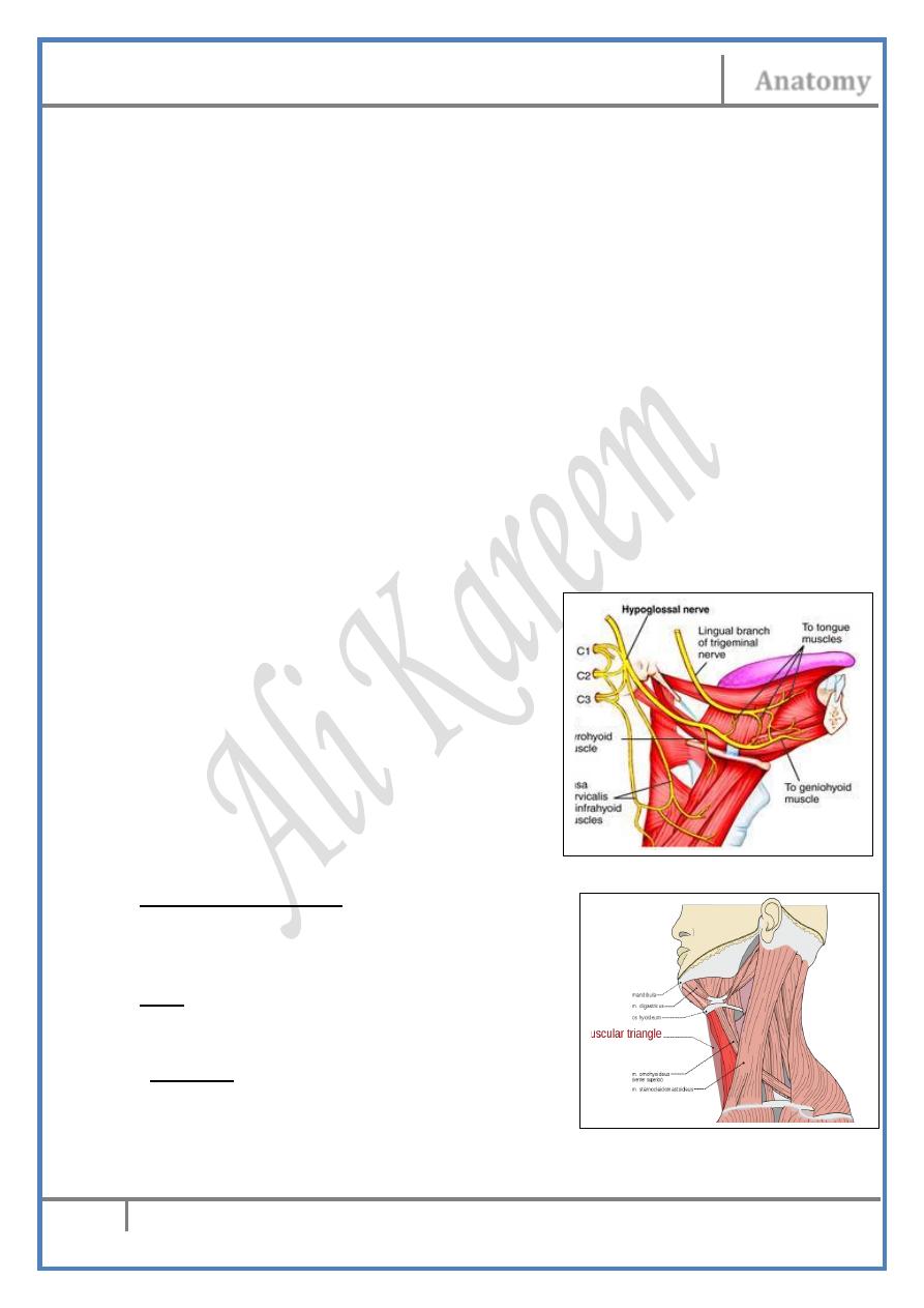
Hypoglossal Nerve :
• Descends in a downward curve on the
lateral aspect of the ECA crossing it at
the point of origin of the lingual artery.
• Enters the floor of the mouth lateral to
hyoglossus.
• In the neck it gives the branches of C1
which hitch-hacked along it.
Deep Cervical Lymph Nodes
The muscular triangle :
- Is the space between midline, superior
belly of omohyoid & the anterior border
of the lower part of SCM
Roof :
- Skin, subcutaneous connective tissue,
platysma and investing fascia.
Contents :
1- Infrahyoid (strap) muscles.
2- Thyroid gland.
3- Structures of the midline (pharynx, larynx, trachea & oesophagus).
Head & Neck Dr. Nawfal K. Al-Hadithi
Anatomy
9
Lec. 4 The Ant. Cervical Triangle
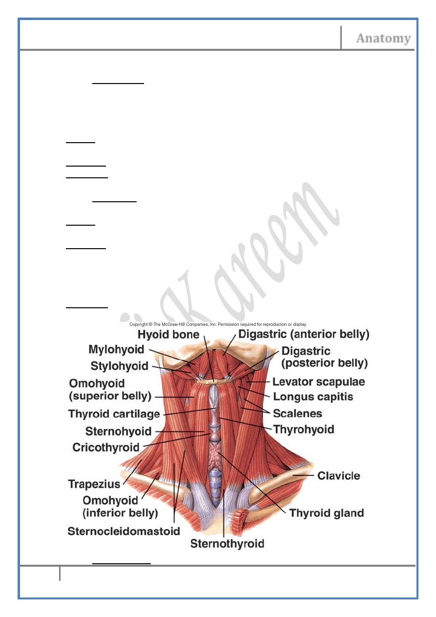
Infrahyoid muscles :
1- Sternohyoid :
- This thin narrow muscle is the most medial of the infrahyoid
muscles
- Separated from its fellow on the opposite side by the midline &
Adam’s apple only
Origin; Posterior end of the sternal end of clavicle & back of manubrium
sterni.
Insertion; The lower border of the body of hyoid bone.
N. supply; C1,2,3 by ansa cervicalis.
2- Omohyoid :
- Formed of 2 bellies & lies lateral to sternoh.
Origin; Inferior belly arises from the scapular ligament & upper border
of scapula
Insertion; Lying in the base of the posterior triangle, the inferior belly
ends in an intermediate tendon behind SCM being held to this
muscle by the deep layer of investing fascia. From this tendon
the superior belly arises & ascends lateral to sternohyoid to the
lower border of the body of the hyoid bone.
N. supply; Ansa cervicalis, C1 to the superior belly & C2,3 to the inferior
one.
3- Sternothyroid :
Head & Neck Dr. Nawfal K. Al-Hadithi
Anatomy
10
Lec. 4 The Ant. Cervical Triangle
- Shorter, broader & deeper than sternohyoid.
Origin; Back of manubrium sterni reaching lower than sternohyoid, &
1st costalcartilage.
Insertion; oblique line of thyroid cartilage.
N. supply; By ansa cervicalis,C2 & 3.
4- Thyrohyoid :
Origin; Oblique line of thyroid cartilage as a continuation of
sternothyroid.
Insertion; Lower aspect of greater horn of hyoid bone.
N. supply; Nerve to thyrohyoid, C1 fibers given as a branch from the XII
nerve.
Action of infrahyoid muscles :
• Together with the suprahyoid muscles, these muscles move &
regulate the movements of the hyoid bone, tongue & larynx
• They depress the hyoid bone either directly or by depressing the
thyroid cartilage.
• Thyrohyoid could elevate the larynx to produce high notes if the
hyoid bone is fixed from above
• Sternothyroid could depress the larynx to produce low notes if the
hyoid bone is fixed from above
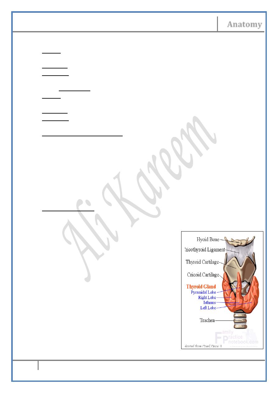
The Thyroid Gland :
Shape & position :
This H-shape gland has two lateral lobes connected in the midline by an
isthmus:
The lateral lobe :
- Is a pyramidal shape lobe with an
upward apex lying deep to thyrohyoid
& a wide base reaches down to the level
of the 6th tracheal ring
- The anterior border is thin & inclines
inferomedially from the apex to reach
the midline
- The rounded posterior border are
separated from each other by the airway
& foodway & touching the carotid sheaths
posterolaterallly
The isthmus : Connects the two lateral lobes in
the midline opposite to the 2nd, 3
rd
& 4th tracheal rings.
The pyramidal lobe :
Head & Neck Dr. Nawfal K. Al-Hadithi
Anatomy
11
Lec. 4 The Ant. Cervical Triangle
- Sometimes arises from the isthmus & directed to one lobe (usually
the left)
- Sometimes it is connected to the hyoid by levator glandulae
thyroidae muscle
Fascia :
- The pretracheal fascia invests the gland within which lies free
inside it except at the region of the isthmus
- The fascia is attached superiorly to the thyroid cartilage therefore
the gland and any related swelling will move upward during
deglutition
Capsule :
The gland has a histological fibrous capsule which sends septa into the
gland dividing it into lobules
Relations :
* Antero-laterally: Infrahyoid muscles (except thyrohyoid).
* Posteriorly: Carotid sheath.
* Medially: Thyroid & cricoid cartilages & the upper 6 tracheal rings
“anteriorly” and thyropharyngeus, cricopharyngeus & upper oesophagus
“ posteriorly”.
Arteries :
1- Superior thyroid artery :
- Branch of ECA
- Pierces the fascia & distributed to the gland by an anterior &
posterior branches.
2- Inferior thyroid artery :
- Branch of the thyrocervical trunk from the 1st part of subclavian a.
- Arches up then down behind the carotid sheath to reach behind the
lower border of the gland where it divides into (5-6) branches
which penetrate the fascia separately & distributed to the gland
- The recurrent laryngeal n. lies in between them.
- It gives tracheal, oesophageal, inferior laryngeal & pharyngeal
branches
3- Thyroidea ima artery :
- Small branch arises from the brachiocephalic trunk or aortic arch &
enters the lower pole
- They are seen n 10% of individuals.
Veins :
1- Superior thyroid vein :
- Formed on the anterolateral aspect of the lateral lobe of the gland
Head & Neck Dr. Nawfal K. Al-Hadithi
Anatomy
12
Lec. 4 The Ant. Cervical Triangle
- Crosses in front of the CCA & receives tributaries corresponding to
the branches of the superior thyroid a.
- It empties in the IJV separately or with the facio-lingual v.
2- Middle thyroid vein :
- Formed at the lateral surface of the gland
- This short wide v. courses laterally to empty in the lower part of
the IJV
3- Inferior thyroid veins :
- Arise from the venous plexus which lies near the lower pole of the
gland & communicates with the upper 2 veins
- They descend down receiving tributaries which correspond to the
branches of the inferior thyroid a.
- Pass behind the manubrium to end in the corresponding
brachiocephalic vein.
- Sometimes the 2 veins unite forming a single vein which empties
in the left brachiocephalic v.
Nerves :
1- Sensory: Recurrent laryngeal n.
2- Sympathetic (vasomotor) : Middle cervical sympathetic ganglion along
the branches of the inferior thyroid a.
Lymph :
Along arteries:
• Superior thyroid: Anterosuperior group of deep cervical nodes
• Inferior thyroid: Posteroinferior group of deep cervical nodes
• A midline neck swelling that moves with swallowing is related to
the thyroid gland until proved otherwise.
• Incisions for thyroidectomy are made horizontally at the lower part
of the neck.
• The recurrent laryngeal nerve is commonly injured during ligation
of the inferior thyroid arteries in thyroid surgery
Pharynx & Larynx will be discussed later
Edited & Published by :
Ali Kareem