ANATOMY
HEAD & NECK
Dr. Nawfal K. Al-Hadithi
Lec . 3
The Neck
By : Ali Kareem
مكتب اشور لالس تنساخ
2013 – 2014
Head & Neck Dr. Nawfal K. Al-Hadithi
Anatomy
2
Lec. 3 The Neck
THE NECK
SURFACE ANATOMY
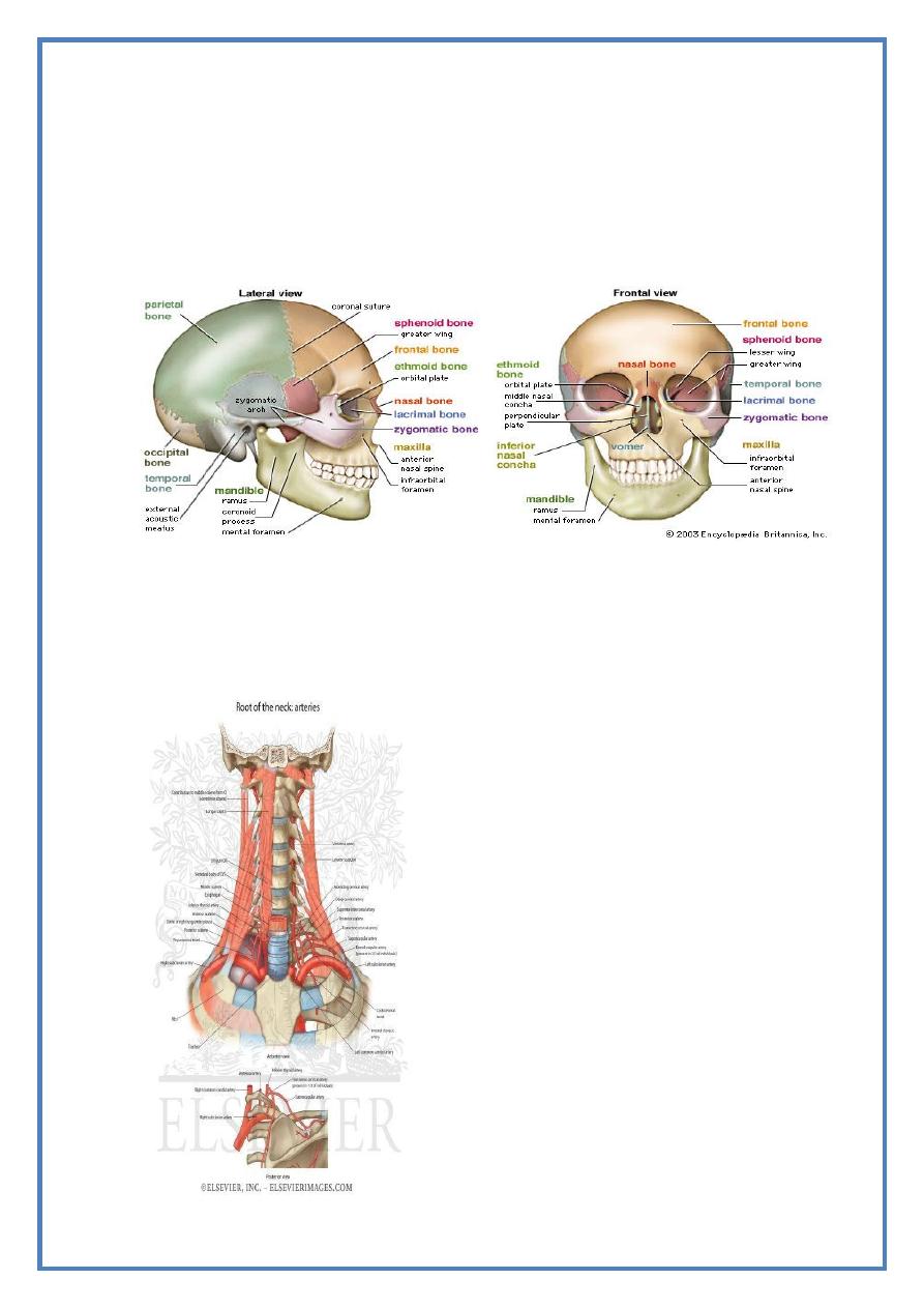
Bony & cartilagenous landmarks :
1- Hyoid bone :
- Lies in the angle between the chin &
the front of the neck
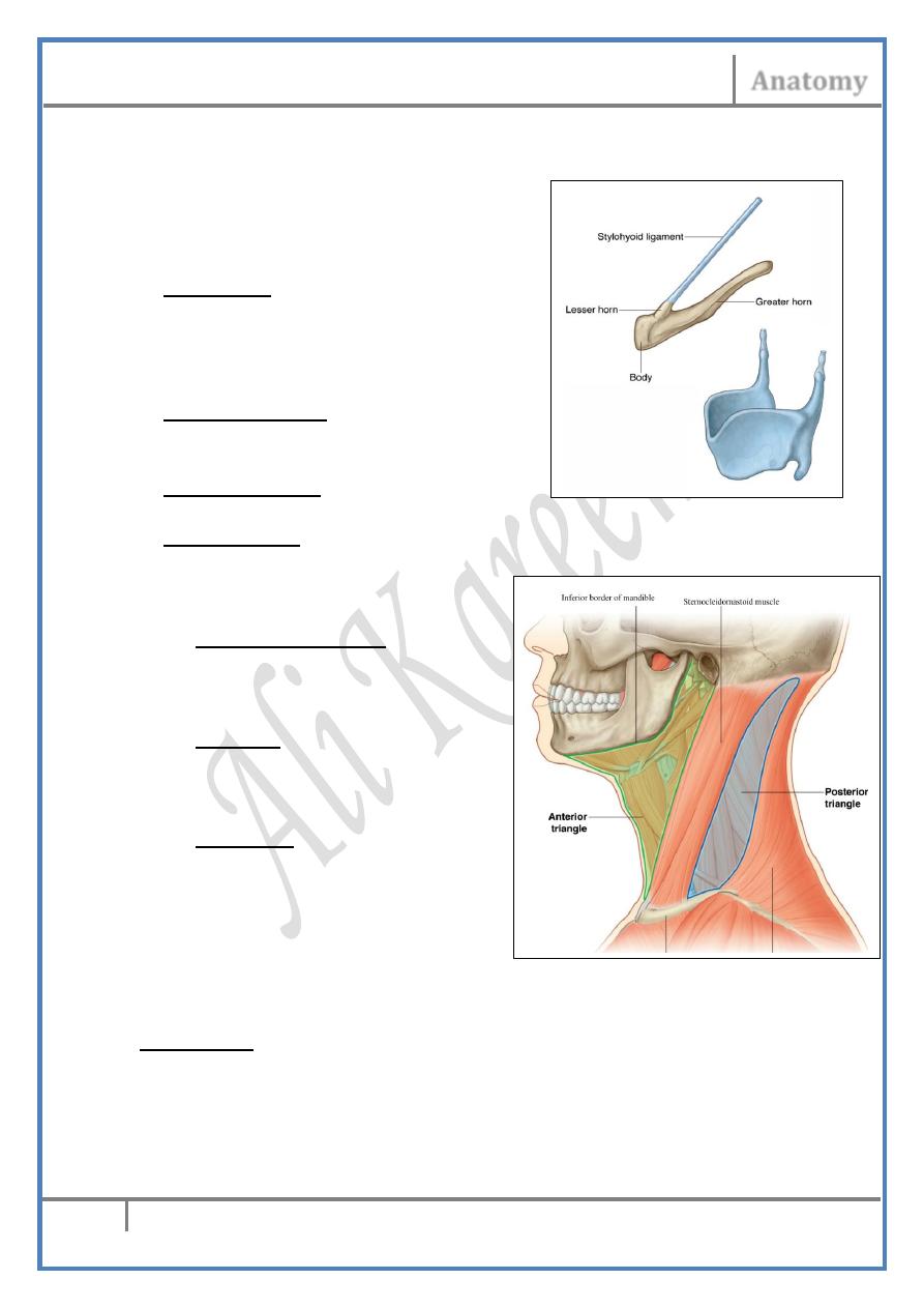
- Formed of body & two
horns (greater & lesser)
2- Thyroid cartilage : Forms the midline
prominence of the larynx 1-2 cm
below the hyoid bone
3- Cricoid cartilage : Just below the thyroid
cartilage separated from it by a sulcus
4- Tracheal rings : Frequently felt below
the cricoid.
Muscular landmarks :
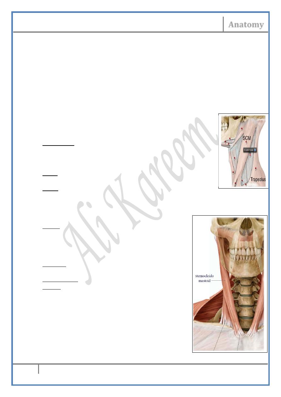
1- Sternocliedomastoid :
Bisect the side of the neck
into anterior & posterior
triangles
2- Digastric : By its two
bellies divide the suprahyoid
compartment intosubmandibular
& submental triangles.
3- Omohyoid : Behind SCM divides
The anterior & posterior triangles
into further smaller ones.
SUPERFICIAL DISSECTION OF THE NECK:
The skin :
Langer lines “lines of cleavage” :
- These lines represent the direction of arrangement of the collagen
bands in the skin
- An incision along Langer lines heals with a minimum scar while an
incision across them leaves
Head & Neck Dr. Nawfal K. Al-Hadithi
Anatomy
3
Lec. 3 The Neck
- an unfavorable scar tissue after healing Langer lines in the neck are
arranged transversely along the circumference of the neck so
incisions should be done in this direction unless contra-indicated
Subcutaneous conn. Tissue :
• Formed of loose areolar & fatty tissues
• Replaced anteriorly by platysma
• Contains the cutaneous vessels & nerves which lie deep to
platysma
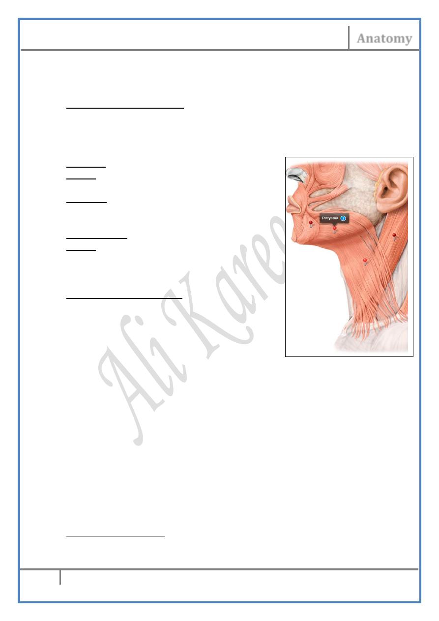
Platysma :
Origin ; Fascia covering pectoralis major
& deltoid
Insertion ; Lower border of the mandible &
skin of lower part of the face
& corners of the mouth
Nerve supply ; Cervical branch of VII
Action ; Depresses the corner of the mouth
& lower border of the mandible
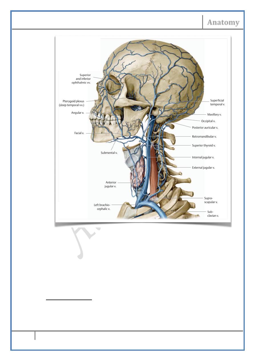
Superficial veins :
The External Jugular Vein :
• Formed behind the angle of the mandible
by union of the posterior auricular v.
& posterior division of retromandibular v.
• Descends vertically between platysma &
SCM towards the middle of the clavicle
• 2 cm above the clavicle it pierces the neck
fascia & drains into the subclavian vein
• Provided with 2 pairs of valves; one at its end in the subclavian v.
& the other 4 cm above
• Tributaries:
- Posterior EJV from the back of neck
- Anterior JV from the anterior part of the neck
- Suprascapular & transverse cervical vv.
Clinical importance of the EJV :
1- Medical: as it is used sometimes as a sign of heart failure & to
measure the central venous pressure ..
2- Surgical: sometimes used in cannulation & i.v therapy …
Anterior Jugular Vein :
• Formed near the hyoid bone by confluence of small submental vv.
•
Head & Neck Dr. Nawfal K. Al-Hadithi
Anatomy
4
Lec. 3 The Neck
• Descends vertically on each side of the midline (sometimes as a
single vein in the midline)
• 3 cm above the manubrium, it perforates the superficial layer of
deep cervical fascia & enters the suprasternal space
• In this space it communicates with its fellow be the jugular venous
arch
• Then it turns laterally to empty in the EJV at its termination
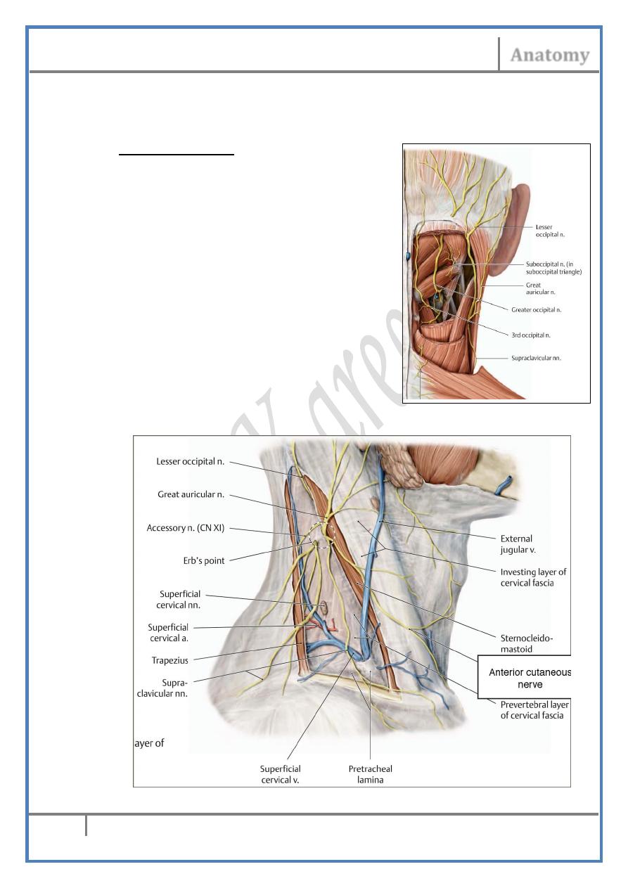
Cutaneous nerves :
I ) Nerves of the back :
- These are the Dorsal Primary Rami of Cervical Nerves
1- Greater Occipital Nerve : C2 Pierces semispinalis capitis &
trapezius at their skull attachments and supplies the back of the
scalp as high as the vertex.
Head & Neck Dr. Nawfal K. Al-Hadithi
Anatomy
5
Lec. 3 The Neck
2- Third Occipital Nerve : C3 Communicates with the great occipital
n. & supplies the upper part of the back of the neck.
3- C4 : Supplies the reminder of the back of the neck.
II ) Nerves of the front :
- These are the cutaneous branches
of cervical plexus
- Nerves approach the surface near
the midpoint of the posterior border
of SCM where they diverge.
1- Lesser occipital Nerve : C2 & 3
- Ascends along the posterior border
of SCM
- Supplies skin & subcutaneous tissue
at the insertion of the muscle &
behind & above the auricle.
2- Great auricular Nerve : C2 & 3
- Hooks below the posterior border of SCM
- Ascends in the direction of the auricle &
angle of the mandible where it supplies :
Skin over the antero-inferior part of mastoid BY mastoid branches
Head & Neck Dr. Nawfal K. Al-Hadithi
Anatomy
6
Lec. 3 The Neck
Auricle, except the upper ½ of lateral surface BY auricular br.
Skin over parotid & angle of mandible BY facial br.
3- Anterior cutaneous nerve : C2 & 3
- Crosses SCM horizontally deep to platysma to reach the anterior
triangle
- It divides into superior & inferior branches penetrating platysma &
supplies skin & subcutaneous tissues of the cylinder of the neck
- Its block in local anaesthesia results in sensory loss in a wide area
of the neck.
4- Supraclavicular nerves : C3 & C4 Descend toward the clavicle
where they divide into 3 main groups :
- Medial; skin over manubrium sterni
- Intermediate; skin over the pectoral region down to the 3rd rib
- Lateral; skin over deltoid as far as the distal 1/3 of muscle
DEEP DISSECTION OF THE NECK:
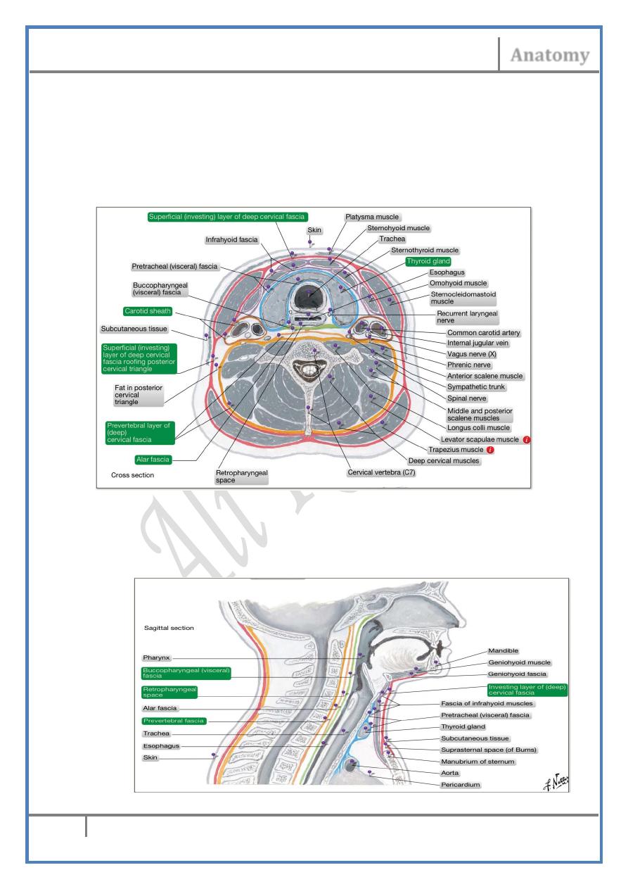
Deep fascia :
FOUR entities of well defined deep fasciae are diagnosed in the neck :
1- The investing cervical fascia
2- The pretracheal fascia
3- The prevertebral fascia
4- The carotid sheath (one on each side)
The Investing Fascia :
• Double layered membrane encloses the whole structures of the
neck like a collar
• It splits at certain areas to enclose 2 muscles & 2 glands on each
side
• Upper attachment (to the skull base) :
Head & Neck Dr. Nawfal K. Al-Hadithi
Anatomy
7
Lec. 3 The Neck
- External occipital protuberance - superior nuchal line (splits for
trapezius & SCM) - mastoid process - splits in 90 degrees :
- Superficial layer (covers the parotid gland) ; Lower border of
zygomatic arch - lower border of mandible
- Deep layer (deep to the parotid gland) ; Lower border of tympanic
plate - fuse with carotid sheath
Lower attachment :
- To the pectoral girdle around the attachment of SCM & trapezius
Fixation points :
To the hyoid bone forming the angle between the chin & the neck
Special derivatives :
Parotid fascia
Submandibular fascia
Stylomandibular ligament: thickening in the deep layer of parotid
fascia between the styloid process & the angle of mandible
Enclosed structures :
Parotid & submandibular glands
SCM & trapezius
Suprasternal space containing the JVA
The Prevertebral Fascia :
• Forms a smaller cylinder inside the large investing one enclosing
the vertebral column & the surrounding muscles
• Superior attachment is to the base of the skull anterior to the
attachments of the prevertebral muscles
• Laterally it is attached to the tips of transverse processes of the
cervical spines
• Then extends laterally to cover the pre- & para-vertebral muscles
reaching ligamentum nuchae & the vertebral spines
• Downward it reaches the lowest limit of longus colli (T4)
• Extends along the axillary artery as the axillary sheath
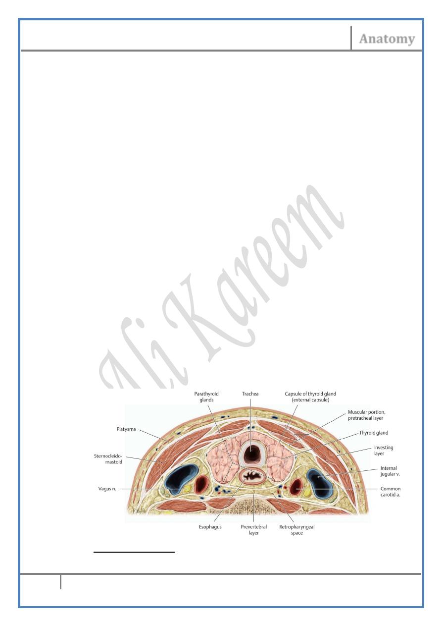
The Pretracheal Fascia :
• Attached superiorly to the hyoid bone & the oblique line of thyroid
cartilage
• Encloses the thyroid gland forming its sheath
• It is responsible for the upward movement of the thyroid gland &
any related swelling during deglutition
• Laterally it reaches the carotid sheath
• Inferiorly it blends with fascial coverings of the aortic arch
• It is pierced by the thyroid vessels
The Carotid Sheath :
• A dense cylindrical condensation of connective tissue surrounding
the CCA, ICA, IJV & vagus nerve
Head & Neck Dr. Nawfal K. Al-Hadithi
Anatomy
8
Lec. 3 The Neck
• Attached superiorly at the margins of carotid canal
• Reaches downward to the aortic arch
• Its deficient around the IJV (for venous expansion)
• Fused laterally with the deep layer of investing fascia & antero-
medially with the pretracheal fascia
• It is loosely attached to the anterior aspect of the prevertebral
fascia along the tips of transverse processes of the cervical spines
Tissue spaces of the neck :
Behind the prevertebral fascia :
The space descend down to reach the lowest attachment of the
fascia at T4, though an abscess there usually points in the posterior
triangle by pathological walling off
Head & Neck Dr. Nawfal K. Al-Hadithi
Anatomy
9
Lec. 3 The Neck
In front of the prevertebral fascia (retropharyngeal space) :
The space extends from the base of the skull down to the posterior
mediastinum
Anterior to the pretracheal fascia :
The space reaches down through the superior to the anterior
mediastinum
Between the pretracheal & prevertebral fasciae :
The space leads down through the superior to the posterior
mediastinum
Posterior cervical triangle :
Is the triangular space which lies in a spiral fashion on the
postero-lateral aspect of the neck.
Boundaries :
- Anterior ; posterior border of SCM
- Posterior ; anterior border of trapezius
- Inferior ; middle 1/3 of clavicle
Roof :
Skin, subcutaneous tissue, platysma, investing fascia
Floor :
Scalene muscles, levator scapulae & splenius capitis covered by
prevertebral fascia.
Sternocliedomastoid :
Origin :
- Tendinous sternal head from the anterior
surface of manubrium sterni
- Fleshy clavicular head from the upper
surface of the medial 1/3 of clavicle
Insertion : Lateral side of mastoid process &
lateral ½ of superior nuchal line
Nerve supply : Accessory nerve
Action :
- Unilateral contraction draws the mastoid
process toward the shoulder (pushes the face
to the opposite side)
- Bilateral contraction flexes the head over
the neck
- With stabilization of the head, it is an
accessory muscle of inspiration
Head & Neck Dr. Nawfal K. Al-Hadithi
Anatomy
10
Lec. 3 The Neck
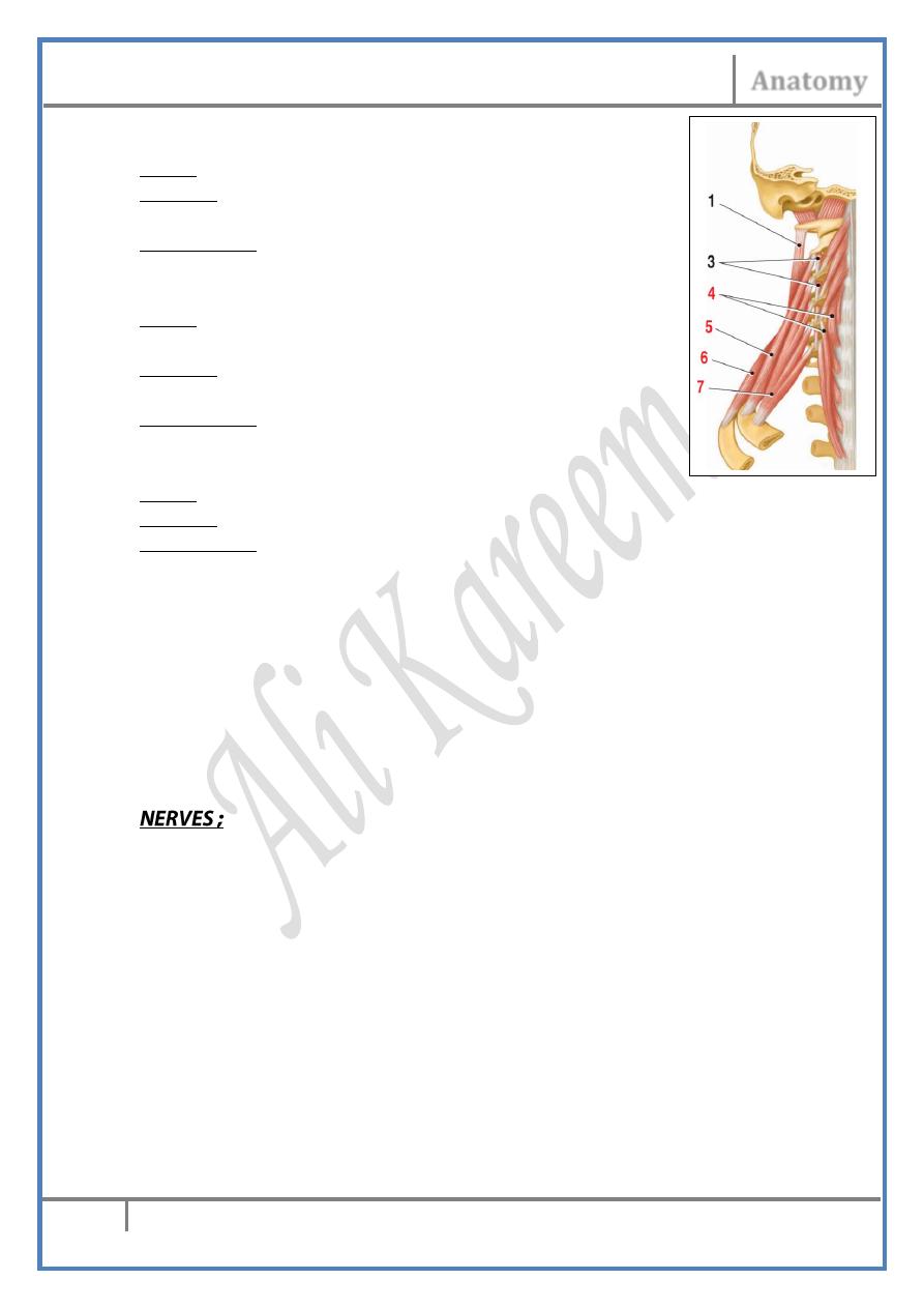
Scalenus anterior : (7)
Origin : Anterior tubercles of transverse processes of C3-C6
Insertion : Scalene tubercle on the upper surface of the 1
st
rib
Nerve supply : C5-C7 anterior primary rami
Scalenus medius : (5)
Origin : Posterior tubercles of transverse
processes of C2-C7
Insertion : Upper surface of the 1st rib behind Subclavian
artery groove
Nerve supply : C3-C8 anterior primary rami
Scalenus posterior : (6)
Origin : Posterior tubercles of transverse processes of C5-C7
Insertion : Lateral surface of the 2nd rib rib
Nerve supply : C7 & C8 anterior primary rami
Contents of the posterior triangle :
I) In the roof (superficial to the investing fascia) :
1-Platysma.
2-EJV.
3-EJ lymph nodes: 1-2 nodes along the EJV receive from the ear &
parotid & send to the superior group of deep cervical nodes.
4-Cutaneous branches of cervical plexus, especially the lesser occipital &
supraclavicular nerves.
II) In the triangle (between the investing & prevertebral fasciae) :
1- The spinal root of accessory nerve :
• Crosses the triangle on the undersurface of investing fascia
• Is directed vertically toward the tip of the shoulder between the
posterior border of SCM & anterior border of trapezius
• It supplies trapezius & SCM
Accessory nerve injury :
Unilateral injury of the accessory nerve results in paralysis of the SCM
muscle on that side with the resultant unopposed action of the
contralateral muscle so the face turns to the injured side & the mastoid
process on the healthy side approaches the shoulder in a “wry neck”
deformity.
2- The cervical plexus :
• Formed by the ventral primary rami of the upper four cervical
nerves inside the substance of the prevertebral muscles.
Head & Neck Dr. Nawfal K. Al-Hadithi
Anatomy
11
Lec. 3 The Neck
• It gives 4 sensory & 4 motor branches.
• The sensory branches were discussed.
• The motor branches:
A) Direct muscular branches to prevertebral muscles:
C1,2; longus capitis & the anterior & lateral recti
C2,3,4; longus capitis & longus colli
C3,4; levator scapulae & the scalene
B) Phrenic nerve : C3 & C4
• Descends vertically across the oblique course of
scalenus anterior from its lateral to medial border
• Lies deep to the prevertebral fascia & crossed by
the transverse cervical & suprascapular arteries
• Enters the thoracic inlet in a variable relation to
the subclavian vein (usually behind it) &
descends in the thorax to the diaphragm.
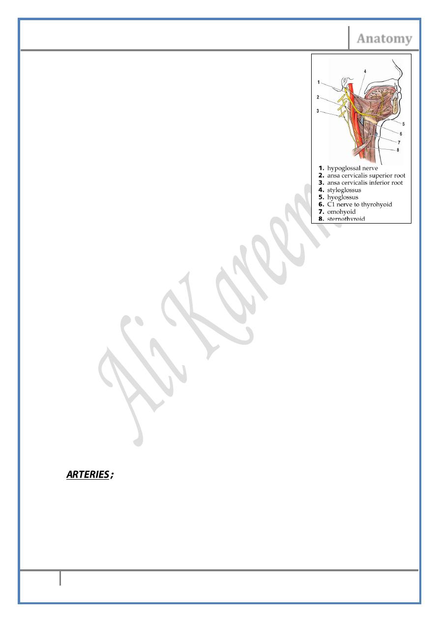
C) C1 fibers to hypoglossal nerve :
• XII nerve receives fibers from the C1 nerve at the anterior condylar
canal
• These fibers leave XII as 4 branches :
1- Meningeal branch to the dura around foramen magnum
2- Superior ramus of ansa cervicalis (descendens hypoglossi)
3- Nerve to thyrohyoid
4- Nerve to geniohyoid
D) Inferior ramus of ansa cervicalis (descendens cervicalis): C2 & C3
• Descends behind the carotid sheath to join the superior ramus
usually lateral to the IJV
• Ansa cervicalis, so formed around the IJV in a Y-shape or loop
pattern, has a variable vertical position
• It supplies the infrahyoid muscles except thyrohyoid.
• Their supply is segmental (upper part of the muscle receives C1,
middle part C2 & the lower part C3)
E) Proprioceptive fibers to SCM & trapezius (C2 & 3).
1- The transverse cervical & suprascapular arteries :
• Are branches of the thyrocervical trunk
• Crosses laterally in the base of the triangle above & parallel to the
clavicle to hide underneath the anterior border of trapezius (TC) &
behind the inferior belly of omohyoid (SS).
• The transverse cervical lies superior to the suprascapular.
2- The occipital artery :
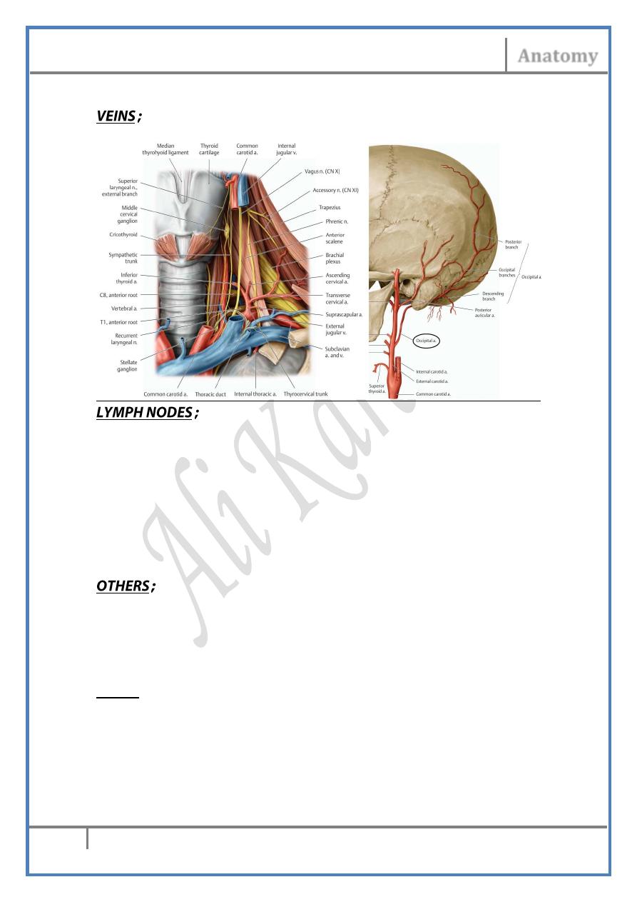
Head & Neck Dr. Nawfal K. Al-Hadithi
Anatomy
12
Lec. 3 The Neck
Runs part of its course in the apex of the triangle in its way to the scalp
Similar to arteries.
1- The supraclavicular lymph nodes :
• Are extension from the postero-inferior group of the deep cervical
nodes along the transverse cervical vessels.
• Receive lymph (in addition to the normal drainage of the postero-
inferior nodes) from the apical axillary nodes, breast & sometimes
from the upper limb & anterior chest wall.
2- The occipital lymph nodes :
• Situated along the occipital vessels in the apex of the triangle.
• Drains the back of the scalp upward to the vertex.
The inferior belly of omohyoid :
• Emerges from behind the lower part of the posterior border of
SCM & enters deep to trapezius.
• Divides the triangle into occipital & supraclavicular ones.
III) In the floor of the triangle (deep to the prevertebral fascia) :
Nerves ;
Brachial plexus :
Branches from the roots & trunks: these are branches have part of their
course in the neck, though they are distributed to muscles in the upper
limb :
a) Dorsal scapular nerve: passes over scalenus medius & posterior &
hides deep to levator scapulae
Head & Neck Dr. Nawfal K. Al-Hadithi
Anatomy
13
Lec. 3 The Neck
b) Long thoracic nerve: leaves scalenus medius by two roots which
unite down to form the nerve
c) Suprascapular nerve:passes above the plexus & hides under
trapezius
Vessels ;
The subclavian artery :
- From the aortic arch (on the left) & brachioceohalic trunk (on the
right), the artery leaves the back of the corresponding
sternoclavicular joint & goes laterally in the root of the neck
- Crosses over the 1st rib behind scalenus anterior which divides it
into 3 part (1st medial, 2nd behind & 3rd lateral to it)
- Around the 1st part of the right artery the right recurrent laryngeal
nerve hooks
- It leaves the outer border of the 1st rib to the axilla as the axillary
artery.
Branches :
I: From the 1st part:
1- Vertebral artery:
- Arises from the dorsosuperior aspect of the subclavian
- Enters the foramen transversarium of C6
- Ascends in the upper 6 cervical vertebrae in front of the emerging
spinal nerves
- Arching behind the atlanto-occipital joint, it enters foramen
magnum & unites with its fellow at the clivus forming the basilar
artery
Branches ;
* muscular (in the neck): to deep neck muscles
* spinal (in spinal canal): radicular arteries
2- Thyrocervical trunk:
- Short thick artery arises from the subclavian a. opposite to the
internal thoracic a.
- Soon after its origin it divides into:
* Transverse cervical a.
* Suprascapular a.
* Ascending cervical a.: passes medial to the phrenic nerve anterior to
scalenus anterior
* Inferior thyroid a.:
- Arches up as high as the cricoid cartilage where it pierces the
prevertebral fascia & enters behind the thyroid gland
- The recurrent laryngeal nerve lies in between its 5-6 branches
which pierce the pretracheal fascia separately & enters the thyroid
gland

Head & Neck Dr. Nawfal K. Al-Hadithi
Anatomy
14
Lec. 3 The Neck
- It gives the inferior laryngeal a. to the lower part of the larynx.
3- Internal thoracic artery:
- Arises from the inferior aspect of the subclavian a.
- Descends behind the subclavian v. & phrenic nerve to enter the
thorax
- It passes on each side of the sternum (1 cm away)
- Gives the anterior intercostal arteries
- Ends by dividing into superior epigastric & musculophrenic
arteries.
II: From the 2nd part:
The costocervical trunk:
- Ascends on the back of cervical pleura & apex of the lung
- Passes between the trunks of brachial plexus
- On the neck of 1st rib it divides into highest intercostal & deep
cervical artery
III: From the 3rd part:
- In 70% , the dorsal scapular a. leaves the 3rd part of the artery &
follows the corresponding nerve after disappearing underneath the
anterior border of trapezius
- In 30%, this artery arises from the transverse cervical artery
The subclavian vein :
- Lies in the root of the neck anterior to scalenus anterior crossing
over the 1st rib & then grooves the apex of the corresponding lung
& cervical pleura
- Joined by the IJV at the medial border of the muscle forming the
brachiocephalic v.
- Receives only the EJV
Edited & Published by : Ali Kareem