1
Baghdad medical college 2015 - 2016
Neuro-ophthalmology
Optic nerve
Applied anatomy:
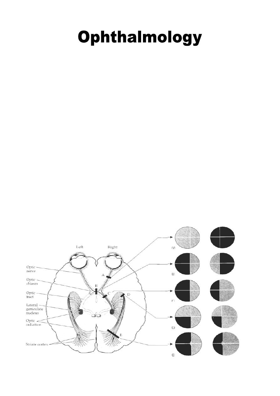
The optic nerve carries about 1.2 million afferent nerve fibers, which are represent
the axons of retinal ganglion cells. Most of these (90%) synapse in the lateral
geniculate body "LGB" (carrying visual stimulation), and the rest (10%) reach other
centers, notably the pre-tectal nuclei in the mid brain carrying light pupillary reflex.
The afferent pathway for light pupillary reflex is the optic nerve while the efferent
pathway is the oculomotor nerve.
The optic nerve is approximately 5 cm long from globe to optic chiasm, where
decussation occurs, the temporal fibers for each nerve pass to ipsilateral optic tract,
while nasal fiber cross to contralateral optic tract (crossing fibers).
Any injury to the axons of ganglion cells before LGB causes optic disc atrophy seen
by fundoscopy (ipsilateral if pre-chiasmal and bilateral if post-chiasmal), while the
injury that occurs after LGB (optic radiation and occipital cortex), will not cause
optic disc atrophy.
Visual center lies mainly on the medial surface of the occipital cortex (Broadman's
area no. 17).
Dr. Najah
Lecture: 21

2
Optic nerve can be subdivided into four segments:
1- Intra-ocular segment (optic disc, nerve head): about 1mm depth & 1.5mm in
diameter.
2- Intra-orbital segment: 3.0 cm, this part has S shape allowing the eye for
movement without nerve stretching (i.e. the distance from the apex of orbit to
the posterior part of eyeball is less than 3.0 cm).
3- Intra-canalicular segment: 1cm.
4- Intra-cranial segment: 6-8 mm, joins the chiasm.
* The optic nerve is surrounded by pia, arachnoid and duramater, so the CSF reaches
up to the posterior sclera around the optic nerve.
Axoplasmic transport:
It is the movement of cytoplasmic organelles within a neuron between the cell
body and the termina+l synapse of Ganglion cells.
Retinal cotton-wool spots are the result of accumulation of cytoplasmic organelles
due to interruption of axoplasmic flow between the retinal ganglion cells and their
terminal synapses.
Papilloedema is similarly caused by hold-up of axoplasmic flow at the lamina
cribrosa ( Small pores present at posterior sclera for exit of optic nerve fibers).
Signs of optic nerve dysfunction:
1- Decreased visual acuity.
2- Diminish light pupillary reflex.
3- Dyschromatopsia (impairment of color vision): affected eye sees the colors less
bright.
4- Diminished light brightness sensitivity.
5- Visual field defect: depends on the type of the pathology, e.g. central scotomas,
centrocaecal scotomas and altitudinal.
Special investigations:
1- Manual kinetic Perimetry (Goldmann) for assessment of peripheral VF.
2- Automated Perimetry for assessment of peripheral and central VF.
3- MRI: detect tumors or degenerative diseases like multiple sclerosis.
4- Visual Evoked Potential (VEP): is a recording of the electrical activity of the
visual cortex by stimulation of the retina (diagnose any damage from ganglion
cell to occipital cortex), while for diseases from receptors to ganglion cell we use
ERG (Electro-Retinography).
5- Fluorescein angiography: to differentiate between optic nerve diseases and
papilloedema, e.g. optic disc drusen and papilloedema, as drusen do not leaking
fluorescein dye while papilloedema leaking the dye.

3
Optic neuritis
It is an inflammatory or demyelinating process affecting the optic nerve.
1- Ophthalmoscopic classification:
a- Retrobulbar neuritis: in which the optic disc appearance is normal, at least
initially, because the optic nerve head is not involved. It is the most frequent type
in adult and is frequently associated with multiple sclerosis.
b- Papillitis: in which the pathological process affects the optic nerve head. It is
characterized by variable hyperemia and edema of the optic disc, which may be
associated with parapapillary flame-shaped hemorrhages. Papillitis is the most
common type of optic neuritis in children, although can also affect adults.
2- Etiological classification:
a- Demyelinating: which is by far most common cause usually young females with
Multiple Sclerosis (MS).
b- Para-infectious: it is follow a viral infection or immunization.
c- Infectious: which may be sinus-related or associated with syphilis, lyme disease,
cat-scratch fever and cryptococcal meningitis. patients with AIDS or Herpes
zoster can be presented with optic neuritis.
d- Autoimmune: may be associated with systemic autoimmune disease.
Treatment of optic neuritis:
is according to the etiology.
if the cause is demyelination in MS, the patient need urgent IV methylprednisolone,
then oral prednisolone because the vision is severely affected.
Optic atrophy
It is an important sign of advanced optic nerve disease. It is of two types:
1- Primary optic atrophy:
It is occurs without antecedent swelling of the optic nerve head. It may be caused
by lesions affecting the visual pathways from the retro laminar (behind lamina
cribrosa) portion of the optic nerve to the lateral geniculate body. Lesions anterior
to the optic chiasm result in unilateral optic disc atrophy, whereas those involving
the chiasm and optic tract will cause bilateral optic disc atrophy.
Causes:
- Retro bulbar neuritis (but not Papillitis, as it is preceded by disc swelling)
- Compressive lesions, such as tumors and aneurysms.
- Hereditary optic neuropathies.
- Toxic and nutritional optic neuropathies.
Signs:
- Pale, flat disc with clearly delineated margin.
- Reduction in number of small blood vessels on the disc surface.

4
2- Secondary optic atrophy:
It is preceded by swelling of the optic nerve head.
Causes:
- Papillitis.
- Chronic papilledema.
- AION (Anterior Ischemic Optic Neuropathy): usually occurs in old age patients, it
is of two types; non arteritic [in diabetes, hypertension] and arteritic .e.g. Giant cell
arteritis.
Signs:
- White or dirty grey, slightly raised disc with poorly delineated margins due to
gliosis.
- Reduction in number of small blood vessels on the disc surface.
Papilledema
It is swelling of the optic nerve head secondary to raised intracranial pressure. It is
nearly always bilateral, although it may be asymmetrical.
All other causes of disc edema in the absence of raised ICP are referred to as "disc
swelling" and usually produce visual impairment.
All patients with bilateral discs swelling should be suspected of having an
intracranial mass until proved otherwise. However, not all patients with raised ICP
(intra cranial pressure) have necessarily developed papilledema.
1- Early features of papilledema:
- Visual symptoms are absent and visual acuity is normal.
- Optic disc shows hyperemia and mild elevation.
2- Established papilledema:
- Transient visual obscurations lasting a few seconds.
- Visual acuity is normal or reduced.
- Optic disc shows severe hyperemia, moderate elevation and indistinct margin.
3- Atrophic papilledema:
- Visual acuity is severely impaired.
- Optic discs are dirty grey color, slightly elevated and indistinct margin.
Other differential diagnosis of bilateral discs swelling:
1- Malignant hypertension.
2- Bilateral simultaneous Papillitis.
3- Bilateral compressive thyroid ophthalmopathy.
4- Bilateral simultaneous AION.
5- Bilateral compromised venous drainage in central retinal vein occlusion or
carotid-cavernous fistula.
5
Abnormal pupillary reaction
Applied anatomy
LIGHT REFLEX
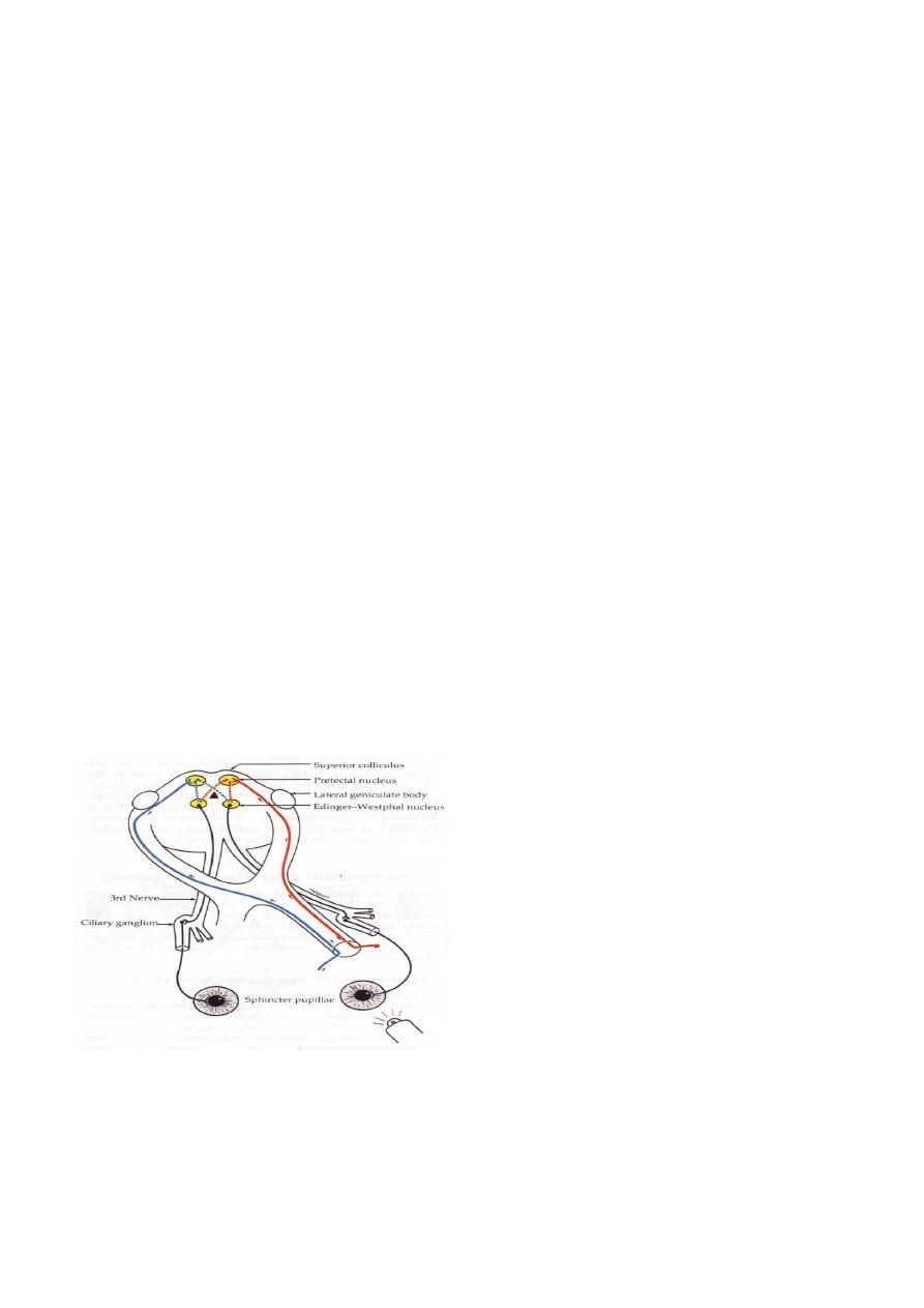
The pupillary light reflex consists of four neurons.
1. The first connects the retina with the pre-tectal nucleus in the mid-brain at the
level of the superior colliculus. The reflex is mediated by the retinal
photoreceptors. Impulses originating from the nasal retina are conducted by fibers
which decussate in the chiasm and pass up the optic tract to terminate in the
contralateral pre tectal nucleus. Impulses originating in the temporal retina are
conducted by uncrossed fibers which terminate in the ipsilateral pre tectal nucleus.
2. The second connects the pre tectal nucleus to both Edinger-Westphal nuclei by
internuncial fibers. This is why a unilateral light stimulus evokes a bilateral and
symmetrical pupillary constriction. Damage to these internuncial neurons is
responsible for light-near dissociation in neurosyphilis and pinealomas.
3. The third connects the Edinger-Westphal nucleus to the ciliary ganglion inside
the orbit. In the orbit, these parasympathetic fibers pass in the inferior division of
the third cranial nerve and reach the ciliary ganglion via the nerve to the inferior
oblique muscle.
4. The fourth leaves the ciliary ganglion and passes with the short ciliary nerves to
innervate the sphincter pupillae. The ciliary ganglion is located within the muscle
cone, just behind the globe. It should be noted that, although the ciliary ganglion
contains other nerve fibers (sensory and sympathetic), only the parasympathetic
fibers synapse there.
NEAR REFLEX
The near reflex triad consists of: (1) increased accommodation, (2) convergence of the
visual axes and (3) constriction of the pupils. The term 'light-near dissociation' refers to
a condition in which the light reflex is absent or abnormal, although the near response is
intact. Vision is not a prerequisite for the near reflex, and there is no clinical condition in
6
which the light reflex is present but the near response absent. Although the final
pathways for the near and light reflexes are the same (i.e. third nerve, ciliary ganglion,
short ciliary nerves), the center for the near reflex is ill-defined. There are probably two
supra nuclear influences: the frontal and occipital lobes. The mid-brain center for the
near reflex is probably located in a more ventral location than the light reflex (in pre-
tectal nucleus) and this may be one of the reasons why compressive lesions such as
pinealomas preferentially involve the dorsal pupillomotor fibers, sparing the ventral
fibers until late.
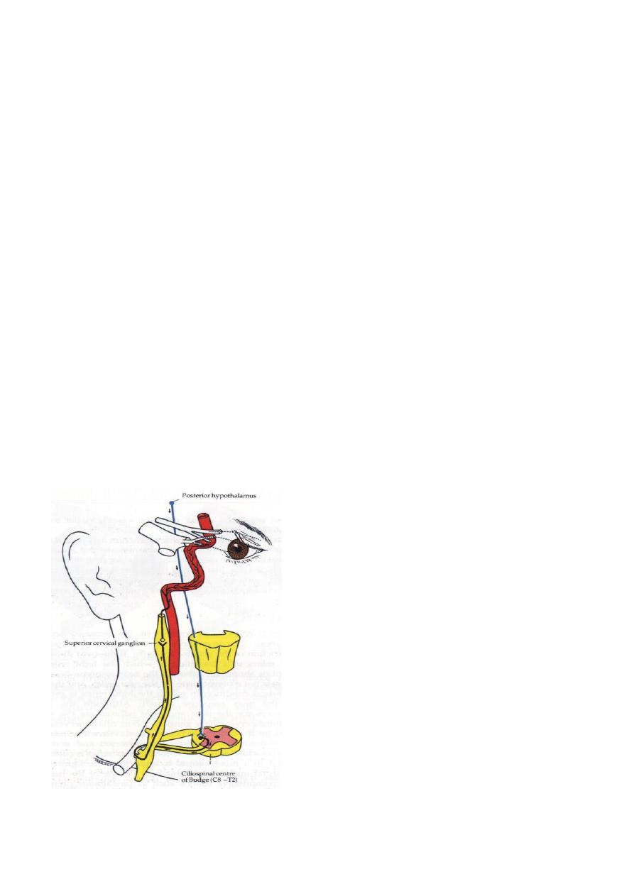
SYMPATHETIC SUPPLY
The sympathetic supply consists of three neurons:
1. The first starts in the posterior hypothalamus and descends, uncrossed, down the
brain stem to terminate in the ciliospinal center of Budge located between C8 and
T2.
2. The second passes from the ciliospinal center of Budge to the superior cervical
ganglion in the neck. During its long course, it is closely related to the apical
pleura where it may be damaged by bronchial carcinoma (Pancoast's tumor) or
during surgery on the neck.
3. The third ascends along the internal carotid artery to enter the skull, where it
joins the ophthalmic division of the trigeminal nerve. The sympathetic fibers are
also passing through ciliary ganglia but without relay and it are reaching the
ciliary body and the dilator pupillae muscle via the nasociliary nerve and the long
ciliary nerves.

7
Afferent pupillary conduction defects
A total afferent pupillary defect (TAPD, Amaurotic pupil) is caused by a complete optic
nerve lesion and is characterized by the following:
1. The involved eye is completely blind (i.e. no light perception).
2. Both pupils are equal.
3. When the affected eye is stimulated neither pupil reacts but when the normal eye
is stimulated both pupils react normally.
4. The near reflex is normal in both eyes.
A relative afferent pupillary defect (RAPD, Marcus Gunn pupil) is caused by an
incomplete optic nerve lesion or severe retinal disease, but not by a dense cataract or
vitreous hemorrhage. The clinical features are those of an Amaurotic pupil but more
subtle. The difference between the pupillary reactions is enhanced by the 'swinging-
flashlight test' in which each pupil is stimulated in rapid succession. When the
abnormal pupil is stimulated it dilates instead of constricting. This paradoxical reaction
of the pupil to light occurs because the dilatation of the pupil, by withdrawing the light
from the normal eye, outweighs the constriction produced by stimulating the abnormal
eye.
Idiopathic intracranial hypertension
Definition: it is elevation of intracranial pressure (ICP) without an identifiable cause,
typical Patients are young adult obese females, and certain medications have also been
implemented such as oral contraceptives and tetracyclines.
Symptoms and signs:
is present in more than 90% of patients, usually in the early morning and may
Headache
awaken the patient from sleep and is exacerbated by coughing or bending, nausea and
g is
localizing sign, disc swellin
-
as a non
6th cranial nerve palsy
projectile vomiting,
bilateral and progressive, vision is initially normal but once papilledema is established
become
enlargement of the blind spot
of vision and
transient ( 30 seconds) obscurations
evident.
Investigations:
tic nerve sheath diameter just behind
can be used to measure the op
scan ultrasound
-
B
the globe.
is used to exclude intracranial mass and typically demonstrates slit like ventricles,
MRI
additionally MRV can be used to exclude cerebral venous thrombosis.

8
Treatment:
ontrolling ICP; this can be achieved by dietary
is very effective in c
Weight loss
intervention and bariatric surgery.
Stopping offensive medications.
are controversial but might be
steroids
( acetazolamide, furosemide ),
diuretics
using
used in sever papilledema.
Lumboperitoneal shunting is reserved for resistant and sight threatening cases.
Follow up and prognosis:
Conjoined management between ophthalmologist and neurologist is essential,
monitoring signs of optic nerve damage is very important, well controlled patients have
good visual prognosis, while 25% of poorly managed patients will have permanent
visual impairment.