Light in medicine
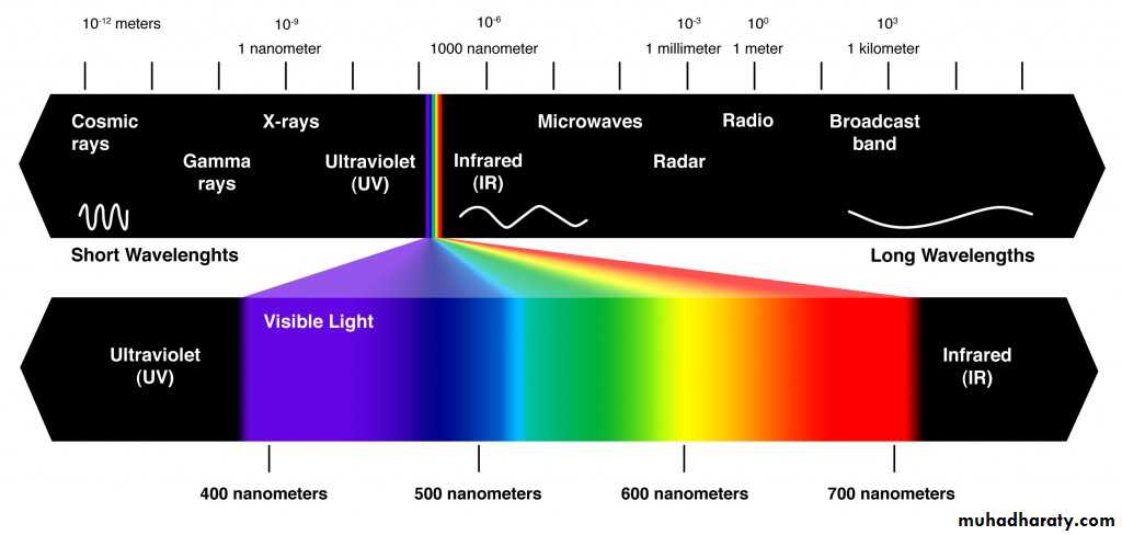
Spectrum of light
The units of light ismicrometer( µ) =10-6
angstrom (Å) =10-8
Nanometer(nm) = 10-9
The units of light is
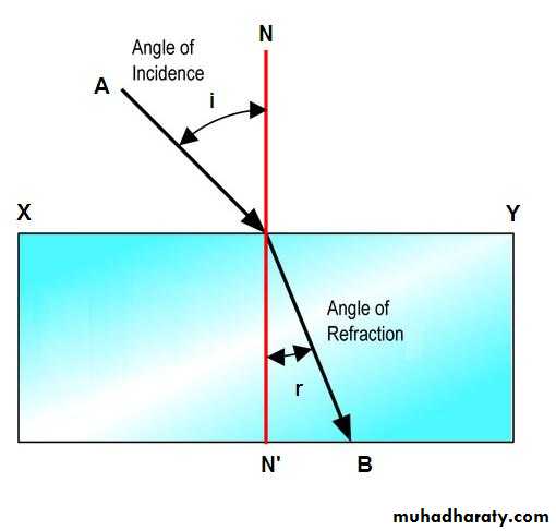
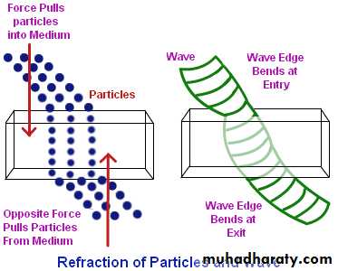
1.The speed of light changes when it goes from one material into another. The ratio of the speed of light in a vacuum to its speed in a given material is called the index of refraction. If a light beam meets a new material at an angle other than perpendicular. It bends, or refracted as shown in figure 1.
The properties of light are:
Figure(1)
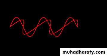
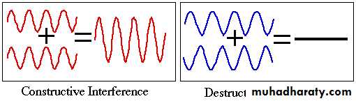
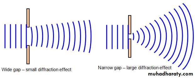
2. Light behaves both as a wave and as a particle(figure2). As a wave it produces interference(figure3) and diffraction(figure4) , which are of minor importance in medicine. As a particle it can be absorbed by a single molecule. When a light photon is absorbed its energy is used in a various ways. It can cause an electrical change.
Figure(2)
Figure(3)
Figure(4)
3. When light is absorbed, its energy generally appears as heat. This property is the basis for the use in medicine.
4. Sometime when a light photon is absorbed ,a lower energy light photon is emitted. This property is known a fluorescence.
5. light is reflected to some extent from all surfaces(figure5). There are two types of reflection(figure 6).
Figure(5)
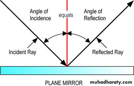
Figure(6)
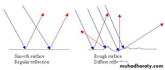
Diffuse reflection occurs when rough surfaces scatter the light in many directions.
Specular reflection is obtained from very smooth shiny surfaces such as mirrors where the light is reflected at an angle that is equal to the angle at which it strikes the surface.Pediatricians use a shine light into the bodies of infants and observe the amount of scattered light produced in order to detect water – head or collapsed lung.
Pediatricians use visible light for treating jaundice in premature infants.
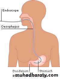
Light source in endoscope uses to see inside the body.
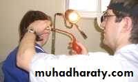
Physician use normal light to examine the skin.
Medical uses of visible light
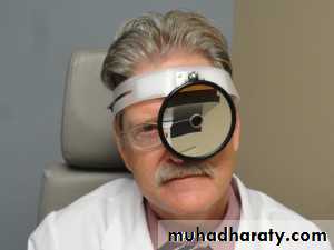
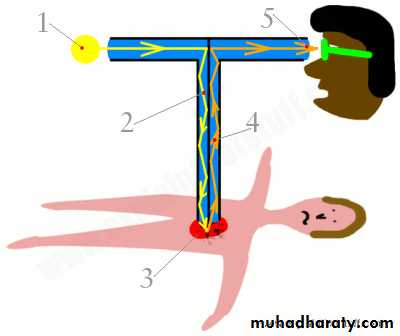
5.The visible light used in the ophthalmoscope for looking into eyes ,and in the otoscope for looking into ears by using a concave mirror to direct light in the body and a hole in the middle of it for the physician to look through(figure 7).
Figure(7)
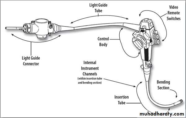
An endoscope works by inserting a long, thin and bendable tube into the body(figure8). On one end is a light source and in most cases also a video camera. The endoscope can be inserted through a natural opening in the body such as the throat or it can be inserted through a cut made in the skin.
Endoscope
An endoscope consists of two or three optical cables. Each cable includes up to 50,000 separate optical fibers that are made from glass or plastic. One or two of these cables will carry light down into the patient’s body, this illuminates where the endoscope has been inserted.
Endoscope
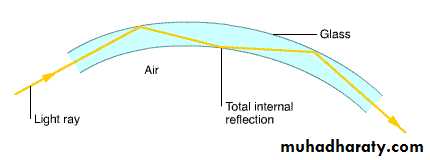
The light is reflected along the walls of the cable into the patient’s body. The light does this due to total internal reflection(figure10), which means that for this to happen the light ray must be at an angle of 82 degrees (which is the angle required for air to glass to be internally reflected).
Figure(10)
The other cable will carry reflected light that shines off the patient’s body, this light is the image of the body. The light bounces off the glass walls as it goes up to the physician’s eyepiece or into a camera. If the reflected light is carried up to a camera it will then be displayed on a TV monitor.
Eye and vision
The Human Eye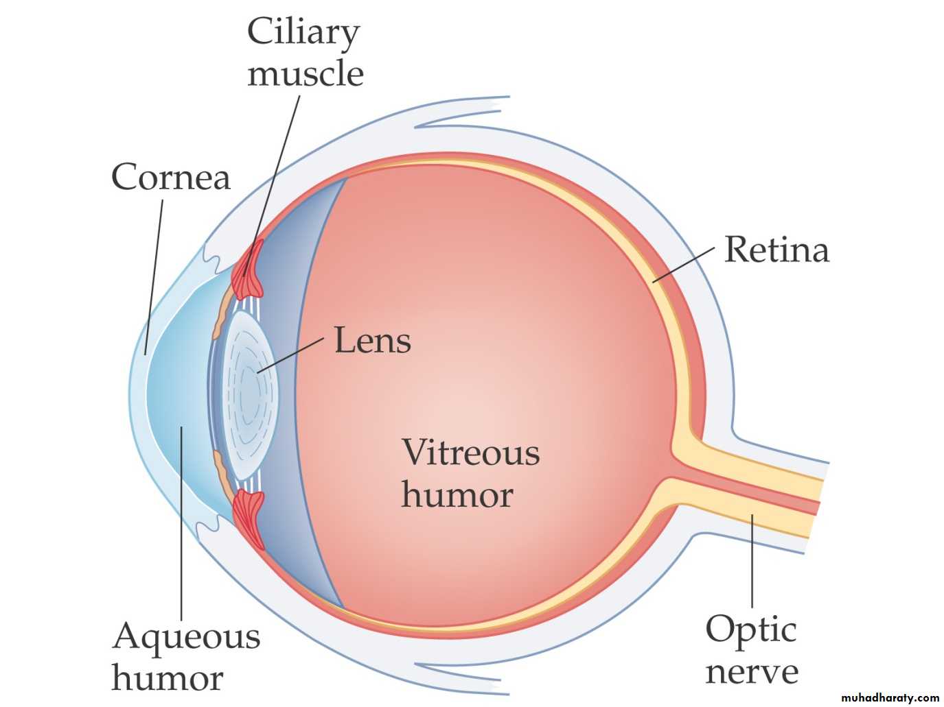
Light passes through the cornea of the human eye and is focused by the lens on the retina. The ciliary muscles change the shape of the lens, so
it can focus at different distances. The vitreous and aqueous humors are transparent. Rods and cones on the retina convert the light into electrical impulses, which travel down the optic nerve to the brain.
The cornea focuses by bending (refracting)
the light rays. The amount of bendingdepends on the curvatures of its surfaces
and the speed of light in the lens compared
with that in the surrounding material.
1-If the cornea is curved too much the eye is
near sighted(myopia).
2-Not enough curvature results in far sight ness (Hyperemia).
3-Uneven curvature produces astigmatism.
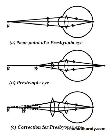
4-As people get older, their lenses lose some accommodation, presbyopia (old sight) results when the lens has lost nearly all of its accommodation
The index of refraction is nearly constant for all corneas, but the curvature varies
considerably from one person to anotherand is responsible for most our defective
vision.
The Human Eye
The eye produces a real, inverted image on the retina. Why don’t things look upside down to us? The brain adjusts the image to appear properly.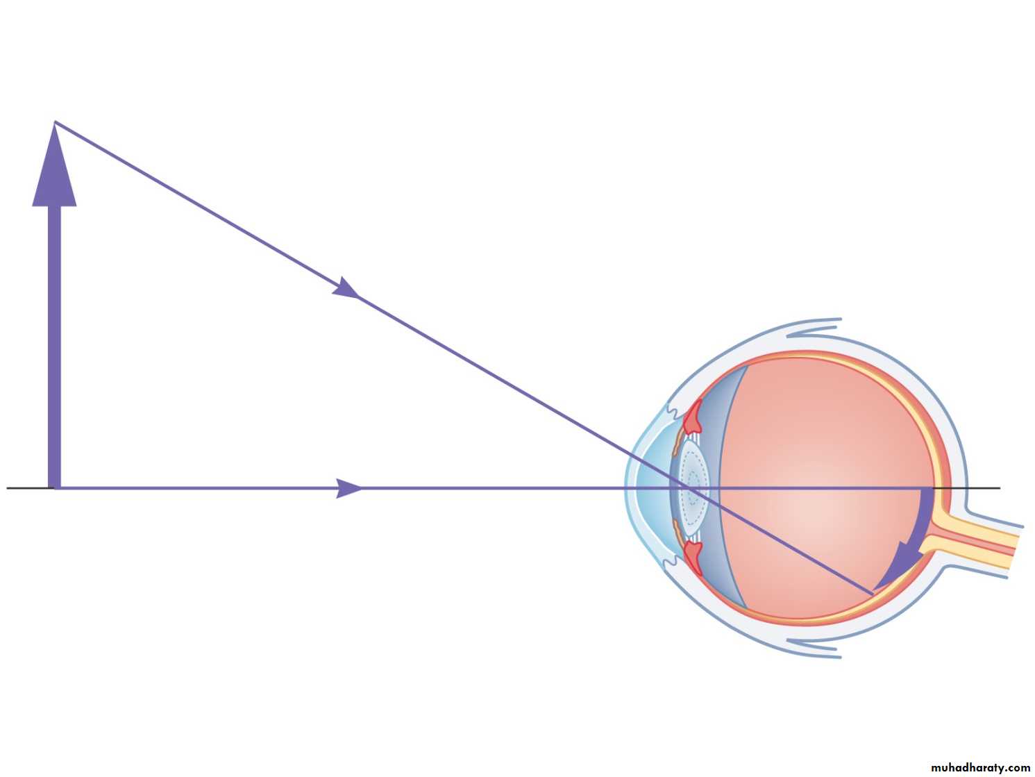
The Human Eye
The ciliary muscles adjust the shape of the lens to accommodate near and far vision.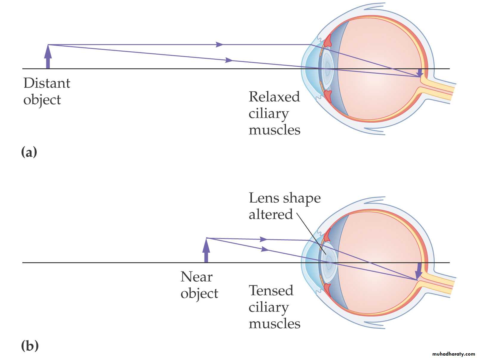
The Human Eye
The near point is the closest point to the eye that the lens is able to focus. For those with normal vision, it is about 25 cm from the eye, but increases with age as the lens becomes less flexible.The far point is the farthest point at which the eye can focus; it is infinitely far away, if vision is normal.
The retina, the light sensitive part of the eye, converts the light images into electrical nerve impulses that are sent to the brain.
The absorption of a light photon in photoreceptor triggers an electrical signal to brain-an action potential.
THE RETINA-THE LIGHT DETECTOR OF THE EYE
The light photon apparently cause a photochemical reaction in the photoreceptor
which in some way initiates the action potential. The photon must be above a minimum energy to cause the reaction.1-Infrared photons have insufficient energy and thus are not seen.
2-Ultraviolet photons have sufficient energy, but absorbed before they reach the retina and also are not seen.The image on the retina is very small. A
convenient equation for determining the size of image on the retina comes from the ratios of the lengths of the sides of similar triangles.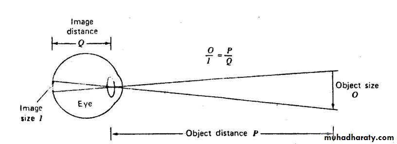
I: is image size
Q: is image distanceO: is object size
P: is object distance
Thus we can write
O/P=I/Q
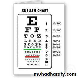
The optometrist usually uses a snellen chart
to test visual acuity. If he tells you that youreyes test normal at 20/20, he means that
you can read detail from 20 ft that person
with good vision can read from 20 ft. If your
eyes test at 20/40, you can just read from 20
ft the line that a person with good vision
can read from 40 ft.
HOW SHARP ARE YOUR EYES
The ability of the eye to recognize separate
lines also depends on the relative"blackness "and "whiteness" , the contrast
between two areas is defined as optical
density OD
OD = Log (Io/I)
Where Io is the light intensity without
absorber and I is intensity with absorber
EXAMPLE: A piece of film that transmits 10% of the incident light has an optical density
OD = Log(1/0.1) = 1.0EXAMPLE: A film that absorbs 99% of the light has an optical density
OD = Log(1/0.01) = 2.0
An OD=3 means that only 0.001 of the light
transmitted.
There a relation between the focal length F ,the object distance P , and the image distance Q of a thin lens.
1/F = 1/P + 1/Q
If F is measured in meters , then 1/F is the strength in Diopters (D),
Detective vision and its corrections
Thus, a positive (converging ) lens with a focal length of 0.1 m has a strength of 10 D .
The focal length F of a negative (divergence) lens is considered to be negative . A negative lens with a focal length of -0.5 m has a strengthof -2D.
The focal length F of a combination of two lenses with focal length F1 and F2 is given by
1/F = (1/F1)+ (1/F2)The strength of combination in diopters is equal to the sum of diopters of the various lenses.
Example
Assume lens A with focal length FA = 0.33m is combined with a lens B with focal length FB =0.25m. What is the focal length of the combination? What is the dioptric strength of the combinations?1/F = 1/ FA + 1/ FB
= 1/0.33 +1/0.25 = 1/0.143Or F = 0.143m. note that lens A is 3 D and lens B is 4 D .
The combination is the sum ,or 7 D.
Lenses in Combination and Corrective Optics
In a two-lens system, the image produced by the first lens serves as the object for the second lens.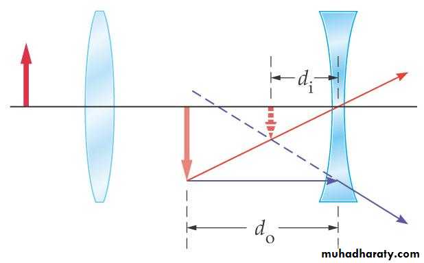
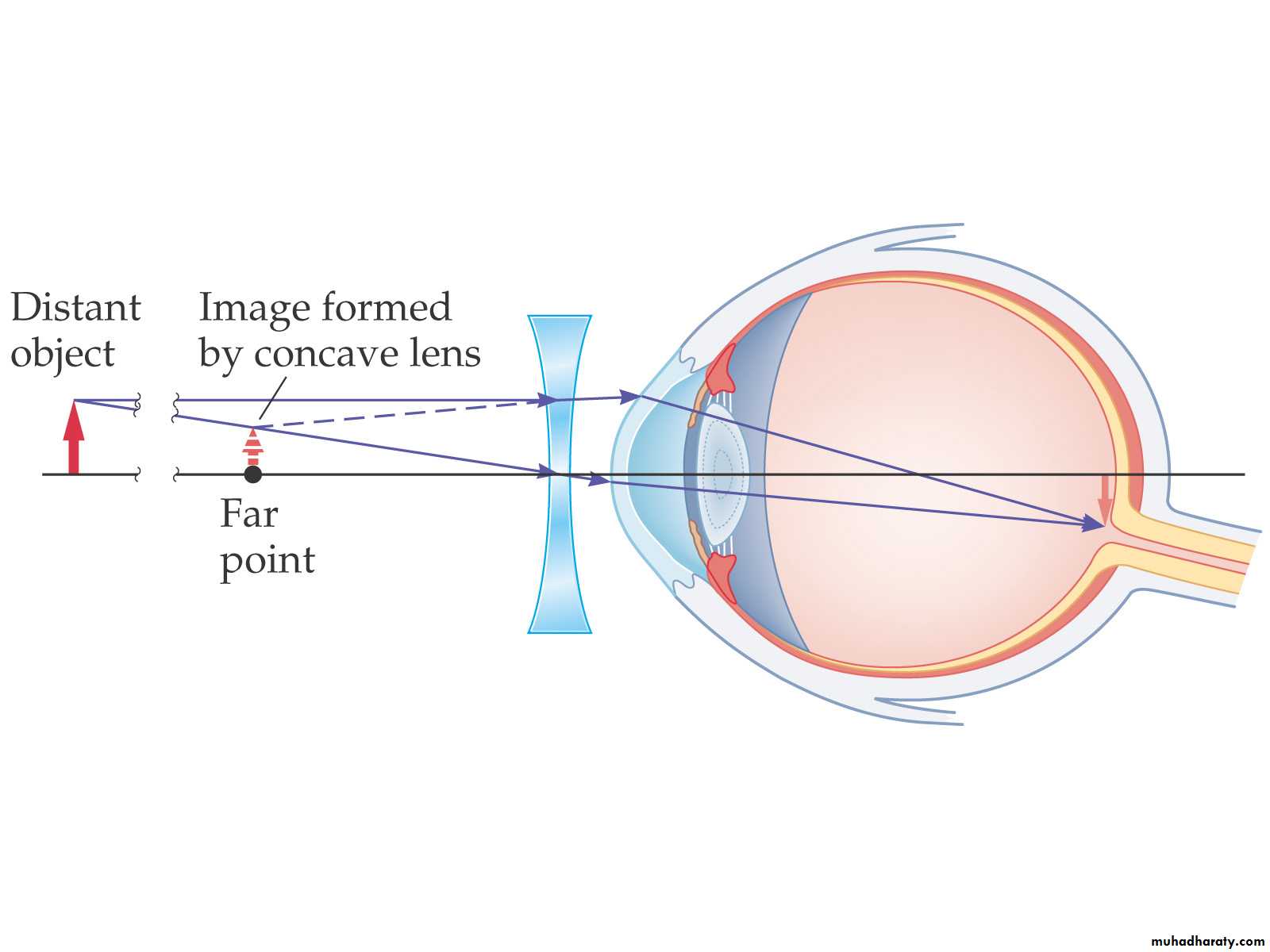
Myopia
A nearsighted person has a far point that is a finite distance away; objects farther away will appear blurry. This is due to the lens focusing too strongly, so the image is formed in front of the retina.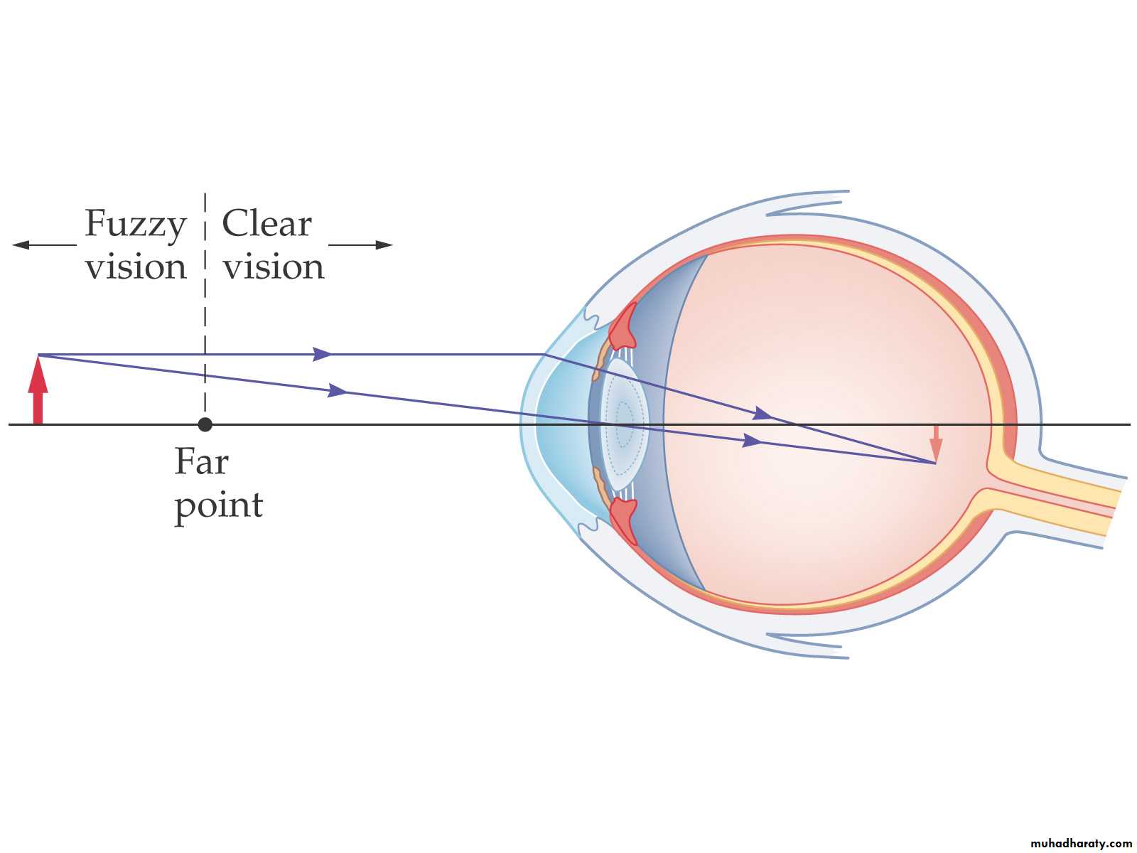
To correct this, a diverging lens is used. Its focal length is such that a distant object forms an image at the far point:
Hyperemia
A person who is farsighted can see distant objects clearly, but cannot focus on close objects – the near point is too far away. The lens of the eye is not strong enough, and the image focus is behind the retina.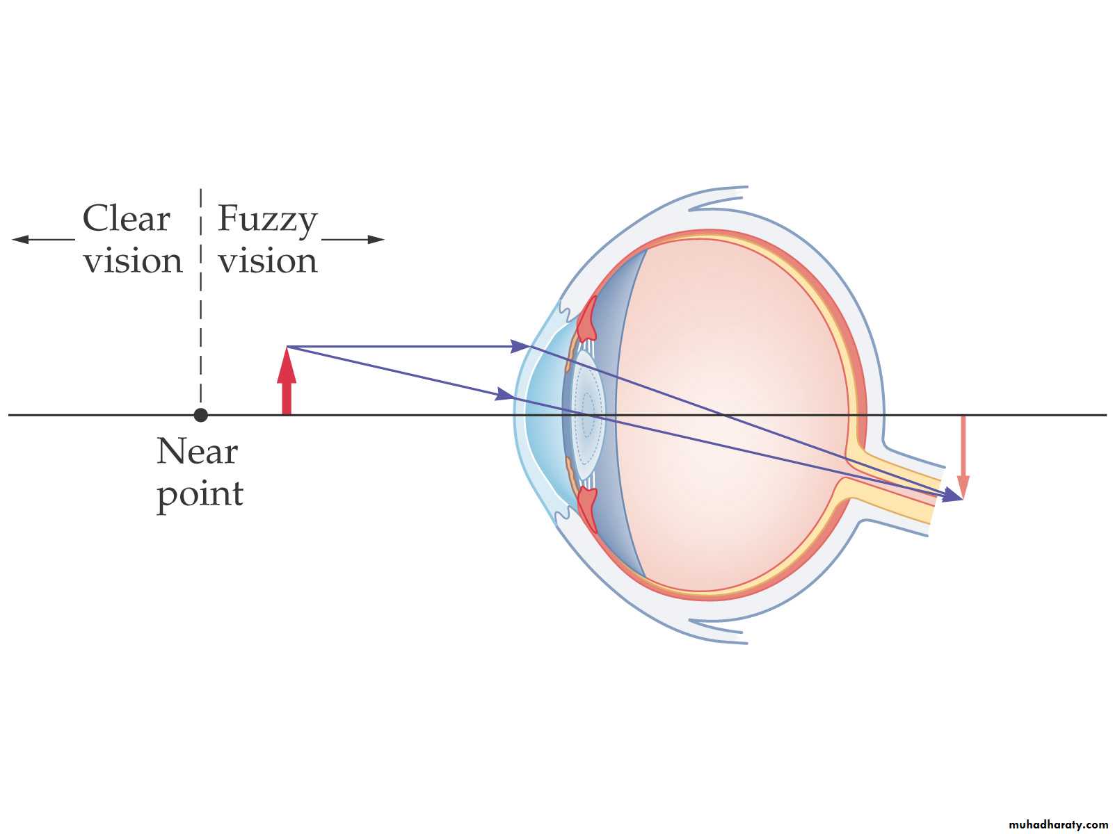
Hyperemia
To correct farsightedness, a converging lens is used to augment the converging power of the eye. The final image is past the near point: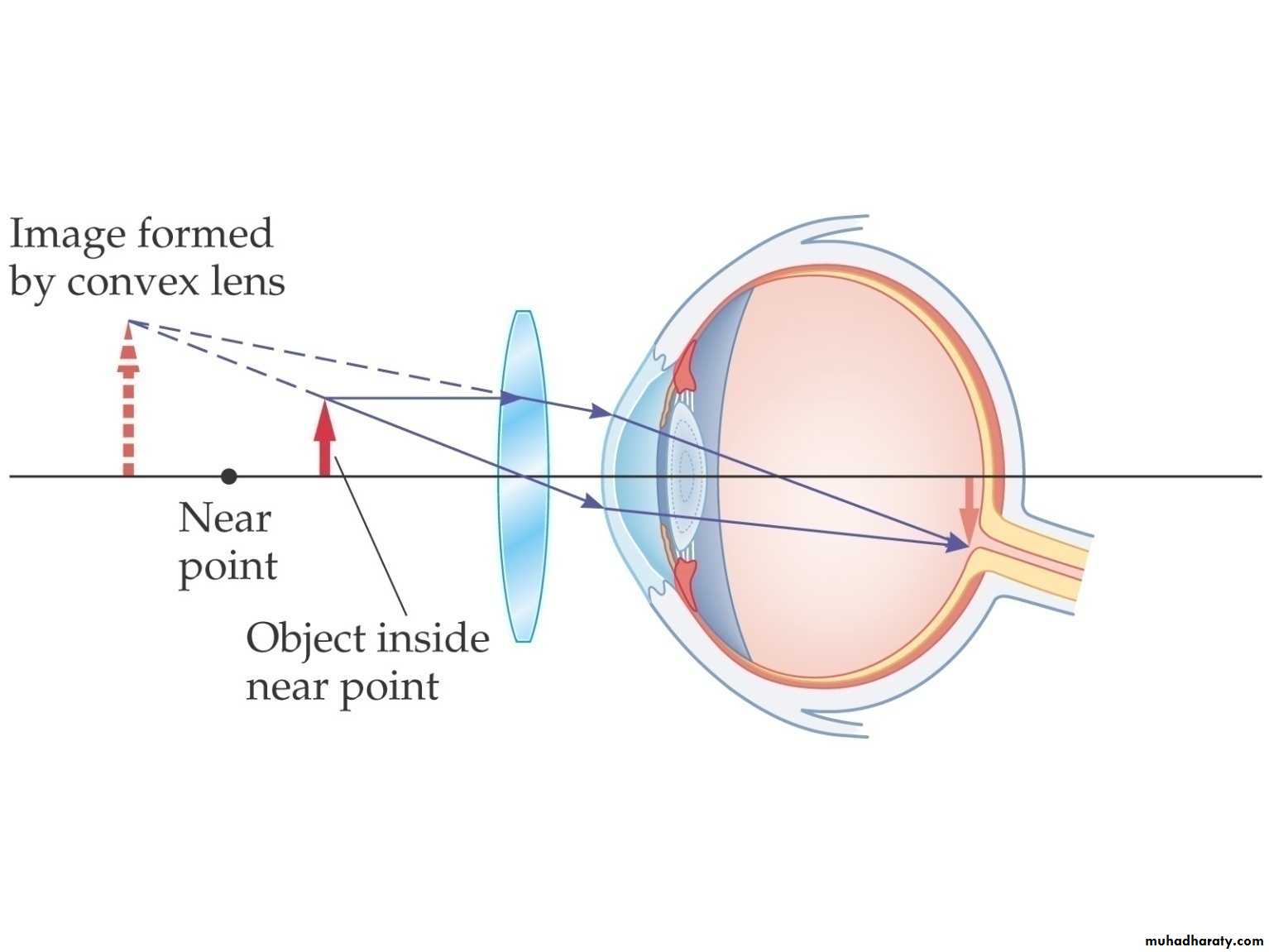
Let us determine the strength of a lens needed to correct a myopic eye with a far point of 1m.
We consider the image distance(lens to retina)to be 2cm(0.02m).
example
A person who is focusing an 0bject at 1m has a lens strength of
1/F =(1/1.0)+(1/0.02) =51 D
An eye able to focus at infinity has a strength of 1/F=(1/∞ )+ (1/0.02) = 50 D
51 D – 50D =1D
Thus a myopic person with a far point 1m has 1 D, and a negative lens of -1.0 D will correct his vision.
Myopia
Let us consider afar sighted eye with a near point of 2.0 m. what power lens will let this person read comfortably at 0.25 m?
The strength of a good eye focused at 0.25 m is given by 1/F=(1/0.25)+ ( 1/ 0.02)
= 4+ 50 =54D.
An eye focused at 2m has a strength of
1/F= (1/2.0) + (1/ 0.02) =0.5 + 50 =50.5 D
A corrective lens of 54-50.5= +3.5 D would prescribed for his eye.
hyperemia
As people get older the cillary muscles weaken and lens losses some of its elasticity.
The power of accommodation diminishes with age. This defect is corrected by two parts of lenses upper half of each lens is diverging and corrects the myopia when the wears is looking ahead at distance objects, the lower half corrects the presbyopia with a suitable converging lens, and the wearer looks through this part when readingPRESBYOPIA( old sight)
Old sight
When astigmatism is present, point objects do not form point images on the retina. This is normally due to the corneas unequal curvature in different directions. If the curvature is greater in a horizontal section than in the vertical section, rays brought to a focus more quickly in the horizontal than in the vertical plane. The defect is corrected by the use of cylindrical spectacle lenses
ASTIGATISM
ASTIGATISM
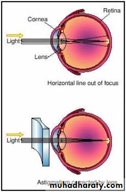
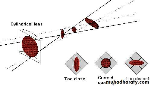
ASTIGATISM
Lens aberrations can distort images.There are two types of lens aberration:
1. Spherical aberration.
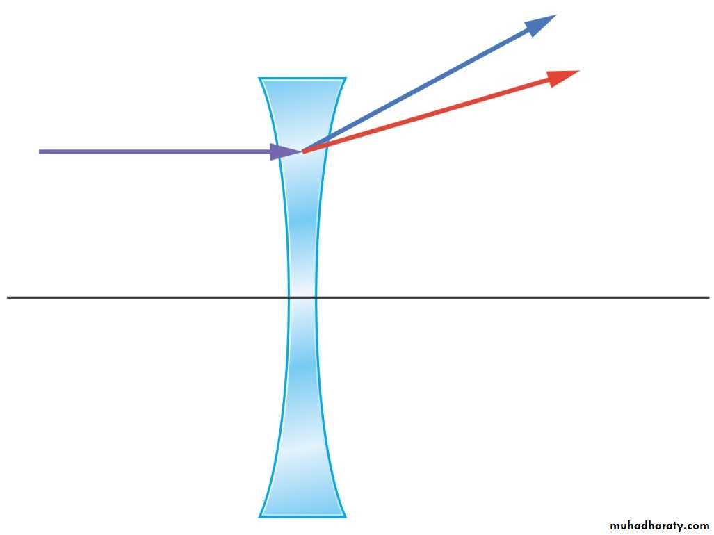
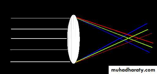
2. Chromatic aberration.
Lens Aberration
Spherical Lens Aberrations
Spherical aberration occurs when light striking the lens far from the axis does not focus properly. It can be fixed by grinding the lens to a precision, non-spherical shape.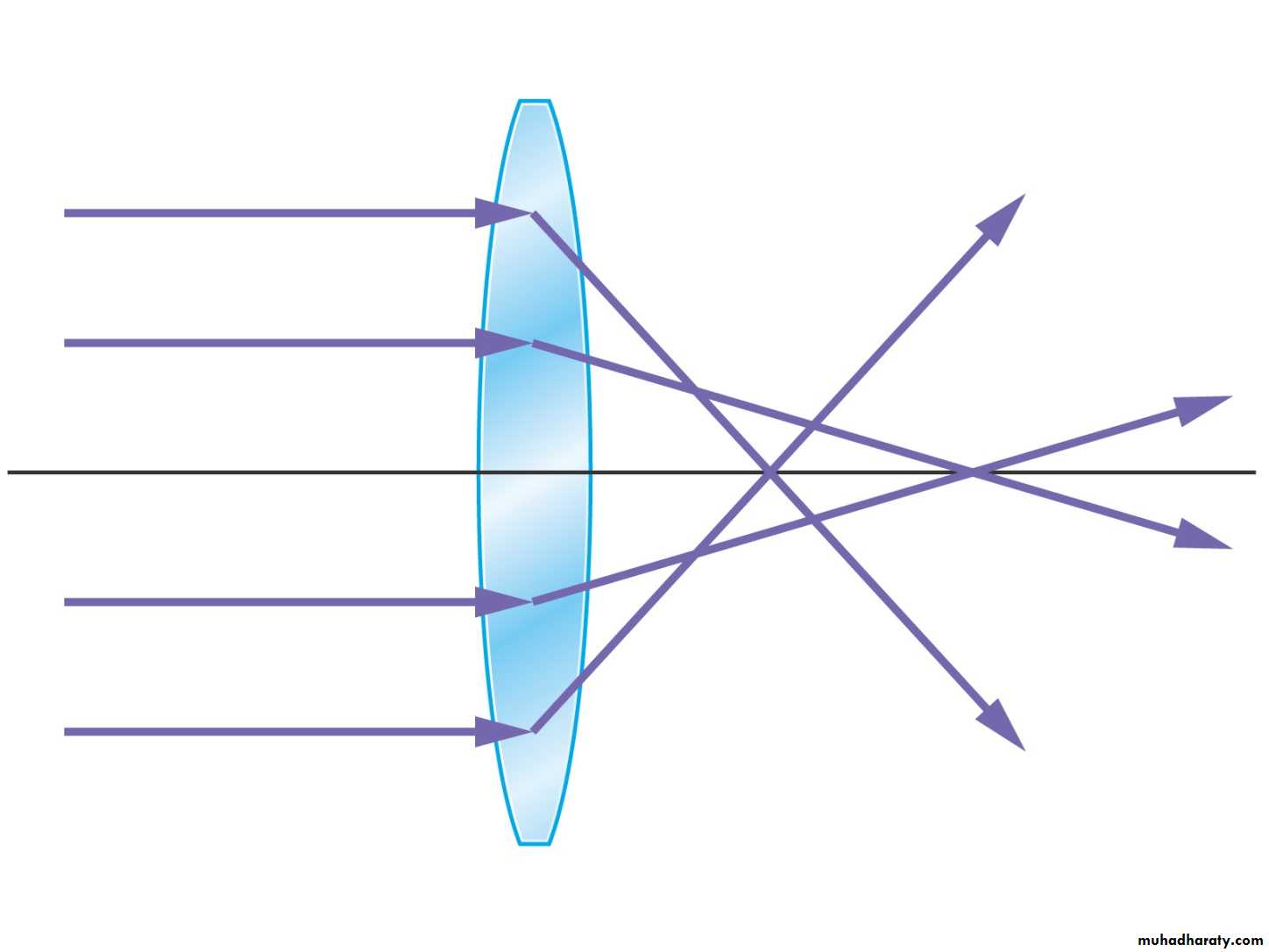
Chromatic Lens Aberrations
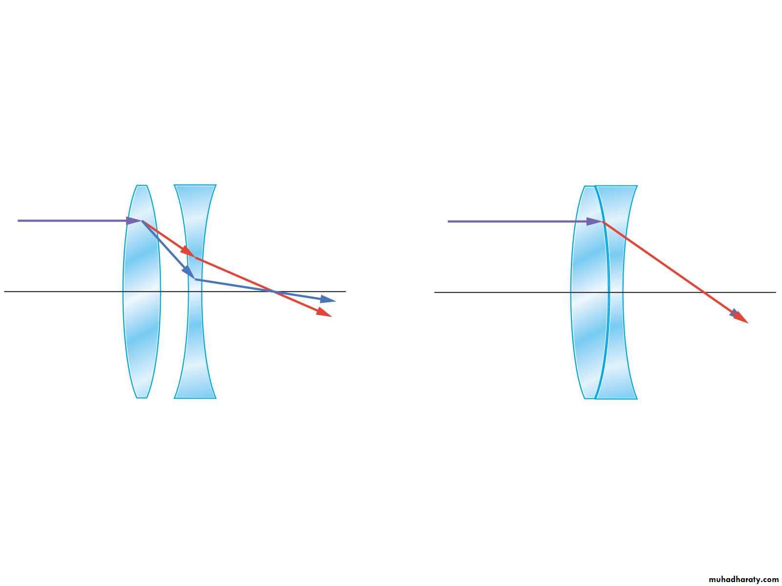
Chromatic aberration occurs when different colors of light focus at different points.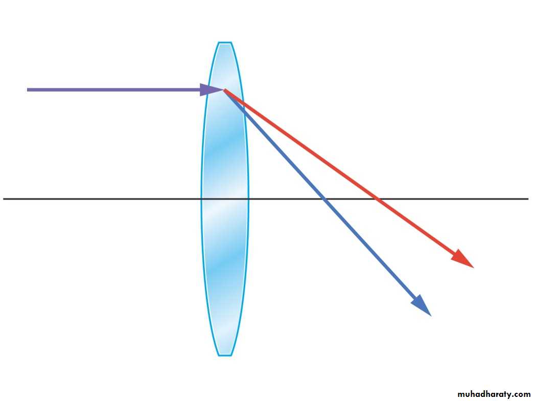
Lens Flaws: Chromatic Aberration
Lens has different refractive indices for different wavelengths.Could cause color fringing:
i.e., lens cannot focus all the colors at the same point.
Lens Aberrations
Chromatic aberration can be improved by combining two or more lenses that tend to cancel each other’s aberrations. This only works perfectly for a single wavelength, however.