
Lecture 5
Thorax
د.رندعبداللطيف
Pericardium
The pericardium is a fibroserous sac that encloses the heart and the roots of the
great vessels lies within the middle mediastinum posterior to the body of the
sternum and the 2nd to the 6
th
costal cartilages.
Fibrous Pericardium
The fibrous pericardium is the strong fibrous part of the sac. It is firmly attached
below to the central tendon of the diaphragm & in front to the sternum by the
sternopericardial ligaments.. It fuses with the outer coats of the great blood
vessels: the aorta, the pulmonary trunk, the superior and inferior venae cavae,
and the pulmonary veins.
Serous Pericardium
The serous pericardium lines the fibrous pericardium and coats the heart. It is
divided into parietal and visceral layers. The parietal layer lines the fibrous
pericardium and is reflected around the roots of the great vessels to become
continuous with the visceral layer of serous pericardium that closely covers the
heart.
The visceral layer is closely applied to the heart and is often called the
epicardium.
The slitlike space between the parietal and visceral layers is referred to as the
pericardial cavity. Normally, the cavity contains a small amount of tissue fluid
(about 50 mL), the pericardial fluid, which acts as a lubricant to facilitate
movements of the heart.
Pericardial Sinuses
1- On the posterior surface of the heart, the reflection of the serous
pericardium around the large veins (four pulmonary veins and the
superior and inferior venae cavae) forms a recess called the oblique
sinus.
2- The transverse sinus, which is a short passage that lies between the
reflection of serous pericardium around the aorta and pulmonary trunk
and the reflection around the large veins.
Nerve Supply of the Pericardium
The fibrous pericardium and the parietal layer of the serous pericardium are
supplied by the phrenic nerves. The visceral layer of the serous pericardium
is innervated by branches of the sympathetic trunks and the vagus nerves.
Heart
Lecture 5
Thorax
د.رندعبداللطيف
The heart is a hollow muscular organ that is somewhat pyramid shaped and lies
within the pericardium in the mediastinum.
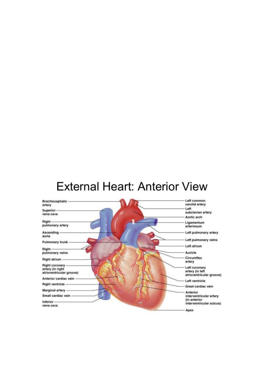
Surfaces of the Heart : The heart has three surfaces:
1- sternocostal (anterior): is formed mainly by the right atrium and the right
ventricle, which are separated from each other by the atrioventricular
groove
2- Diaphragmatic (inferior): is formed mainly by the right and left
ventricles.
3- The base (posterior): is formed mainly by the left atrium
The apex of the heart, formed by the left ventricle, is directed downward,
forward, and to the left. It lies at the level of the fifth left intercostal space, (9
cm) from the midline. In the region of the apex, the apex beat can usually be
seen and palpated in the living person.
Borders of the heart:
The right border is formed by the right atrium.
The left border is formed by the left ventricle and part of the left auricle.
Chambers of the Heart

Lecture 5
Thorax
د.رندعبداللطيف
The heart is divided into four chambers: the right and left atria and the right and
left ventricles. The 2 atria are separated from the 2 ventricles by
Atrioventricular groove(Coronary sulcus).The 2 ventricles separated by anterior
interventricular groove contains the great cardiac vein & anterior
interventricular artery.The walls of the heart are composed of cardiac muscle,
the myocardium; covered externally with serous pericardium, the epicardium;
and endocardium which lined internally with a layer of endothelium.
Each Atria leads to its corresponding ventricle by Atrioventricular Valve(the
right one is Tricuspid consists of 3 cusps connected to 3 papillary muscles on
the inside of the right ventricle via thread like structur known as Chorda
tendinae, while the left valve is Bicuspid (Mitral ) has 2 cusps connected to 2
papillary muscles via chorda tendinae.
The R.A consists of 2 part a smooth (sinus venosus) and rough part ( Musculae
pectinatae) separated by Crista terminalis. It receives the openings of superior
vena cava (from above), inferior vena cava ( from below) and opening of the
coronary venous sinus..
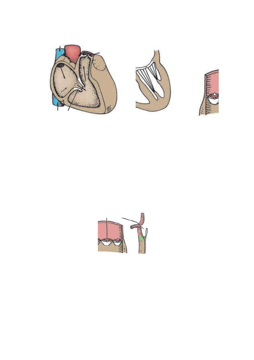
Right Ventricle
The walls of the right ventricle are much thicker than those of the right
atrium and show several internal projecting ridges formed of muscle
bundles known as trabeculae carneae. The papillary muscles, are attached
by their bases to the ventricular wall, their apices are connected by
fibrous chords (the chordae tendineae) to the cusps of the tricuspid valve.
The moderator band, crosses the ventricular cavity from the septal to the
anterior wall. It conveys the right branch of the atrioventricular bundle.
The tricuspid valve guards the right atrioventricular orifice and consists
of three cusps formed by a fold of endocardium with some connective
tissue enclosed: anterior, septal, and posterior cusps. The bases of the
cusps are attached to the fibrous ring of the skeleton of the heart,whereas
their free edges and ventricular surfaces are attached to the chordae
tendineae. The chordae tendineae connect the cusps to the papillary
muscles.
When the ventricle contracts, the papillary muscles contract and prevent
the cusps from being forced into the atrium and turning inside out as the
intraventricular pressure rises. To assist in this process, the chordae
tendineae of one papillary muscle are connected to the adjacent parts of
two cusps.
The right ventricle pumps the deoxygenated blood to the pulmonary trunk via
the pulmonary valve to reach the lungs for exchange of gasses ( Minor
circuit),while the L.V pumps the oxygenated blood to the Aorta via Aortic valve
to be distributed to different parts of the body as Systemic circulation
Lecture 5
Thorax
د.رندعبداللطيف
The pulmonary valve guards the pulmonary orifice and consists of three
semilunar cusps formed by folds of endocardium with connective tissue. No
chordae or papillary muscles are associated with these valve cusps; the
attachments of the sides of the cusps to the arterial wall prevent the cusps from
prolapsing into the ventricle. At the root of the pulmonary trunk are three
dilatations called the sinuses, and one is situated external to each cusp.
Left Atrium
Similar to the right atrium, the left atrium consists of a main cavity and a left
auricle.
The interior of the left atrium is smooth, but the left auricle possesses
muscular ridges as in the right auricle. The left atrium is smooth& receives the
opening of 4 pulmonary veins ( Oxygenated blood from the lungs),2 left & 2
right pulmonary veins.The 2 atria are separated by a septum showing the fossa
Tricuspid valve

Lecture 5
Thorax
د.رندعبداللطيف
ovalis ( Remnant of foramen ovale during fetal life). The left atrioventricular
orifice is guarded by the mitral valve.
Left Ventricle
The walls of the left ventricle are three times thicker than those of the right
ventricle.
In cross section, the left ventricle is circular; the right is crescentic.
The part of the ventricle below the aortic orifice is called the aortic vestibule.
The mitral valve guards the atrioventricular orifice. It consists of two cusps
similar to that of the cusps of the tricuspid valve, one anterior and one posterior,
the anterior cusp is the larger.
The aortic valve guards the aortic orifice and is precisely similar in structure
to the pulmonary valve. One cusp is anterior and two are posterior.
Behind each
cusp, the aortic wall bulges to form an aortic sinus.
The anterior aortic sinus
gives origin to the right coronary artery, and the left posterior sinus gives origin
to the left coronary artery.
Structure of the Heart
The walls of the heart are composed of a thick layer of cardiac muscle, the
myocardium, covered externally by the epicardium and lined internally by the
endocardium. The two atria are separated by interatrial septum. The two
ventricles are separated by interventricular septum.
The upper part of the septum is thin and membranous and attached to the
fibrous skeleton. The lower part of the septum is thick and formed of muscle.
The skeleton of the heart
It consists of fibrous rings that surround the atrioventricular, pulmonary, and
aortic orifices and are continuous with the membranous upper part of the
ventricular septum. The fibrous rings around the atrioventricular orifices
separate the muscular walls of the atria from those of the ventricles. They
support the bases of the valve and prevent them from stretching and becoming
incompetent. The skeleton of the heart forms the basis of electrical discontinuity
between the atria and the ventricles.
Conducting System of the Heart
The normal heart contracts rhythmically at about 70 to 90 beats per minute in
the resting adult. The conducting system of the heart consists of specialized
cardiac muscle present in the sinuatrial node, the atrioventricular node, the
atrioventricular bundle and its right and left terminal branches, and the
subendocardial plexus of Purkinje fibers (specialized cardiac muscle fibers that
form the conducting system of the heart).
Sinuatrial Node

Lecture 5
Thorax
د.رندعبداللطيف
The sinuatrial node is located on the anterior wall of the right atrium in the
upper part of the sulcus terminalis just to the right of the opening of the
superior vena cava. It spontaneously initiates rhythmic electrical impulses that
spread in all directions through the cardiac muscle of the atria and cause the
muscle contraction.
Atrioventricular Node (AV node)
The AV node is placed on the lower part of the atrial septum to the left side of
the opening of Coronary venous sinus. The cardiac impulse is conducted from
AV node to the ventricles by the atrioventricular bundle. It is
stimulated by the
excitation wave from atrial myocardium.
Atrioventricular Bundle (AV bundle)
The atrioventricular bundle (bundle of His) is the only route along which the
cardiac impulse can travel from the atria to the ventricles. The bundle descends
in the membranous part of the ventricular septum and to the muscular part of
septum where it divides into two branches, one for each ventricle.
The right one toward the apex of the heart to inter the moderater band & finally
reaches the anterior papillary muscle and breaks into a number of Purkinje
fibers to all parts of the Right ventricular wall. The left bundle branch goes to
bases of papillary muscles & breaks into a number Purkinje fibers in the wall of
left ventricle.
The conducting system of the heart is responsible for generating rhythmic
cardiac impulses & conducting these impulses rapidly throughout the
myocardium & the different chambers contract in a coordinated and efficient
manner.
The Arterial Supply of the Heart
The arterial supply of the heart is provided by the right and left coronary
arteries, which arise from the ascending aorta & run within subepicardial
connective tissue.
The right coronary artery arises from the anterior aortic sinus and runs
forward between the pulmonary trunk and the right auricle. It descends
vertically in the right atrioventricular groove, and at the inferior border of the
heart it continues posteriorly along the atrioventricular groove to anastomose
with the left coronary artery in the posterior interventricular groove.
It supplies the right atrium and right ventricle and parts of the left atrium and
left ventricle and the atrioventricular septum. It gives the following branches:
1-Marginal

Lecture 5
Thorax
د.رندعبداللطيف
2-Posterior interventricular
3-S.A nodal
4-Anterior atrial &ventricular branches
The left coronary artery is usually larger than the right coronary artery,
supplies the major part of the heart, including the greater part of the left atrium,
left ventricle, and ventricular septum. It arises from the left posterior aortic
sinus and passes forward between the pulmonary trunk and the left auricle. It
then enters the atrioventricular groove and divides into an anterior
interventricular branch and a circumflex branch.
Branches
1-Circumflex
2-Anterior interventricular
3-L.Marginal
4-L. conus
5-L.ventricular&atrial
The collateral circulation of the heart: the terminal branches of the right
and left coronary arteries Anastomoses between each other, but they are
usually not large enough to provide an adequate blood supply to the cardiac
muscle if one of the large branches become blocked by disease. A sudden
block of one of the larger branches of either coronary artery usually leads to
myocardial death (myocardial infarction), although sometimes the collateral
circulation is enough to sustain the muscle.
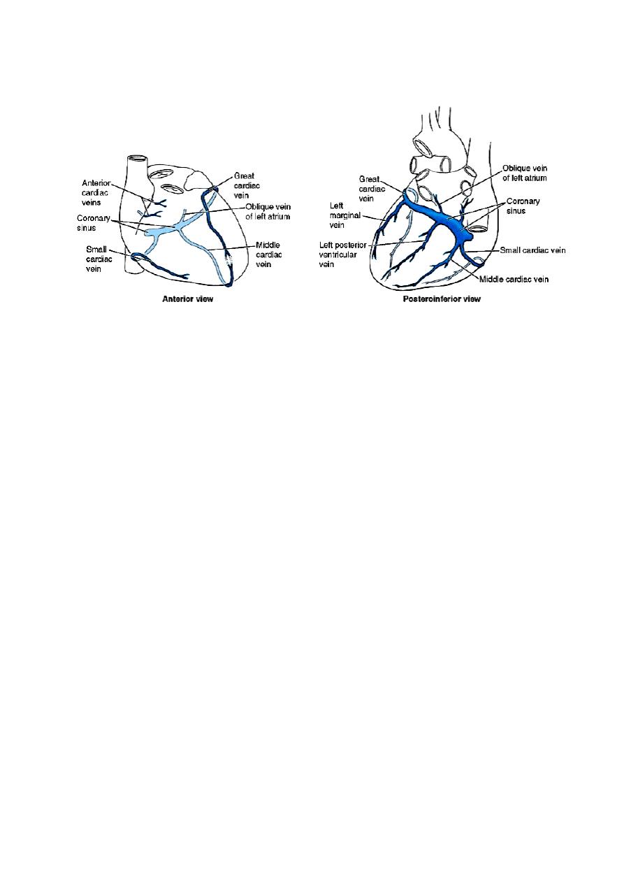
Venous Drainage of the Heart
Is mainly via Coronary venous sinus ( direct continuity of great cardiac vein).
The coronary sinus receives the following tributaries before opening into
the R.A:
1-Great cardiac V
2-Middle cardiac
3-Small cardiac
4-Oblique V of L.A
Lecture 5
Thorax
د.رندعبداللطيف
5-Post. V of L.V .
Nerve Supply of the Heart
Is achieved by Autonomic nervous system( sympathetic component coming
from upper 4-5 thoracic segments of the spinal cord) ,while parasympathetic
component via cardiac branches of R & L vagus nerves.Fibers from both
sympathetic & parasympathetic communicates to form superficial & deep
cardiac plexuses which goes directly to supply the heart & coronary arteries.