Surgery
Renal parenchymal Neoplasms
Dr. Saad Dakhil
Lec. 31
ADENOCARCINOMA OF THE KIDNEY (RCC)
RCC accounts for roughly 2.8% of adult cancers and constitutes
approximately 85% of all primary malignant renal tumors.
RCC occurs most commonly in the fifth to sixth decade and has a male-
female ratio of 2:1.
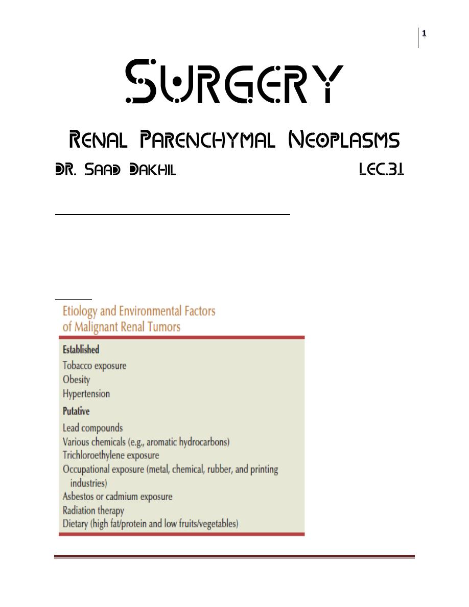
Etiology
Surgery
Renal parenchymal Neoplasms
Dr. Saad Dakhil
Lec. 31
Other Risk Factors:
Von Hippel-Lindau disease ;
(Cerebellar hemangioblastoma, retinal angiomata, and bilateral clear cell RCC.)
Hereditary papillary renal carcinoma; multiple (bilateral renal tumors with a
papillary histologic appearance).
Acquired cystic disease of the kidneys; is a well-recognized entity of
multiple bilateral cysts in the native kidneys of uremic patients(>30 times
higher).
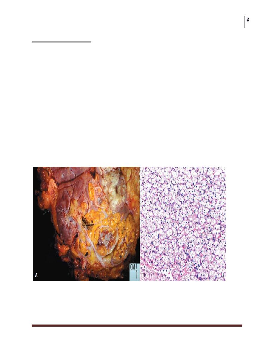
Pathology:
A, Clear cell renal cell carcinoma (RCC) with typical golden yellow color.
B, Low-power view of typical microscopic
appearance of a low-grade clear cell RCC demonstrating a delicate vascular
network interspersed within homogeneous nests of cells with clear cytoplasm.
Surgery
Renal parenchymal Neoplasms
Dr. Saad Dakhil
Lec. 31
Pathogenesis
RCCs are vascular tumors that tend to spread either by ;direct invasion
through the renal capsule into perinephric fat and adjacent visceral structures
or by direct extension into the renal vein. Approximately 25–30% of
patients have evidence of metastatic disease at presentation. The most
common site of distant metastases is the lung.
Surgery
Renal parenchymal Neoplasms
Dr. Saad Dakhil
Lec. 31
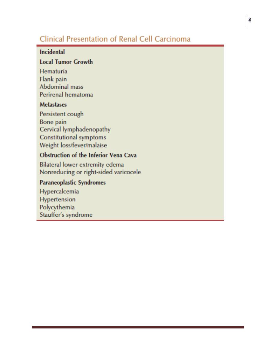
C. LABORATORY FINDINGS
In addition to the laboratory abnormalities associated with the various RCC
paraneoplastic syndromes, anemia, hematuria, and an elevated sedimentation
rate are frequently observed.
Anemia occurs in about 30% of RCC patients.
Gross or microscopic hematuria can be seen in up to 60% of patients
presenting with RCC.
An elevated erythrocyte sedimentation rate is also commonly seen, with a
reported incidence as high as 75%.
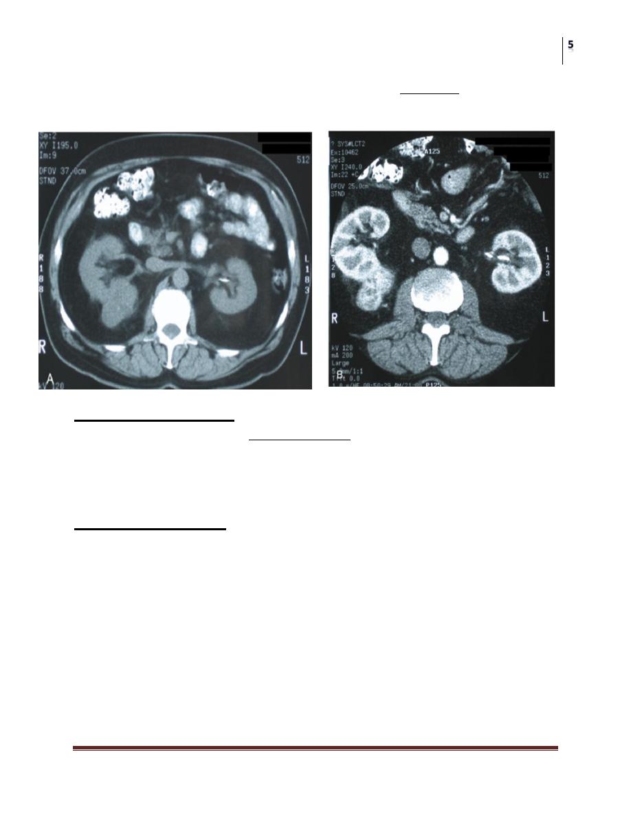
D. Radiological FINDINGS
CT scanning remains the primary technique with which others must be
compared.
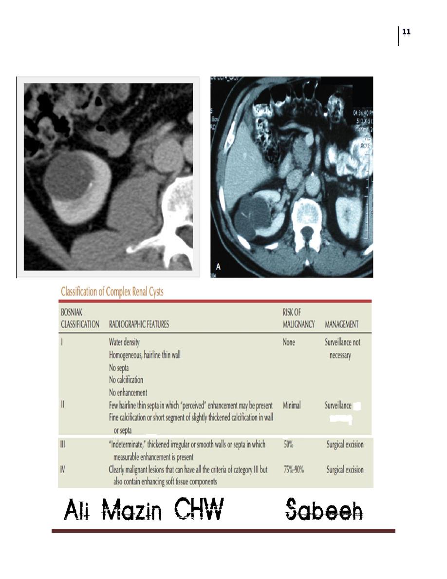
ULTRASONOGRAPHY; It is approximately 98% accurate in
distinguishing simple cysts from solid lesions.
Surgery
Renal parenchymal Neoplasms
Dr. Saad Dakhil
Lec. 31
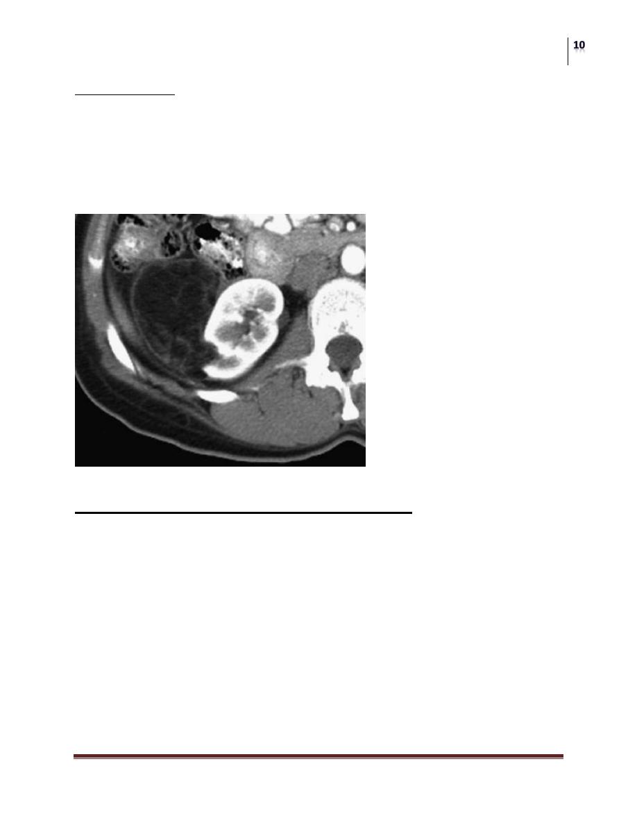
A typical finding of RCC on CT is a mass that becomes enhanced with the use of
intravenous contrast media.
Fine-needle aspiration;
Primary indications for needle aspiration or biopsy of a renal mass are when
a renal abscess or infected cyst is suspected
When RCC must be differentiated from metastatic malignant disease or
renal lymphoma .
Differential Diagnosis
The great majority of renal masses are simple cysts.
A renal abscess may be strongly suspected in a patient presenting with fever,
flank pain, pyuria, and leukocytosis, and an early needle aspiration and
culture should be performed.
Other benign renal masses include granulomas and arteriovenous
malformations.
Renal lymphom , transitional cell carcinoma of the renal pelvis, adrenal
cancer, and metastatic disease (most commonly from a lung or breast cancer
primary).
Surgery
Renal parenchymal Neoplasms
Dr. Saad Dakhil
Lec. 31
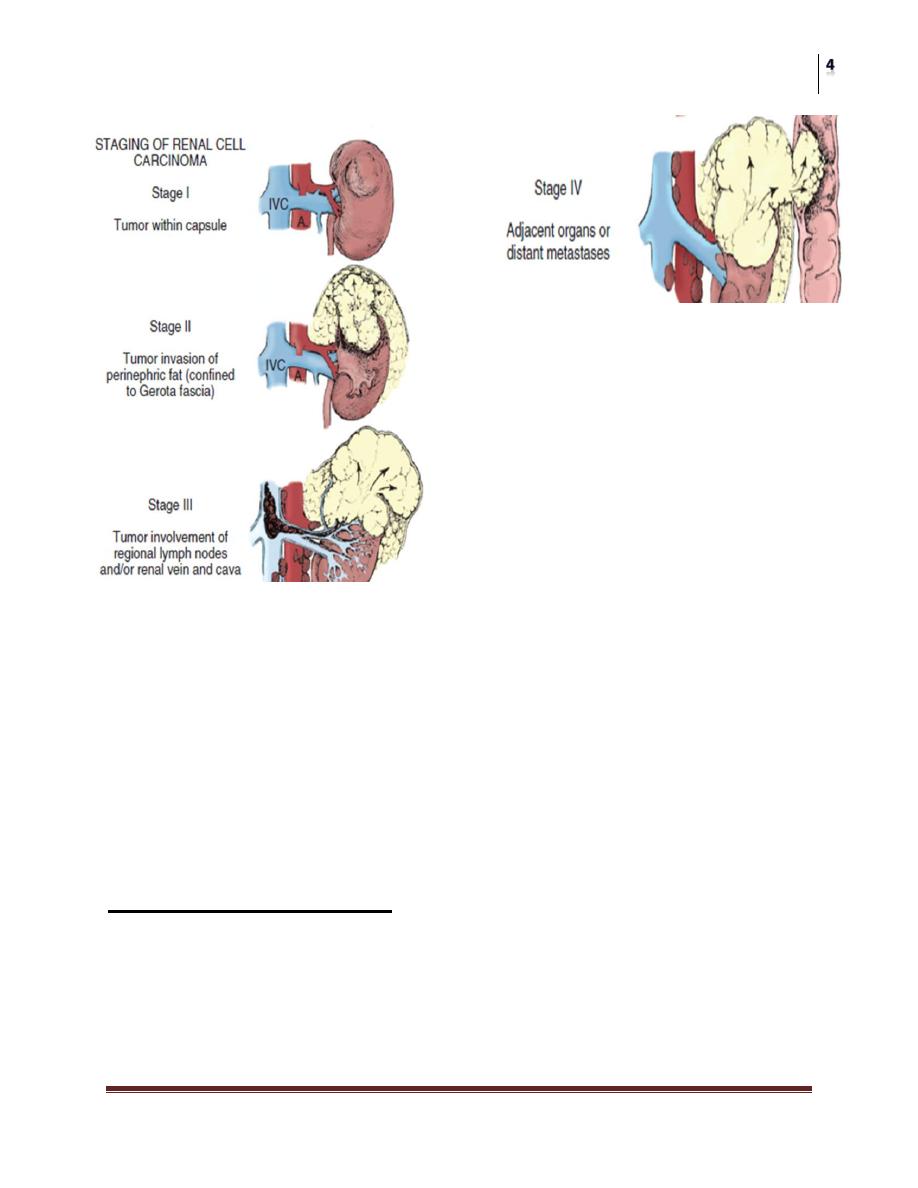
Treatment
1. Localized disease;
Surgical removal of the early-stage lesion remains the only potentially
curative therapy available for RCC patients.
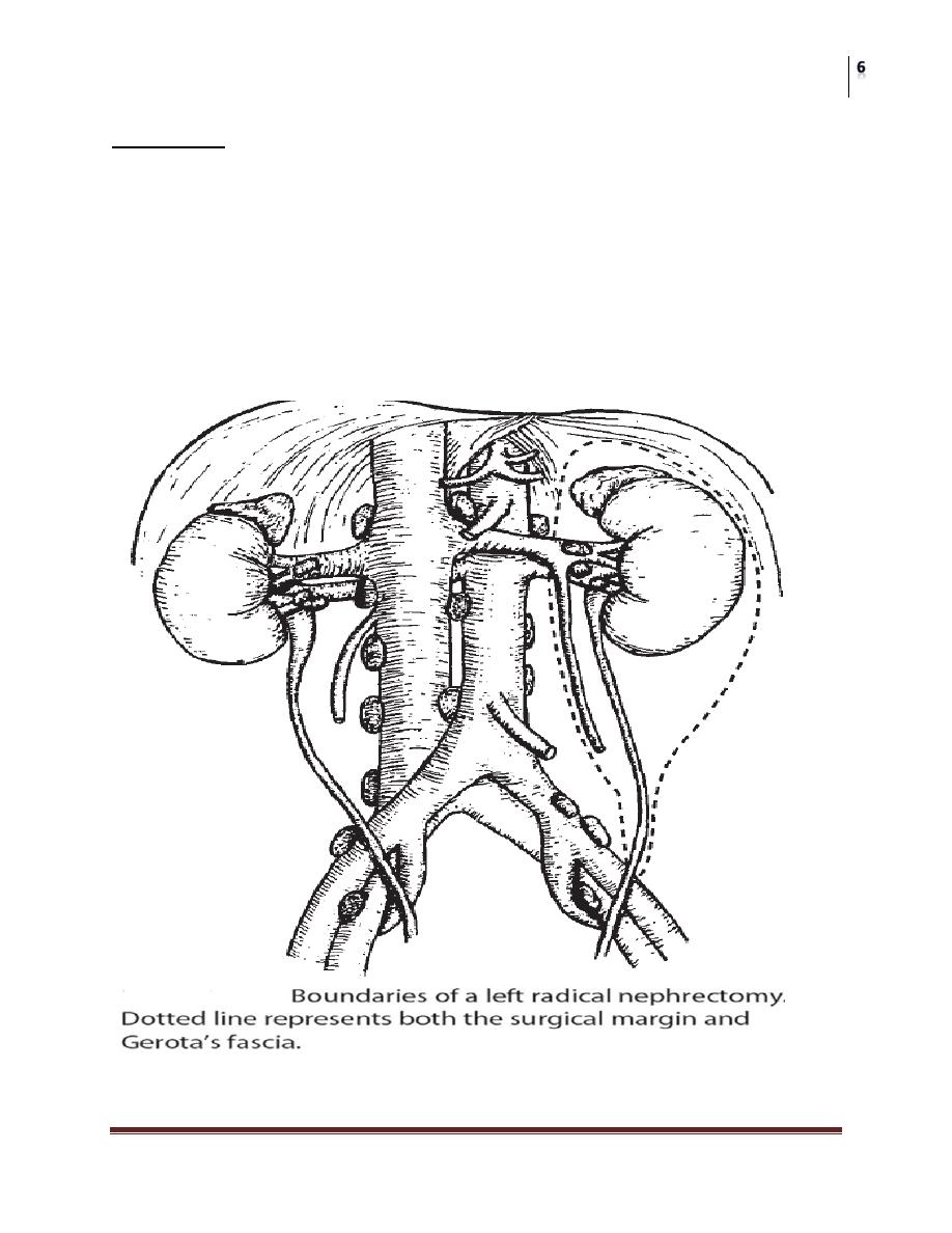
Radical nephrectomy is the primary treatment for localized RCC.
Radical nephrectomy entails en bloc removal of the kidney and its
enveloping fascia (Gerota’s) including the ipsilateral adrenal, proximal one-
half of the ureter, and lymph nodes up to the area of transection of the renal
vessels
Surgery
Renal parenchymal Neoplasms
Dr. Saad Dakhil
Lec. 31
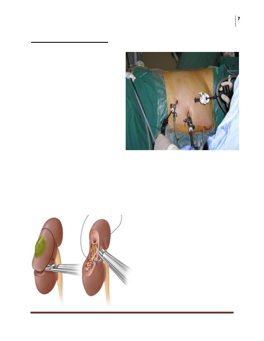
Laparoscopic surgery
Nephron-sparing
surgery
considered for patients with a localised tumour less than 4 cm in diameter,
An anatomical or functional solitary kidney and patients with a contralateral
kidney affected by a condition that might impair renal function in future .
It involves partial nephrectomy with preservation of as much functioning
kidney tissue as possible.
The recovery from a laparoscopic
nephrectomy is remarkably swift
with many patients back to full
activities within 3 weeks. They
also suffer a lot less from the
smaller scars than the conventional
surgical scar.

Surgery
Renal parenchymal Neoplasms
Dr. Saad Dakhil
Lec. 31
Minimal invasive surgery
Used in patient who are not candidates for surgery ;
Ablation
Kidney tumors be removed (ablated) with intense heat or cold, in which
special needles are inserted through the skin, guided by imaging from a CT
scan and ultrasound.
Angioemoblization.
2. Disseminated disease
Approximately 30% of patients with RCC will present with advanced
disease.
Metastatic RCC has a natural history that is typically aggressive and rapidly
progressive, with 5-year survival rates typically <10%.
A. Surgery: Nephrectomy
For managing patients with severe hemorrhage or unremitting pain.
Patients presenting with a solitary metastatic site particularly in the lung that
is amenable to surgical resection may be candidates for combined
nephrectomy and removal of the metastatic foci.
Nephrectomy in the presence of metastatic disease (cytoreductive
nephrectomy) Before Immunotherapy.
B. Radiation therapy
Effective palliation of metastatic disease to the brain, bone, and lungs is
reported in up to two-thirds of patients.
C. Biologic response modifiers
More recently, recombinant interferon-alpha (r-IFN-α). Various doses and
schedules of r-IFN-α have demonstrated reproducible overall response rates of 10–
15% in advanced renal cancer.
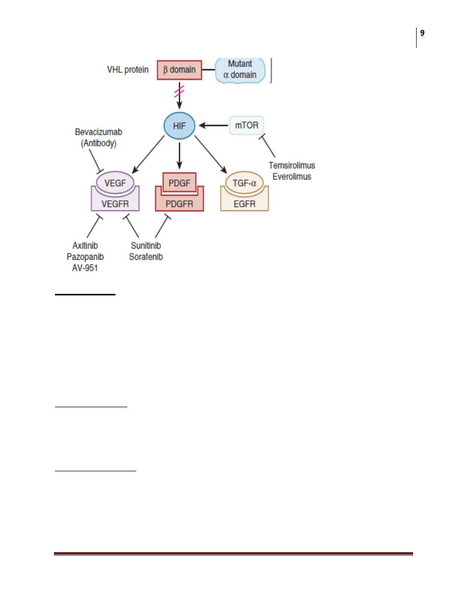
D-TARGETED MOLECULAR AGENTS
Antagonists of the Vascular Endothelial Growth Factor Pathway.
Inhibitors of the Mammalian Target of Rapamycin.
Surgery
Renal parenchymal Neoplasms
Dr. Saad Dakhil
Lec. 31
Prognosis
5-year survival rates for patients with stage T1-T2 disease in the 80–100%
range, with stage T3 in the 50–60% range. Patients presenting with
metastatic disease have a poorer prognosis, with only a 16–32% 5-year
survival rate.
BENIGN TUMORS
Renal Adenomas
The adenoma is the most common benign renal parenchymal lesion . These are
small, well-differentiated glandular tumors of the renal cortex. They are typically
asymptomatic and usually identified incidentally.
Renal Oncocytoma
Renal oncocytoma has a spectrum of behavior ranging from benign to
malignant.
An estimated 3–5% of renal tumors are oncocytomas.
The diagnosis of oncocytoma is predominantly pathologic because there are
no reliable distinguishing clinical characteristics.
Surgery
Renal parenchymal Neoplasms
Dr. Saad Dakhil
Lec. 31
Angiomyolipoma
(Renal Hamartoma)
Angiomyolipoma is a rare benign tumor of the kidney .
Angiomyolipomas are found in approximately 45–80% of patients with
tuberous sclerosis and are typically bilateral and asymptomatic.
Patients with lesions >4 cm with moderate or severe symptoms (bleeding or
pain) should undergo renal-sparing surgery or renal arterial embolization.
SECONDARY RENAL TUMORS
The kidney is a frequent site for metastatic spread of both solid and
hematologic tumors.
The most frequent primary site of cancer was lung (20%), followed by breast
(12%), stomach (11%), and renal (9%).
invasion by lymphoma to be 0.5–7%, with the rates of Hodgkin’s and non-
Hodgkin’s lymphoma distributed equally.
Surgery
Renal parenchymal Neoplasms
Dr. Saad Dakhil
Lec. 31
Renal Cyst