
Periocular lesion in an infantPICTURE QUIZ
BMJ 28 November 2011د. حسين محمد جمعه
اختصاصي الامراض الباطنة
البورد العربي
كلية طب الموصل
2012
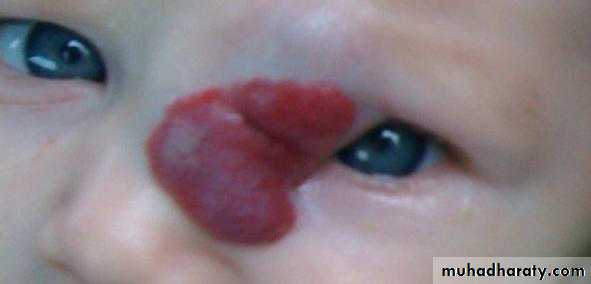
A 3 month old girl presented to the dermatology department with an enlarging lesion over the left lateral bridge of her nose (figure). The lesion started to develop three weeks after birth
as a small red patch and subsequently grew in size. The lesion extended to the left upper eyelid, with partial obscuration of theline of sight. The girl was born at full term after a normal pregnancy and delivery. She was otherwise well and not taking any regular drugs.
Questions
1 What is the diagnosis?
2 What is the natural course of this condition?
3 Should this lesion be treated and, if so, why?
4 What are the treatment options?
Answers
What is the diagnosis?Short answer
The development of a superficial capillary haemangioma (strawberry naevus) with associated deep haemangioma in the first few weeks after birth is consistent with an infantile haemangioma.
Long answer
The patient has a superficial haemangioma (strawberry naevus) combined with a deep haemangioma that is causing elevation.Haemangiomas are a type of vascular tumour.
Infantile haemangiomas are the most common soft tissue tumours of infancy, occurring in 4-10% of infants under 1 year, and they
generally present as a flat red lesion in the first few weeks after birth. The most likely diagnosis in this case is therefore an infantile haemangioma.
Congenital haemangiomas (present at birth) are less common and are defined as rapidly involuting congenital haemangiomas or non-involuting congenital haemangiomas. Neonates with
multiple haemangiomas may have diffuse neonatal
haemangiomatosis, in which haemangiomas may be present systemically in the viscera, eyes, neurological system, and mouth.
Suspected cases require further investigation, most
commonly with conventional ultrasound or Doppler ultrasound.Haemangiomas that occur in a segmental distribution are more commonly associated with extracutaneous anomalies, such as PHACE syndrome (posterior fossa malformations, haemangiomas, arterial anomalies, coarctation of aorta, and eye anomalies) and PELVIS syndrome (perineal haemangioma with any of the following: external genital malformations,
lipomyelomeningocoele, vesicorenal abnormalities, imperforate anus, or skin tag).
What is the natural course of this condition?
Short answer
Infantile haemangiomas follow a variable period of rapid growth for three to nine months but then stabilise and begin to involute.
Regression occurs at approximately 10% a year but can be variable, with some haemangiomas showing much slower regression.
Long answer
Infantile haemangiomas are developmental vascular tumours that are related to the date of conception of the infant rather than their postnatal age. They consist of a clonal expansion of endothelial cells, with the proliferative rapid growth phase driven by growth factors such as vascular endothelial growthfactor and basic fibroblast growth factor.
As the lesions start to involute the concentrations of these growth factors fall and apoptosis of endothelial cells increases. In most cases (80%),
the lesion completely regresses, but some patients may be left with residual telangiectasia, atrophy, scarring, and depigmentation.
Should this lesion be treated and, if so,why?
Short answerThe indications for treatment are lesions at high risk sites (such as subglottic lesions), local complications (such as skin ulceration or bleeding), threat to function, and cosmetic disfigurement. Our patient’s periocular haemangioma presents a serious threat to function because partial obscuring of the visual axis may lead to the development of amblyopia. The haemangioma is also cosmetically disfiguring and should be treated.
Long answer
The best way to treat infantile haemangiomas is to allow them to resolve naturally, with no intervention, because this avoids the risk of systemic or local complications from treatment.However certain haemangiomas will need treatment:
• Lesions at high risk sites: haemangiomas occurring in the subglottic region may cause stridor and airway obstruction
• Lesions that pose a threat to function: facial haemangiomaspose the greatest risk to function; for example, our patient’s lesion posed a threat to the developing visual system (amblyopia)
• Lesions that cause local complications: ulcerating
haemangiomas may be painful or may bleed
• Lesions that cause cosmetic disfigurement: these may be associated psychological morbidity.
Periocular haemangiomas are the most common tumour of the eyelid in infants, and the incidence of secondary ophthalmic complications has been reported to be 46-80%.9 The main risk
is amblyopia (lazy eye) through lack of visual stimulation in the developing visual system.
This may be caused by visual deprivation as a result of obscuration of the visual axis, strabismic amblyopia through an induced squint, or refractive
amblyopia via mechanically induced corneal astigmatism. One group reported that a lesion of 1 cm or more in diameter is an important predictor of amblyopia. Patients should therefore be referred to an ophthalmologist for a careful assessment of vision, ocular motility, refractive error, and fundus.
What are the treatment options?
Short answerTreatment options include drugs, laser treatment, and local excision. The most commonly used drugs are oral steroids and more recently oral propranolol.
Long answer
Treatment options for infantile haemangiomas include drugs, laser treatment, and local excision (which may be performed to debulk a large haemangioma). Paediatric services should be involved in the care, particularly if systemic treatments areindicated.
First line drugs include propranolol and steroids. Second line agents include interferon alfa (the use of which must be balanced against the risk of neurotoxicity), vincristine (which requires central venous access and input from a paediatric oncologist or haematologist), thalidomide, and imiquimod.
Until recently, oral steroids were the mainstay of
treatment—usually prednisolone at a dose of 1-4 mg/kg. Results are good—data from a meta-analysis indicate a response rate of 84% over a mean of 1.8 months. However, oral steroids are associated with serious side effects in the growing infant including hypertension, growth suppression, candida infection,gastric irritation, and immunosuppression (live vaccines should not given). Intralesional steroids can be used and were previously the treatment of choice for superficial periocular haemangiomas.
Propranolol was incidentally found to cause resolution of infantile haemangioma when used in infants for other clinical conditions, and this was first reported in 2008. In both index cases the authors observed rapid softening and colour changes
in the superficial haemangiomas and ultimately complete regression. Propranolol is now often the first line choice for infantile haemangioma, including periocular lesions, where most cases (50-59%) report a reduction in size of 50% or more.
Non-selective β adrenoceptor inhibition has been
suggested to reduce vasodilation, reduce angiogenesis, and increase apoptosis within the infantile haemangioma. Potential side effects include bradycardia, hypotension, bronchospasm,and neonatal hypoglycaemia. Some centres have published local protocols for the use of propranolol in infantile haemangioma, but no national guidelines have been produced to date.
Laser treatment in the form of pulsed dye laser is effective in superficial lesions and may be beneficial if the infantile haemangioma is ulcerated. Pulsed dye laser is also effective for the residual telangiectasia of a resolved infantile haemangioma
Patient outcome
Our patient was started on oral propranolol at a dose of 1 mg/kg in divided doses. The dose was increased to 2 mg/kg in divided doses after one week with no systemic side effects. After two weeks of treatment a dramatic reduction was seen in the size and colour of the haemangioma. Treatment was continued until the effect reached a plateau, after which the dose was tapered and then the drug stopped.