1
Fifth stage
Radiology
Lec-6
د.محمد
2/3/2016
Chest imaging
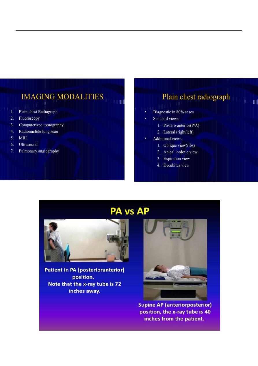
2
3
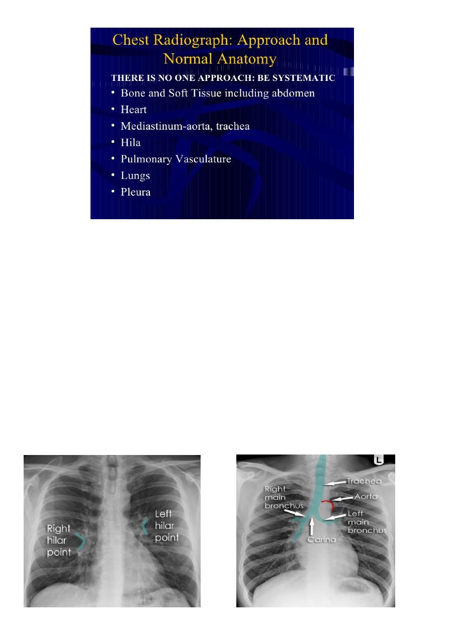
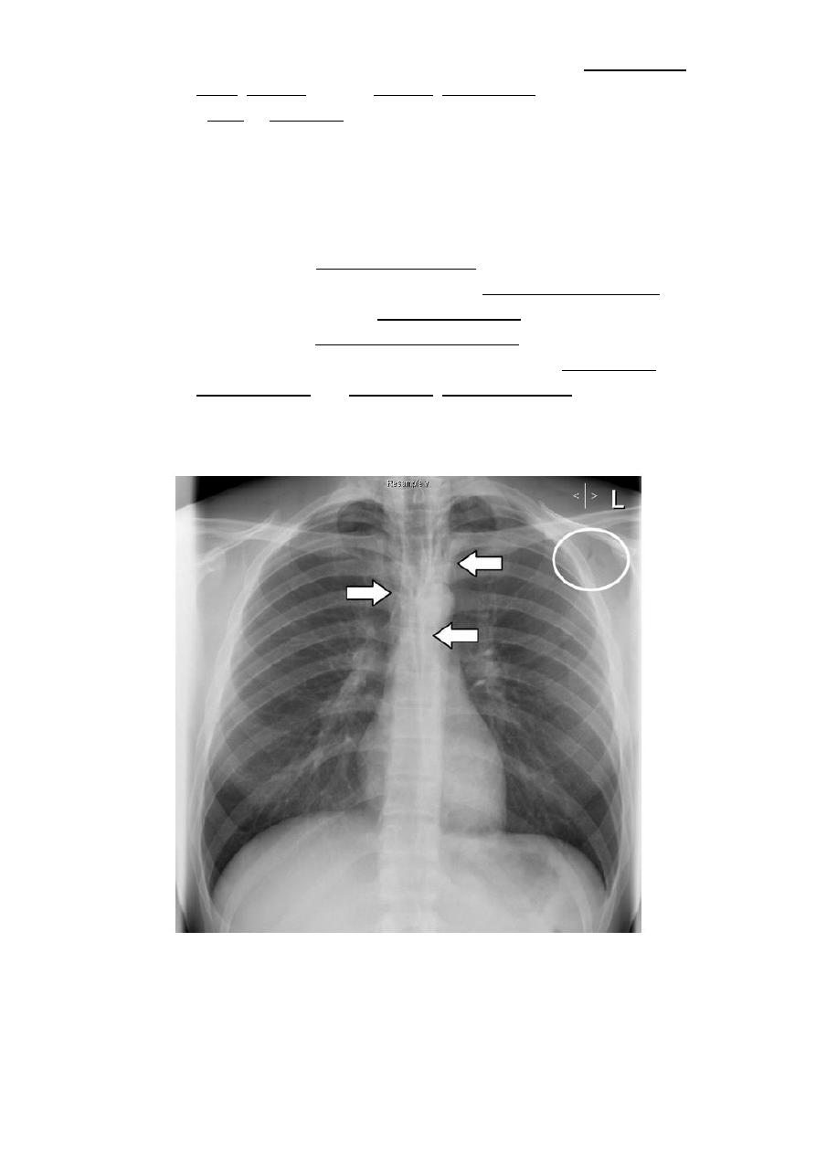
Hilar structures
The hila (lung roots) are complicated structures mainly consisting of the major bronchi and
the pulmonary veins and arteries. These structures pass through the narrow hila on each
side and then branch as they widen out into the lungs. The hila are not symmetrical but
contain the same basic structures on each side.
Key points
Each hilum contains major bronchi and pulmonary vessels
There are also lymph nodes on each side(not visible unless abnormal)
The left hilum is often higher than the right
Both hila should be of similar size and density. If either hilum is bigger and more dense, this
is a good indication that there is an abnormality.
4
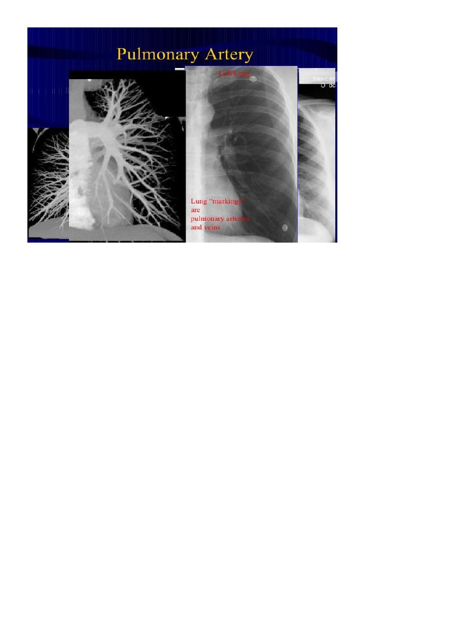
Lung markings
reflects
pulmonary
vasculature
Soft tissues
The soft tissues are often overlooked when viewing a chest x-ray, however, abnormalities
of the soft tissue may give important clues to a diagnosis. Whenever you look at a chest x-
ray, have a look at the soft tissues, especially around the neck, the thoracic wall, and the
breasts.
Soft tissue fat
This close-up demonstrates a normal fat plane between layers of muscle. Fat is less dense
than muscle and so appears blacker.
Note that the edge of fat is smooth. Irregular areas of black within the soft tissues may
represent air tracking in the subcutaneous layers. This is known as surgical emphesyma.
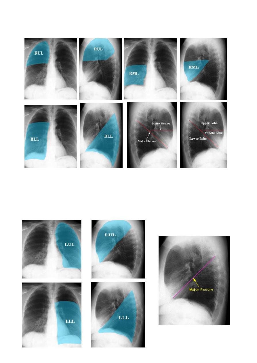
The left
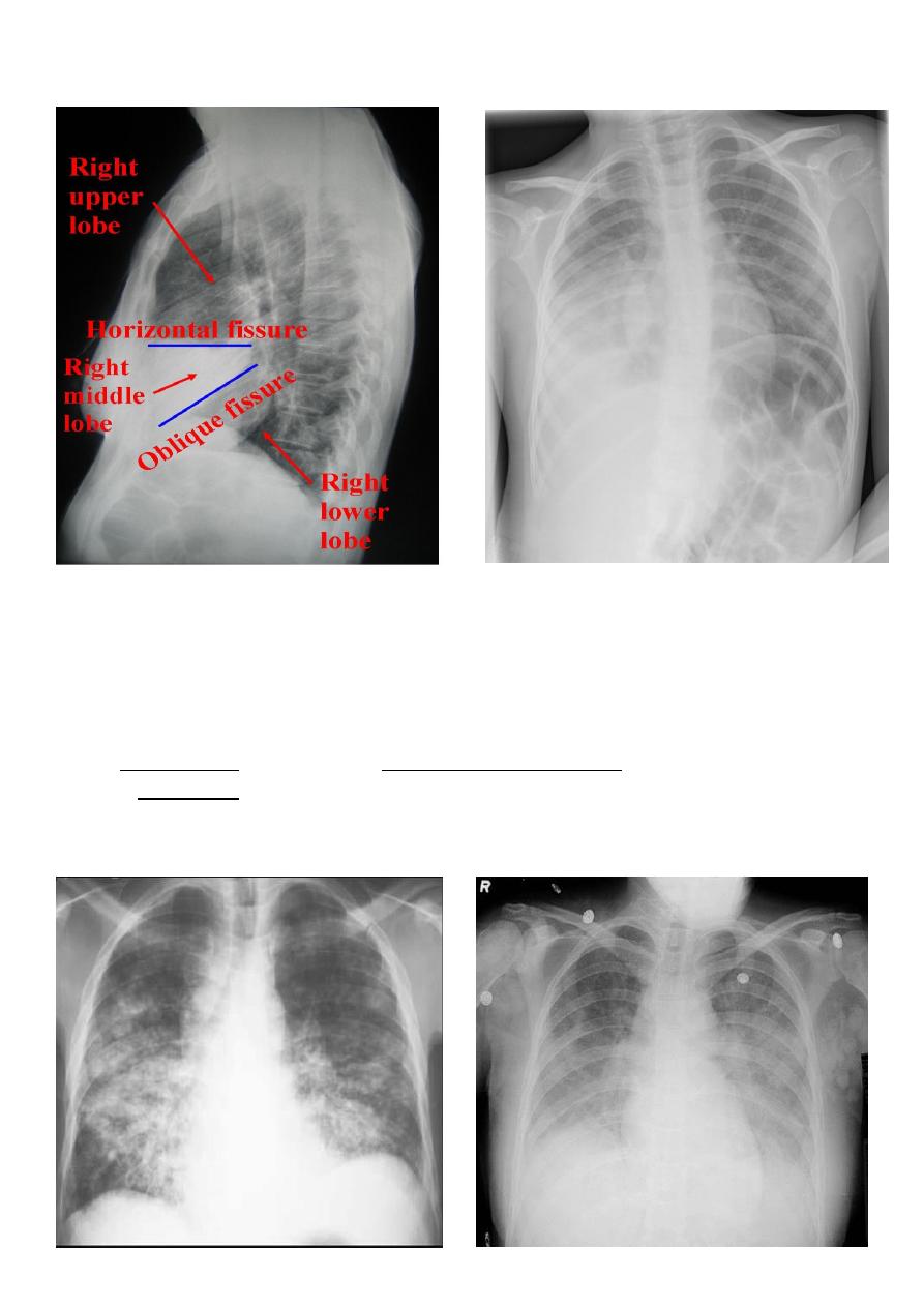
The left lung has two lobes and the right has three
Each lobe has its own pleural covering
The horizontal fissure (right) is often seen on a normal frontal view
The oblique fissures are often seen on a normal lateral view .
Lobes and fissures
This cut-out of a lateral chest x-ray shows the positions of the lobes of the right lung
On the left the oblique fissure is in a similar position but there is usually no horizontal
fissure, and so there are only two lobes on the left.
5
Radiologic anatomy of the RT lung lobes
Radiologic anatomy of the LT lung lobes

6
Corner stone
1. Patch
a. . Consolidation
b. . Collpase
2. Mass
a. single. CA bronchus
b. multiple .metastases
c. multiple Hydatid cysts
3. Cavity
a. . Abscess
b. . Ruptured hydatid cyst
c. . TB cavity
Consolidation is a radiological sign that refers to non-specific air-space opacification on a
chest radiograph or chest CT. Many things can fill the alveolar spaces, including fluid (heart
failure), pus (pneumonia), blood (pulmonary haemorrhage) and cells (lung cancer)
Radiographic features
Consolidated areas are radio opaque on chest radiograph and chest CT compared to
normally air filled lung tissue.
Lobar consolidation
Where increased density/opacity is seen in individual lung lobes. Sharp delineation can be
seen when consolidation reaches a fissure, since it does not cross. Air bronchograms can
also be seen due to bronchi becoming visible against the dense diseased tissue. Volume
loss is usually not seen..
Multi-focal consolidation
Multiple areas of opacity seen throughout the lung most often is due to
bronchopneumonia, starting from bronchi and spreading outwards. Usually ill defined with
peripheral distribution. Neoplasms such as a primary malignancy or metastasis can also
cause this picture.
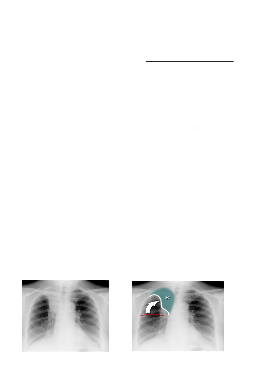
Right upper lobe consolidation
RUL consolidation will be seen as an increased opacity within the right upper lobe. Opacity
may be sharply bordered by the horizontal fissure
Some loss of outline of the upper right heart border may be apparent

7
Radiological sign in chest radiograph
1. Dense opacity seen above the horizontal fissure.
2. Air-bronchogram line
3. The lower border of the consolidation is sharply delinated by the horizontal fissure
suggesting it lies in the anterior segment of the RUL
Right middle lobe consolidation
The right middle lobe is bordered superiorly by the horizontal fissure, and medially by the
right heart border. Any abnormality, which increases density of this lobe, may therefore
obscure the right heart border, or be limited superiorly by the horizontal fissure.
Radiographic features
1. Features of right middle lobe (RML) consolidation on CXR include:
2. opacification of the RML abutting the horizontal fissure
3. indistinct right heart border
4. loss of the medial aspect of the right hemidiaphragm
5. air bronchograms
Right lower lobe consolidation
manifests as airspace shadowing that abuts the right hemidiaphragm,
obliterating the crisp margin of the hemidiaphragm and normal aerated lung.
bulging fissure sign refers to lobar consolidation where the affected portion of the lung is
expanded. It is now rarely seen due to the widespread use of antibiotics.
The most common infective causative agents are :
Klebsiella pneumoniae
The films in the next page
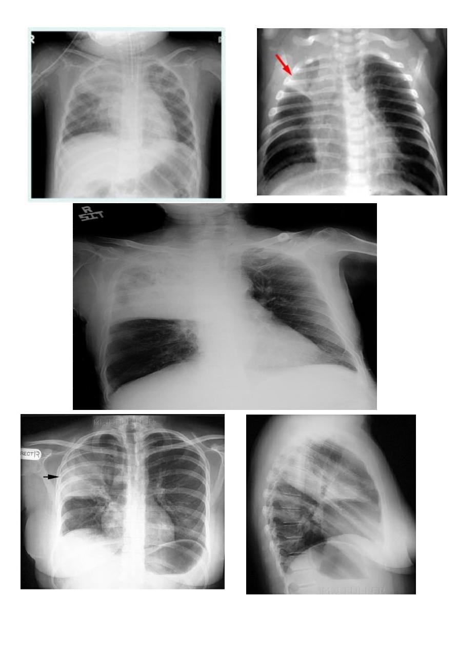
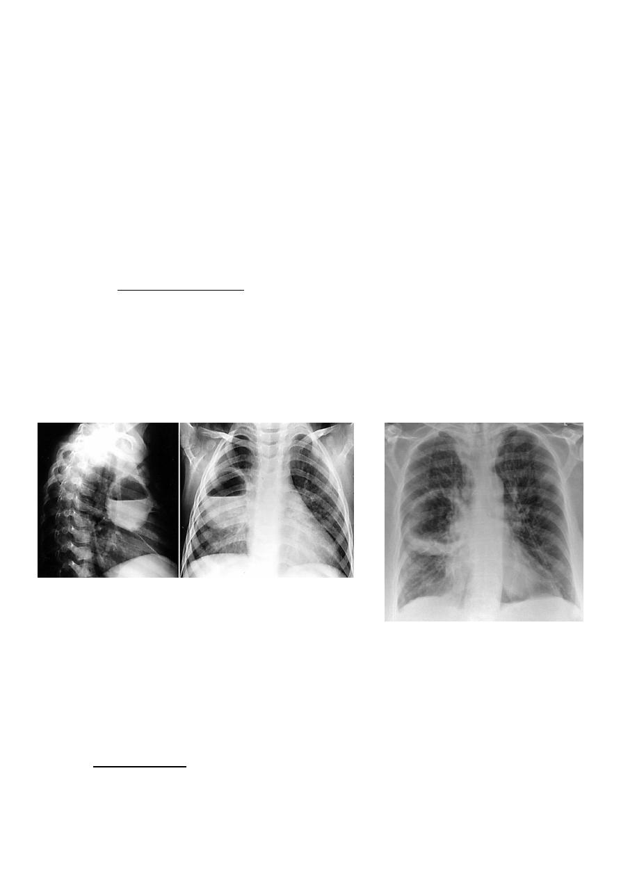
8
RTULConsolidation
9
Klebsiella (Friedlander's) pneumonia: the bulging fissure sign.
bulging fissure sign
RT middle lobe consolidation
10
RML consolidation RT lower lobe consolidation
Bronchopneumonia
Bronchopneumonia (also sometimes known as lobular pneumonia ) is a radiological
pattern associated with suppurative peribronchiolar inflammation and subsequent
patchy consolidation of one or more secondary lobules of a lung in response to a
bacterial pneumonia.

11
Lobar lung collapse
Lobar collapse refers to the collapse of an entire lobe of the lung. As such it is a
subtype of atelectasis (although collapse is not entirely synonymous is
atelectasis), which is a more generic term for 'incomplete expansion'. Individual
lobes of the lung may collapse due to obstruction of the supplying bronchus.
Causes include:
1. luminal
a. aspirated foreign material
b. mucous plugging
2. mural
3. extrinsic
a. compression by adjacent mass
Radiographic features
Radiograph
The appearance on chest x-ray varies according to the lobe involved and are
discussed separately:
Some features, however, are generic markers of volume loss and are helpful in
directing ones attention to the collapse, as well as enabling distinction from
opacification of the lobe without collapse (e.g. lobar pneumonia). These
features include
5
:
1. elevation of the ipsilateral hemidiaphragm
2. crowding of the ipsilateral ribs
3. shift of the mediastinum towards the side of atelectasis
4. crowding of pulmonary vessels or air bronchograms

12
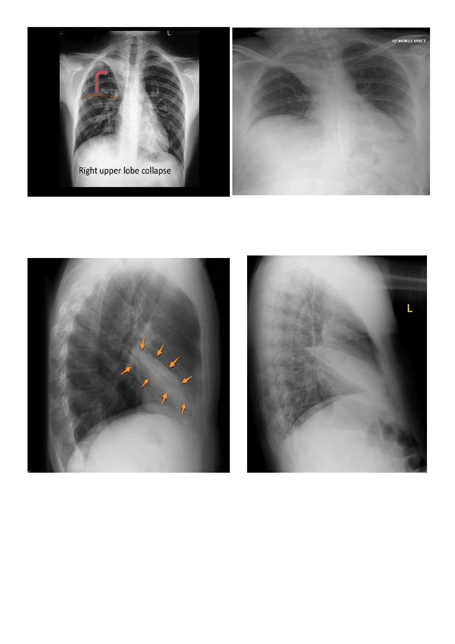
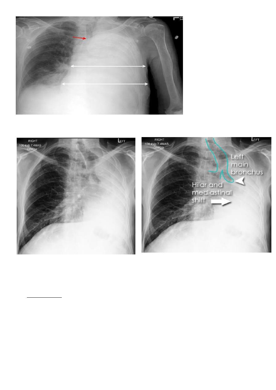
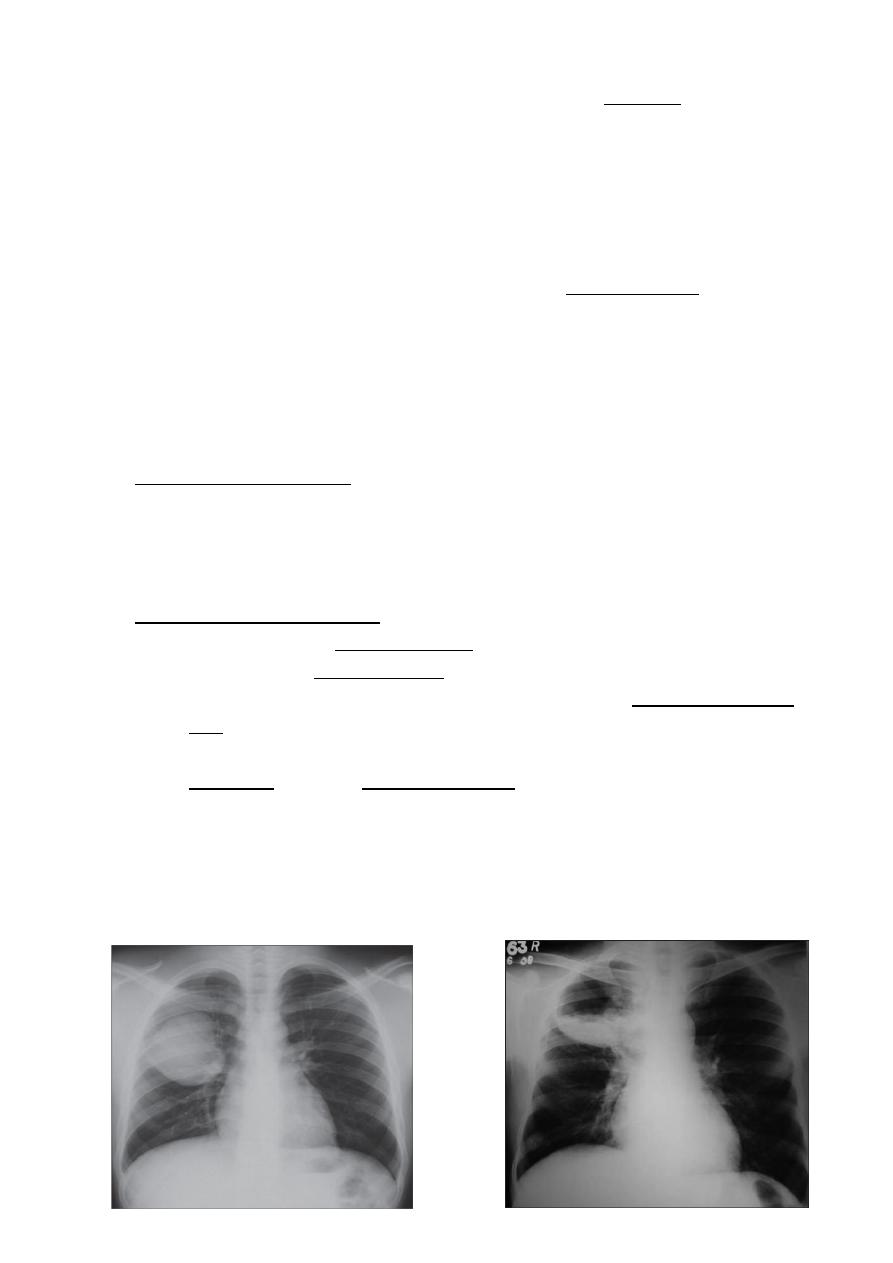
Right upper lobe collapse
has distinctive features, and is usually easily identified on
frontal chest radiographs .
Radiographic features
Chest radiograph
1. Collapse of the right upper lobe is usually relatively easy to identify on
frontal radiographs. Features consist of :
2. increased density in the upper medial aspect of the right hemithorax
3. elevation of the horizontal fissure
4. loss of the normal right medial cardiomediastinal contour
5. elevation of the right hilum
6. hyperinflation of the right middle and lower lobe result in increased
translucency of the mid and lower parts of the right lung
7. right juxtaphrenic peak
8. A common cause of lobar collapse is a hilar mass. When a right hilar
mass is combined with collapse of the right upper lobe, the result is an S
shape to elevated horizontal fissure. This is known as Golden S sign .
9. Non-specific signs indicating right sided atelectasis are also usually
present including:
10. elevation of the hemidiaphragm
11. crowding of the right sided ribs
12. shift of the mediastinum and trachea to the right
Right middle lobe collapse
has distinctive features, and is usually relatively easily
identified.
Radiographic features
Chest radiograph
Frontal chest XR showing opasity cause obscuration of the RT cardiac border
Lateral chest XR film the opacity is tongue like shape
versus (triangular in shape) in RT middle lobe consolidation seen in lateral chest XR film

13
RT lower lobe collapse
usually the medial aspect of the dome of right hemidiaphragm is lost.
the right hilum is depressed
It is important to note that the right heart border, which is contacted by the right
middle lobe remains well seen.
Non-specific signs indicating right sided atelectasis may also be present (although
due to the small size of the right middle lobe they may well be subtle). They include:
elevation of the hemidiaphragm
crowding of the right sided ribs
shift of the mediastinum to the right
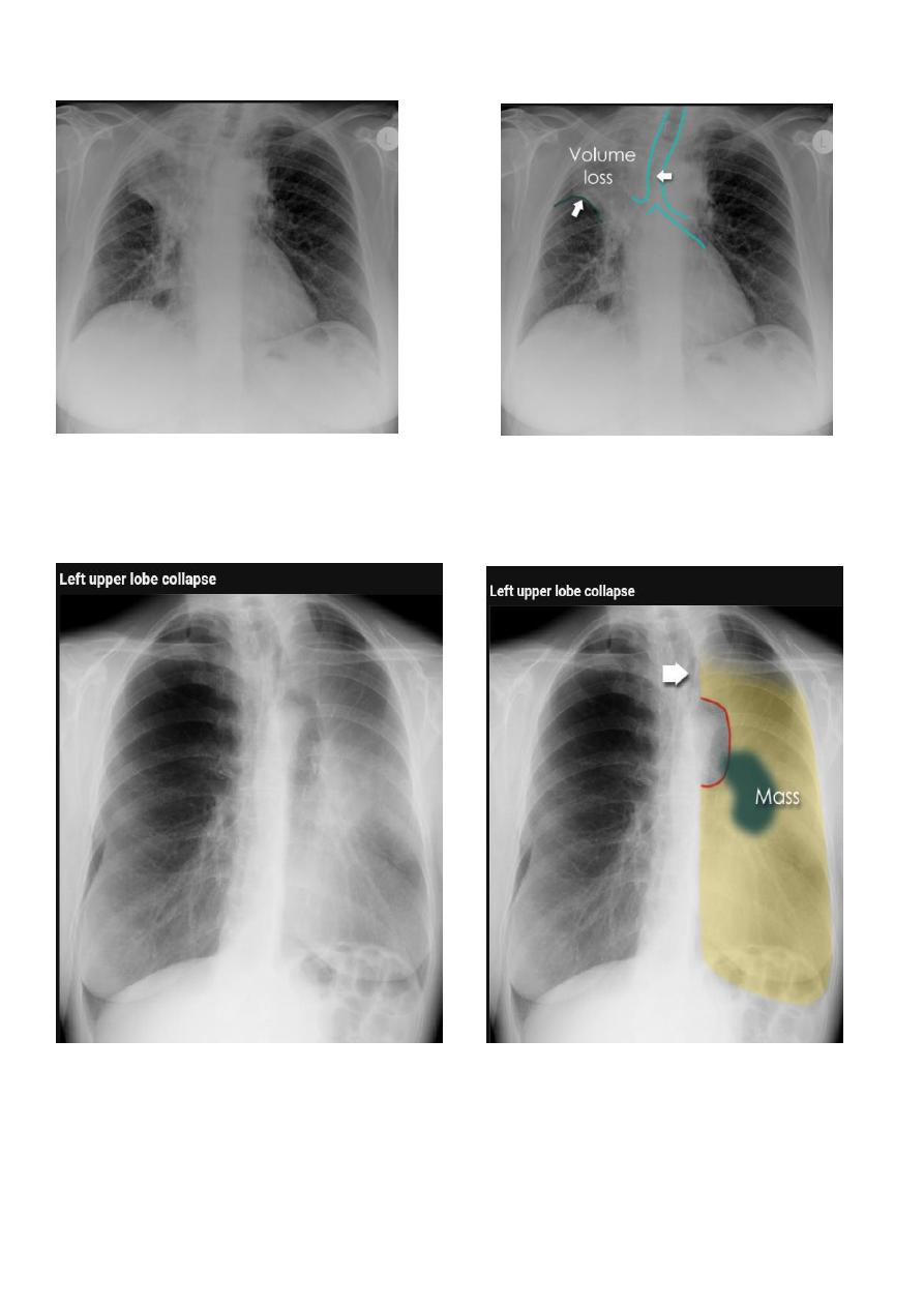
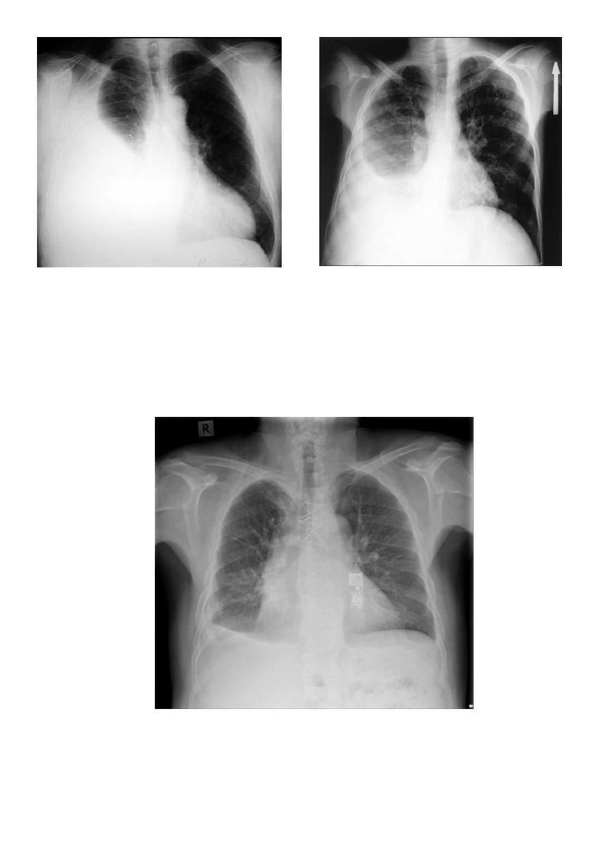
Left upper lobe collapse has distinctive features but can be challenging to identify on chest
radiographs by the uninitiated.
Radiographic features
1. The left upper lobe collapses anteriorly becoming a thin sheet of tissue apposed to
the anterior chest wall, and appears as a hazy or veiling opacity extending out from
the hilum and fading out inferiorly . It thus reverses the normal slight increase in
radiographic density seen as you move down the lung (due to increased thickness of
the chest soft tissues).
2. Parts of the normal cardiomediastinal contour may also be obliterated where the left
upper lobe, particularly the lingula abut the left heart border. The anterior parts of
the aortic arch are also often obliterated from view.
3. In some cases the hyperexpanded superior segment of the left lower lobe insinuates
itself between the left upper lobe and the superior mediastinum, sharply silhouetting
the aortic arch and resulting in a lucency medially. This is known as the luftsichel
sign.
4. The left hilum is also drawn upwards, resulting in an almost horizontal course of the
left main bronchus and vertical course of the left lower lobe bronchus.
5. Non-specific signs indicating left sided atelectasis will also be present, including:
6. elevation of the hemidiaphragm
7. 'peaked' or 'tented' hemidiaphragm: juxtaphrenic peak sign
8. crowding of the left sided ribs
9. shift of the mediastinum to the left
14
10. On lateral projections the left lower lobe is hyperexpanded and the oblique fissure
displaced anteriorly. There is associated increase in the retrosternal opacity.
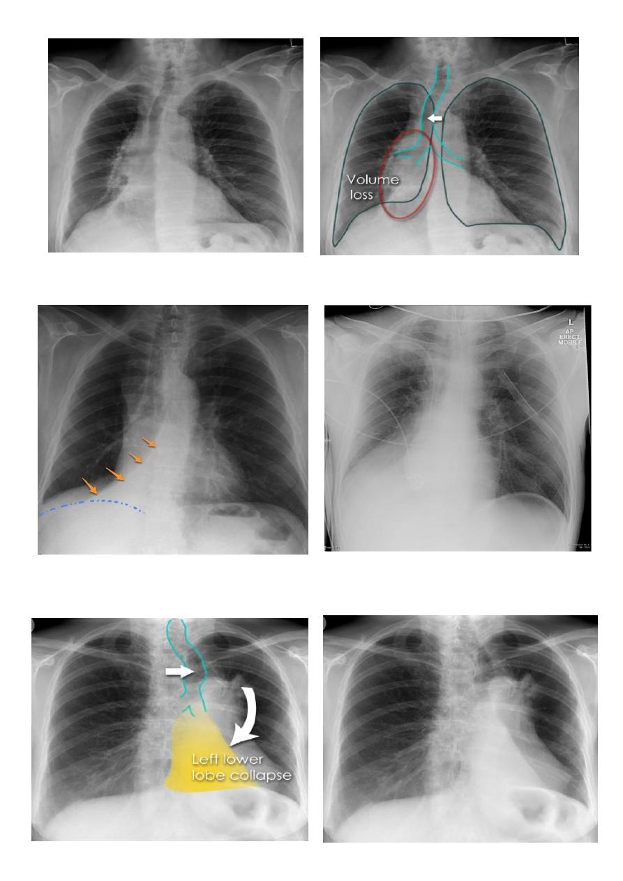
Left lower lobe collapse has distinctive features, and can be readily identified on frontal
chest radiographs, provided attention is paid to the normal cardiomediastinal contours.
The shadow cast by the heart does however make it harder to see than the right lower lobe
collapse
Radiographic features
Left lower lobe collapse
is readily identified in a well penetrated film of a patient with normal sized heart, but can
be challenging in the typical patient with collapse, namely unwell patients, with portable
(AP) often under-penetrated films, often with concomitant cardiomegaly. Features to be
observed include
:
1. triangular opacity in the posteromedial aspect of the left lung
2. edge of collapsed lung may create a 'double cardiac contour'
3. left hilum will be depressed
4. loss of the normal left hemidaphgragmatic outline
5. loss of the outline of the descending aorta
6. Non-specific signs indicating left sided atelectasis are usually also be present
including:
7. elevation of the hemidiaphragm
8. crowding of the left sided ribs
9. shift of the mediastinum to the left
10. On lateral projection the left hemidiaphragmatic outline is lost posteriorly and the
lower thoracic vertebrae appear denser than normal (they are usually more
radiolucent than the upper vertebrae) .
15
RT ULC
LT L L collapse
16
RT middle lobe collapse
17
RT L L collapse
LT L L collapse
18
Total lung collapse
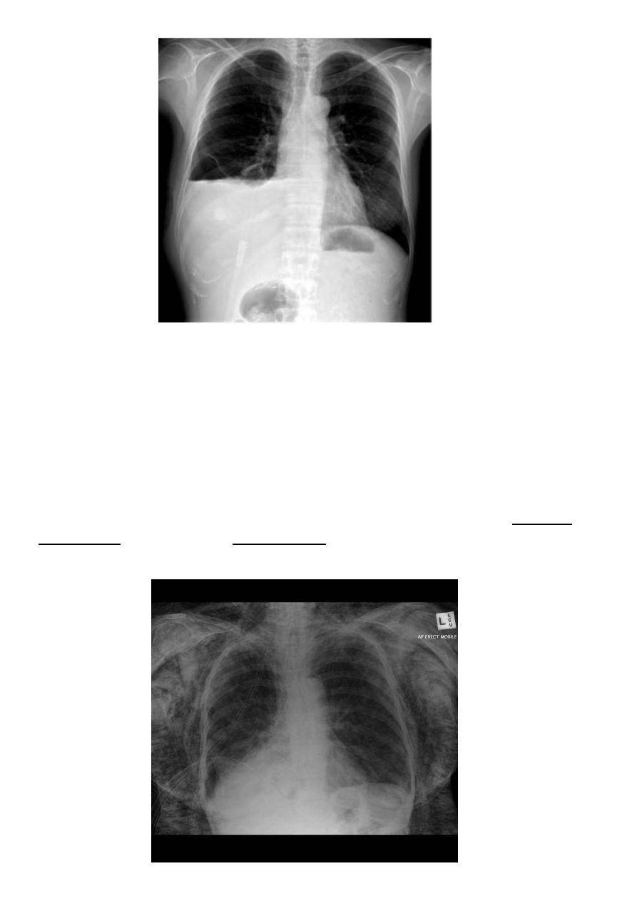
Pleural effusion
Pleural effusion tends to be used as a catch-all term denoting a collection of fluid within
the pleural space. This can be further divided into exudates and transudates depending on
the biochemical analysis of aspirated pleural fluid. Essentially it represents any pathological
process which overwhelms the pleura's ability to reabsorb fluid.
Radiographic appearances
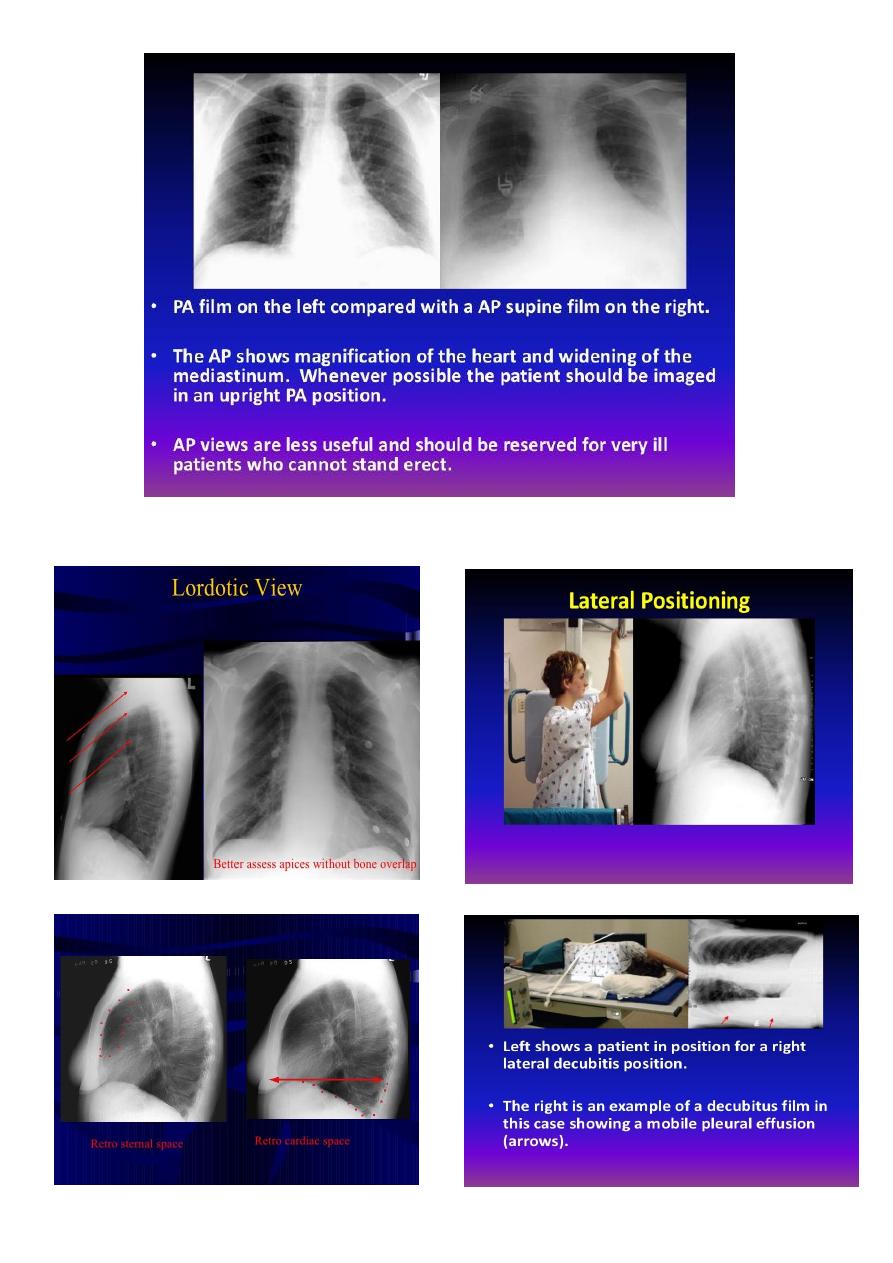
Plain radiograph
Chest radiographs are the most commonly used examination to assess for presence of a
pleural effusion, however it should be noted that on a routine erect chest x-ray as much as
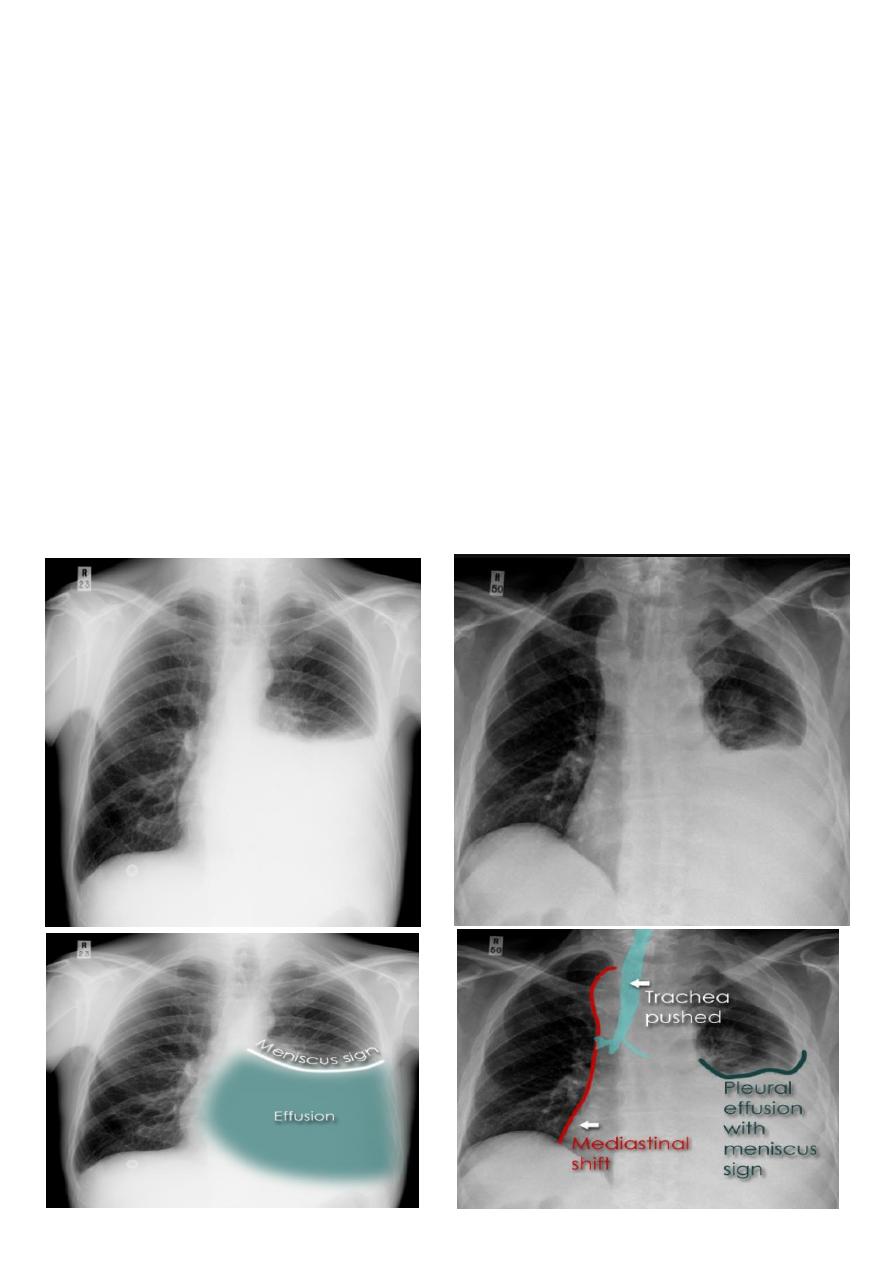
19
250-600 ml of fluid is required before it becomes evident
6
. A lateral decubitus film is most
sensitive, able to identify even a small amount of fluid. At the other extreme, supine films
can mask large quantities of fluid.
CXR (lateral decubitus)
A lateral decubitus film (obtained with the patient lying on their side, effusion side down,
with a cross table shoot through technique) can visualise small amounts of fluid layering
against the dependent parietal pleura.
CXR (erect)
Both PA and AP erect films are insensitive to small amounts of fluid. Features include:
blunting of the costophrenic angle
blunting of the cardiophrenic angle
fluid within the horizontal or oblique fissures
eventually a meniscus will be seen, on frontal films seen laterally and gently sloping
medially
20
A subpulmonic effusion (infrapulmonary effusion) may be seen when there is previously
established pulmonary disease, but can also be encountered in normal lungs , They are
more common on the right, and usually unilateral
21
with large volume effusions, mediastinal shift occurs away from the effusion (note: if
coexistent collapse dominates then mediastinal shift may occur towards the effusion)
An empyema can resemble a pleural effusion
and can mimic a peripheral pulmonary abscess, although a number of features usually
enable distinction between the two Features that help distinguish a pleural effusion from
an empyema include:
Shape and location
Empyemas usually:
form an obtuse angle with the chest wall
unilateral or markedly asymmetric whereas pleural effusions are (if of any significant size)
usually bilateral and similar in size .
lenticular in shape (bi-convex), whereas pleural effusions are crescentic in shape (i.e
concave towards the lung)
Empyema
22
Lung abscess
is a circumscribed collection of pus within the lung, is are potentially life
threatening. They are often complicated to manage and difficult to treat
Lung abscesses are divided according to their duration into acute (< 6 weeks) and
chronic (> 6 weeks) .
A primary abscess is one which develops as a result of primary infection of the lung. They
most commonly arise from aspiration, necrotising pneumonia or chronic pneumonia, e.g.
pulmonary tuberculosis
Some organisms are particularly prone to causes significant necrotising pneumonia
resulting in cavitation and abscess formation. These include :
Staphylococcus aureus
Klebsiella sp: Klebsiella pneumonia
Pseudomonas sp
Plain film
The classical appearance of a pulmonary abscess is a cavity containing an air-fluid level. In
general abscesses are round in shape, and appear similar in both frontal and lateral
projections.
Very important
Empyema vs pulmonary abscess
1. relationship to adjacent bronchi / vessels
a) abscesses will abruptly interrupt bronchovascular structures
b) empyema will usually distort and compress adjacent lung
2. split pleura sign thickening and separation of visceral and parietal pleura is a sign
of empyema
3. abscesses have thick irregular walls
empyema are usually smoother
23
4. angle with pleura
a) abscesses usually have an acute angle (claw sign)
b) empyema have obtuse angles
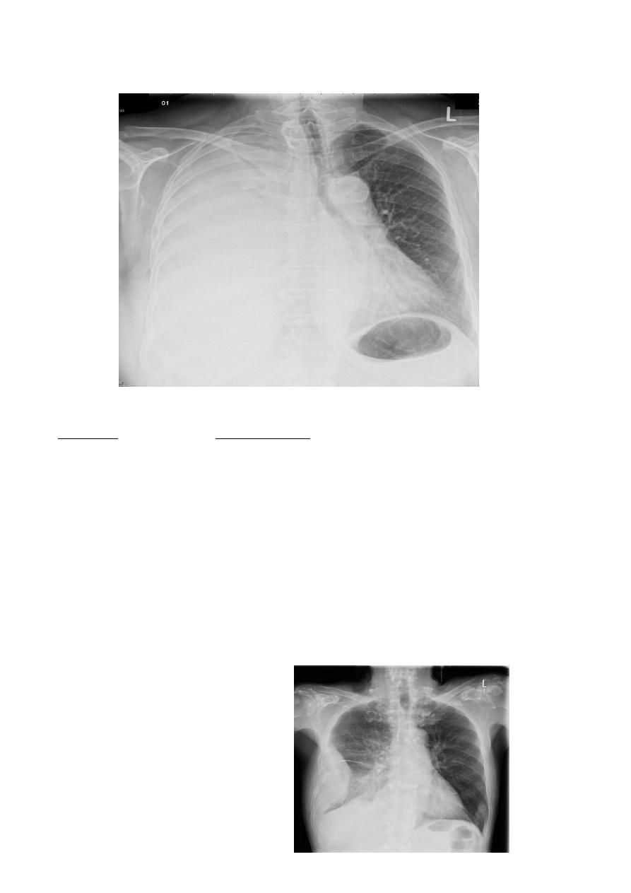
Hydatid cysts result from infection by the Echinococcus, and can result in cyst formation
anywhere in the body. Humans are accidental host and the infection occurs by ingesting
food contaminated with Echinococcus eggs ,
Pulmonary hydatid infection is a common manifestation of hydatid disease.
The lung is the second most common site of involvement
with echinococcosis granulosus in adults after the liver (10-30% of cases), and the most
common site in children. The coexistence of liver and lung disease is present in only 6% of
patients .
Chest XR features include :
1. Non-complicated hydatid
a. multiple or solitary rounded opacity
b. diameter of 1-20 cm
c. unilateral or bilateral
d. predominantly found in the lower lobes
2. Complicated cysts may show:
a. meniscus sign or air crescent sign
b. cumbo sign or onion peel signThe onion peel sign (also called the cumbo
sign) is a feature seen with complicated pulmonary hydatid
cyst in which air lining between the endocyst and pericyst has the
appearance of an onion
c. water-lily is seen in hydatid infections when there is detachment of the
endocyst membrane which results in floating membranes within the
pericyst that mimic the appearance of a water lily.
d. Consolidation adjacent to the cyst (ruptured cyst)
Simple H.C Ruptured H.C
24
Pneumothorax
Pneumothorax refers to the presence of gas (air) in the pleural space. When this collection
of gas is constantly enlarging with resulting compression of mediastinal structures it can be
life-threatening and is known as a tension pneumothorax
It is useful to divide pneumo thoraces into three categories :
primary spontaneous: no underlying lung disease marfan syndrum , Elher danus syndrome
alpha-1 antitrypsin deficiency
secondary spontaneous: underlying lung disease is present
iatrogenic/traumatic
Radiographic features
Chest radiograph
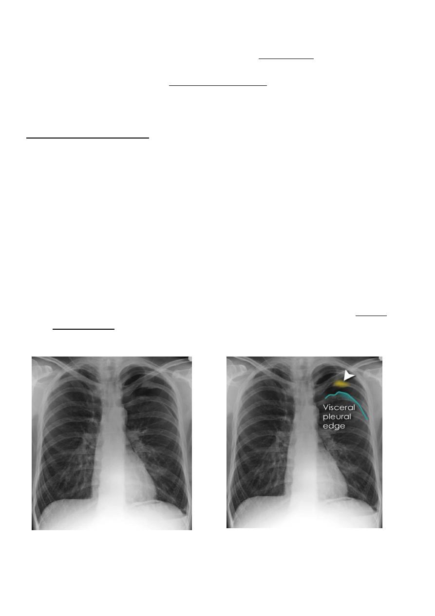
1. A pneumothorax is, when looked for, usually relatively easily appreciated. Typically
they demonstrate:
2. visible visceral pleural edge see as a very thin, sharp white line
3. no lung markings are seen peripheral to this line
4. the peripheral space is radiolucent compared to adjacent lung
5. the lung may completely collapse
6. the mediastinum should not shift away from the pneumothorax unless a tension
pneumothorax is present
25
A tension pneumothorax
A tension pneumothorax occurs when intrapleural air accumulates progressively in such a
way as to exert positive pressure on mediastinal and intrathoracic structures. It is a life
threatening occurrence requiring rapid recognition and treatment is required if
cardiorespiratory arrest is to be avoided.
Radiographic features
1. A pneumothorax will have the same features as a run-of-the-mill pneumothorax with
a number of additional features, helpful in identifying tension. These additional signs
indicate over expansion of the hemithorax:
2. ipsilateral increased intercostal spaces
3. shift of the mediastinum to the contralateral side
4. depression of the hemidiaphragm
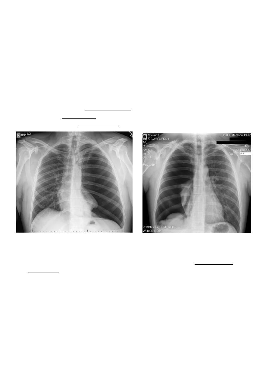
Hydro pnuemothorax
Hydropneumothorax is a term given to the concurrent presence of a pneumothorax as well
as a hydrothorax (i.e. air and fluid) in the pleural space.
Plain radiographs
On an erect chest radiograph, recognition of hydropneumothorax can be rather easy - and
is clasically shown as an air-fluid level. On the supine radiograph, this may be more
challenging where a sharp pleural line is bordered by increased opacity lateral to it within
the pleural space may sometimes suggest towards the diagnosis
The film in the next page
26
Hydropnemothorax
Subcutaneous Emphysema
Subcutaneous emphysema, strictly speaking, refers to air in the subcutaneous tissues. But
the term is generally used to describe any soft tissue emphysema of the body wall or limbs,
since the air often dissects into the deeper soft tissue and musculature along fascial planes .
Radiographic appearance
Plain film
If affecting the anterior chest wall, subcutaneous emphysema can outline the pectoralis
major muscle, giving rise to the ginkgo leaf sign , dissecting air along tissue fat planes
appears as multiple lines of lucency.
27
Pneumomediastinum is the presence of extraluminal gas within the mediastinum. Gas may
originate from the lungs, trachea, central bronchi, oesophagus, and track from the
mediastinum to the neck or abdomen
Radiographic features
1. Small amounts of air appear as linear or curvilinear lucencies outlining mediastinal
contours such as:
2. subcutaneous emphysema
3. air anterior to pericardium: pneumopericardium
4. air around pulmonary artery and main branches: ring around artery sign
5. air outlining major aortic branches: tubular artery sign
6. air outlining bronchial wall: double bronchial wall sign
7. continuous diaphragm sign: due to air trapped posterior to pericardium
8. air between parietal pleura and diaphragm: extrapleural sign