
Lecture 3
Thorax
د.رندعبداللطيف
Diaphragm
The diaphragm is a thin muscular and tendinous septum, dome shaped that
separates the chest cavity above from the abdominal cavity below. It is the most
important muscle of respiration. It consists of a peripheral muscular part, which
arises from the margins of the thoracic opening, and a centrally placed tendon.
The origin of the diaphragm can be divided into three parts:
1. Sternal part arising from the posterior surface of the xiphoid process.
2. Costal part arising from the deep surfaces of the lower six ribs and their
costal cartilages.
3. Vertebral part arising by vertical columns or crura and from the arcuate
ligaments.
The right crus arises from the sides of the bodies of the first three lumbar
vertebrae and the intervertebral discs; the left crus arises from the sides of the
bodies of the first two lumbar vertebrae and the intervertebral disc.
Lateral to the crura the diaphragm arises from the medial and lateral
arcuate ligaments
The two crura are connected by a median arcuate ligament
The diaphragm is inserted into a central tendon, which is shaped like three
leaves. Some of the muscle fibers of the right crus pass up to the left and
surround the esophageal orifice in a slinglike loop. These fibers appear to act as
a sphincter and possibly assist in the prevention of regurgitation of the stomach
contents into the thoracic part of the esophagus.
Nerve Supply of the Diaphragm
1. Motor nerve supply: The right and left phrenic nerves (C3, 4, 5).
2. Sensory nerve supply: The parietal pleura and peritoneum covering the
central surfaces of the diaphragm are from the phrenic nerve and the
periphery of the diaphragm is from the lower six intercostal nerves.
Openings in the Diaphragm:
i.
Caval: T8 , on the Rt of the central tendon, It transmits the inferior
vena cava and terminal branches of the right phrenic nerve.
ii.
Oesophageal: T10, in the fibers of the Lt crus. It transmits the
esophagus, the right and left vagus nerves, the esophageal branches of
the left gastric vessels, and the lymphatics from the lower third of the
esophagus.
iii.
Aortic: T12, in between the 2 crura. It transmits the aorta, the
thoracic duct, and the azygos vein.

Lecture 3
Thorax
د.رندعبداللطيف
Mediastinum
The mediastinum is divided into superior and inferior mediastina by an
imaginary plane passing from the sternal angle anteriorly to the lower border of
the body of the T4 posteriorly. The inferior mediastinum is subdivided by
pericardium into the middle mediastinum, which consists of the pericardium
and heart; the anterior mediastinum, which is a space between the pericardium
and the sternum; and the posterior mediastinum, which lies between the
pericardium and the vertebral column.
For purposes of orientation, it is convenient to remember that the major
mediastinal structures are arranged in the following order from anterior to
posterior:
1- Superior Mediastinum
(a) Thymus, (b) large veins, (c) large arteries, (d) trachea, (e) esophagus and
thoracic duct, and (f) sympathetic trunks.
2- Inferior Mediastinum
(a) Thymus, (b) heart within the pericardium with the phrenic nerves on each
side, (c) esophagus and thoracic duct, (d) descending aorta, and (e) sympathetic
trunks.
Thymus:
Is a flattened, bilobed gland related to the immune system of the body, well
developed in children &rudimentary in adults. It may extends to the superior
mediastinum. The thymus continues to grow until puberty but thereafter
undergoes involution. It has a pink, lobulated appearance and is the site for
development of T lymphocytes. It is supplied by inferior thyroid A. & internal
thoracic artery.
Trachea:
It is midline structure begins in the neck as a continuation of the larynx at the
lower border of the cricoid cartilage at the level of the 6th cervical vertebra and
ends below at the carina by dividing into right and left principal (main) bronchi
at the level of the sternal angle (opposite the disc between T4 and T5) n adults,
the trachea is about (11.25 cm) long and 1 inch (2.5 cm) in diameter. Its wall is
reinforced by C-shaped cartilagenous rings which open posteriorly, the gap is
being closed by a muscle strip (trachealis m.)
In the chest it is related anteriorly to the back of sternum, thymus gland,
L.Brachiocephalic v., arch of Aorta & L. Subclavian &left common carotid
arteries.
Posteriorly to the esophagus& L.recurrent laryngeal nerve ,azygos V.& Vagi
nerves.
Lecture 3
Thorax
د.رندعبداللطيف
Blood Supply of the Trachea
The upper two thirds are supplied by the inferior thyroid arteries and the lower
third is supplied by the bronchial arteries.
Lymph Drainage of the Trachea
The lymph drains into the pretracheal and paratracheal lymph nodes and the
deep cervical nodes.
Nerve Supply of the Trachea
The sensory nerve supply is from the vagi and the recurrent laryngeal nerves.
Sympathetic nerves supply the trachealis muscle.
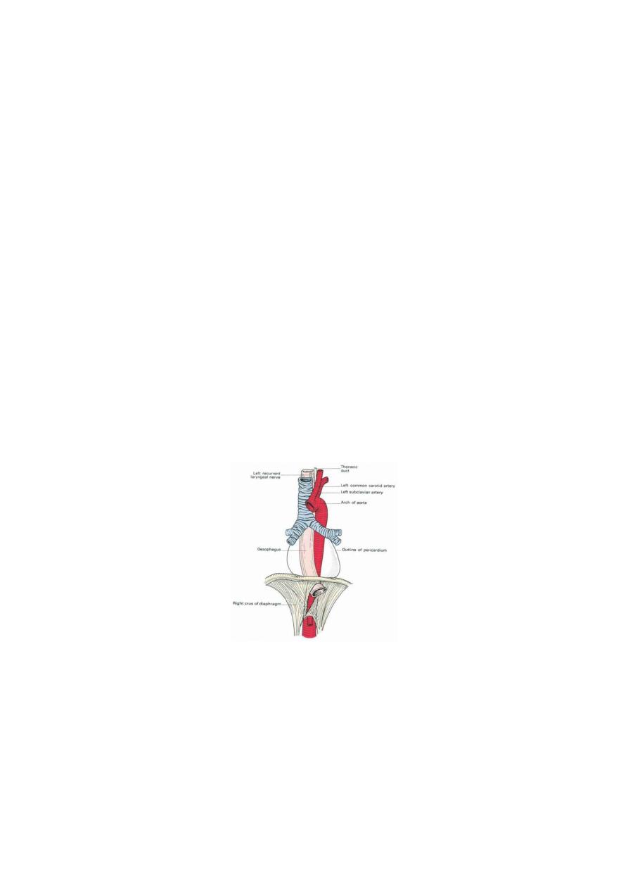
The esophagus:
It is muscular tube, 25 cm long, lies in the midline with tendency to incline to
the left side as it descends. It pierces the diaphragm to the left of the midline. It
starts at C6 level, and passes through the diaphragm at the level of the T10 to
join the stomach. It has 3 main constrictions, at C6, where the left main
bronchus passes in front of it compressing it & at the diaphragmatic orifice. The
esophagus is related anteriorly to trachea, left recurrent laryngeal n. It lies on the
Rt of the aortic arch & descending thoracic aorta, as the esoph. tends to attain Lt
position & the aorta tends to take midline position, the esopha. will pass anterior
to the thoracic aorta crossing it at the level of T8 vertebra.
Blood Supply of the Esophagus
The upper third of the esophagus is supplied by the inferior thyroid artery, the
middle third by esophageal branches from the bronchial arteries & descending
thoracic aorta, and the lower third by branches from the left gastric artery. The
veins from the upper third drain into the inferior thyroid veins, from the middle
third into the azygos veins, and from the lower third into the left gastric vein, a
tributary of the portal vein.
Lymph Drainage of the Esophagus
Lymph vessels from the upper third of the esophagus drain into the deep
cervical nodes, from the middle third into the superior and posterior mediastinal
Lecture 3
Thorax
د.رندعبداللطيف
nodes, and from the lower third into nodes along the left gastric blood vessels
and the celiac nodes.
Nerve Supply of the Esophagus
The esophagus is supplied by parasympathetic and sympathetic fibers via the
vagi and sympathetic trunks and the esophageal nerve plexus.
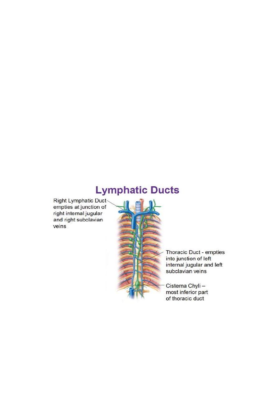
Thoracic Duct
The thoracic duct begins in the abdomen as a dilated sac, the cisterna chyli
which is formed in the posterior abdominal wall at L2 level by union of the
lumbar & intestinal lymph trunks. It ascends through the aortic opening in the
diaphragm, on the right side of the descending aorta. It then runs upward to
enter the root of the neck. Here, it bends laterally behind the carotid sheath and
in front of the vertebral vessels. It crosses the subclavian artery to enter the
beginning of the left brachiocephalic vein. At the root of the neck, the thoracic
duct receives the left jugular, subclavian, and bronchomediastinal lymph trunks,
although they may drain directly into the adjacent large veins. The thoracic duct
drains the lymph from the whole body except the right side above the
diaphragm which drain by Rt lymphatic duct.