Anatomy
For
Lower limbs
http://goo.gl/rjRf4F
I
LOKA
©
http://www.muhadharaty.com/anatomy-lower
I
Content
Topics:
Page:
Front of the thigh
3
Gluteal region
17
The back of the thigh
22
Hip joint
27
Front of the leg and dorsum of the foot
29
Dorsum of the foot
40
Knee Joint
48
Ankle joint
50

Part1
: Front of the thigh
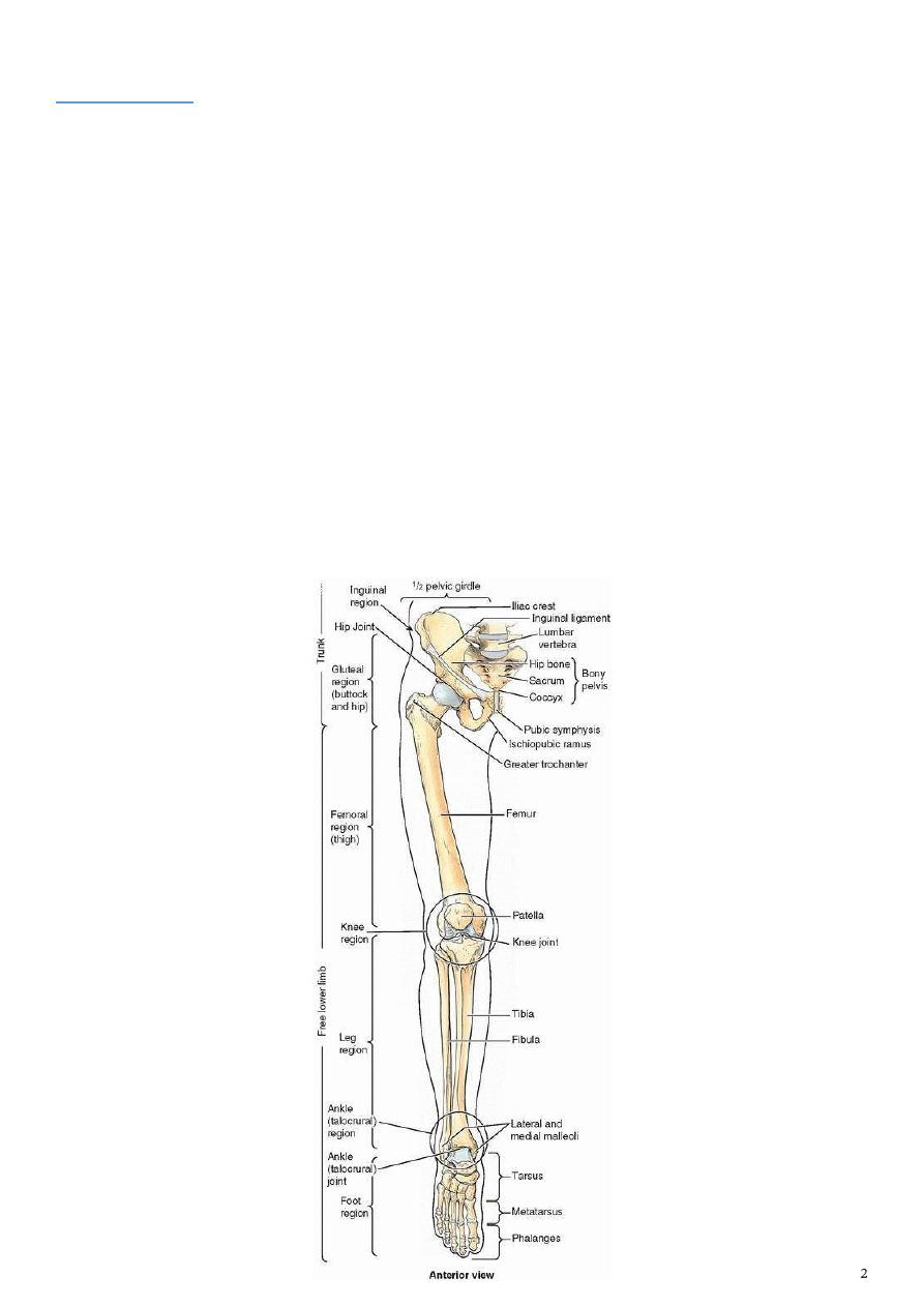
- The region of the thigh includes the area from the iliac crest to the knee
- The bones of the thigh are the hip and the femur.
Consist of : 1- a thick fatty superficial layer
2- and a deeper membranous layer.
a- The superficial layer is a loose fatty layer continuous with the similar layer of the
abdomen, the back, perineum, and the leg .
In the gluteal region the fat deposited in a thick layer which form the transverse fold of
the buttock.
b- The membranous layer of the superficial fascia of the anterior abdominal wall extends
into the thigh and is attached to the deep fascia (fascia lata) about finger breadth below
the inguinal ligament Superficial nerves, superficial vessel, & superficial inguinal lymph
nodes are present between these two layers
- the thigh is surrounded by thick strong fascia, like stock.
- It is thin anteriorly and medially while its thick laterally.
- the most thickened part is attached to iliac crest above and to the lateral tibial condyle
below known as the ilio-tibial tract .
- fascia lata fuses above to the inferior concave border of the inguinal ligament. ,
Fascia lata has three characteristic features :
1- laterally it forms a thick band extend from the iliac tubercle to the lateral tibial chondyle
called the iliotibial tract; Two muscles inserted in this tract, the greater part of the
gluteus maximus posteriorly and tensor fasciae latae anteriorly.
2- medially it forms an opening called saphenous opening
3- sends intermuscular septa to the linea aspera of the femur these septa separate the
thigh into 3 compartments
:
• Anteriorly and laterally the extensor compartment. Supplied by the femoral nerve.
• Medially the adductor compartment. Supplied by the obturator nerve.
• Posteriorly the muscles compartment (Hamstring muscles) supplied by the sciatic nerve.
The Superficial fascia
The deep fascia ( fascia lata)
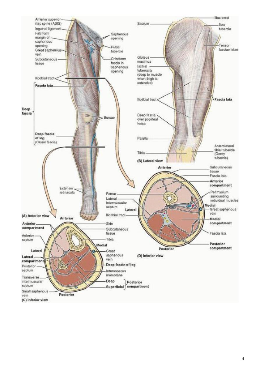

Saphenous opening
1- It is an oval opening in the deep fascia(fascia lata) situated in the front of the upper part
of the thigh, the great saphenous vein passes through it to terminates in the femoral
vein.
2- its center is located 4cm below the inguinal ligament lateral to the pubic tubercle,
3- The opening is covered by a thin and perforated fascia called ciribriform fascia.
4- the lateral margin of the opening is sharp called falciform margin. It transmit:
The great saphenous vein
The superficial arteries from the femoral artery.
Efferent vessels from the superficial inguinal lymph nodes.
the Superficial veins of the leg are great and small saphenous veins and their tributaries
Great saphenous vein
- It is the longest and thickest walled superficial vein in the body.
1- It begins at the junction of the medial end of the dorsal venous arch and the medial
dorsal vein of the great toe
2- runs upwards and backwards anterior to the medial malleolus accompanied by the
saphenous nerve in the medial side of the leg
3- then ascend to the posteromedial surface of the knee
4- it inclines anterolaterally in the thigh to enter the femoral vein through the
saphenous opening
- It receives :
a- tributaries from the dorsum of the foot, the heel, the leg and the calf
b- It also receives the large accessory saphenous vein which drains the medial and
posterior parts of the thigh
c- just before it pass through the saphenous opening the great saphenous vein
receives the superficial epigastric, superficial circumflex iliac and superficial
external pudendal veins.
- The valves of the vein is varied between 10-20, these valves assist in the support of the
column of the blood that fills the vein.
divided into two groups
1-horizontal group lies just below and parallel to the inguinal lig.
2-vertical group lies along the terminal part of
great saphenous vein
The efferent lymphatic vessels from Superficial inguinal lymph nodes pass through the
saphenous opening and join the deep inguinal nodes.
Superficial veins:
Superficial inguinal lymph nodes :
- It is the lower free border of the aponeurosis of the external oblique muscle of the
abdomen.
- It extends
• from the pubic tubercle medially
• to the anterior superior iliac spine laterally.
- Fascia lata attached to the external surface of the ligament .
Inguinal ligament

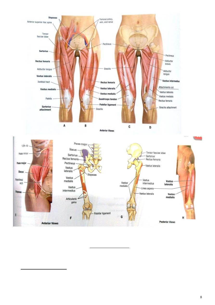
The anterior compartment of the thigh
Muscles of the anterior compartment of the thigh are:
1- Sartorius muscle
2- the quadriceps femoris muscle which include :
a- rectus femoris muscle
b- vastus lateralis muscle
c- vastus medialis muscle
d- vastus intermedialis muscle
3- the articularis genu muscle
Name of muscle
Origin
insertion
Rectus femoris
a- Straight head from ant. Inf. Iliac
spine
b- Reflected head from just above
acetabulum
Common insertion via
guadriceps tendone which
attaches to circumference of
patella and then via patellar
ligament to tibial tuberosity
Vastus medialis
-
From lower part of intertrochantric
line
-
medial lip of linea aspira
Vastus lateralis
-
From upper part of intertrochantric
line
-
lateral lip of linea aspira
Vastus
intermedialis
From ant. And lat. Surfaces of the
shaft of femur
sartorius
From ant. Sup. Iliac spine
Upper part of medial surface
of shaft of tibia
iliacus
From iliac fossa of hip bone
Common tendon inserted ito
lesser trochanter
Psoas major
Transverse process , bodies and
intervertebral discs of T12-L5
Note :
1- all are supplied by the femoral nerve
2- Iliacus + psoas are main flexor at hip joint they also flexes thigh on abdomen
3- Guadriceps are main extensor at knee joint
4- Sartorius are abduct and laterally rotate at hip joint.
Femoral artery
1- It is the direct continuation of the external iliac artery of the abdomen
2- it enters the thigh below the inguinal ligament at the mid-inguinal point medial to the
femoral nerve and lateral to the femoral vein
3- in the femoral triangle the artery covered only by skin and fascia.
4- Then it descends to the adductor canal at the apex of the femoral triangle anterior to
the adductor longus m.
5- it become the popliteal artery by passing through the adductor hiatus.
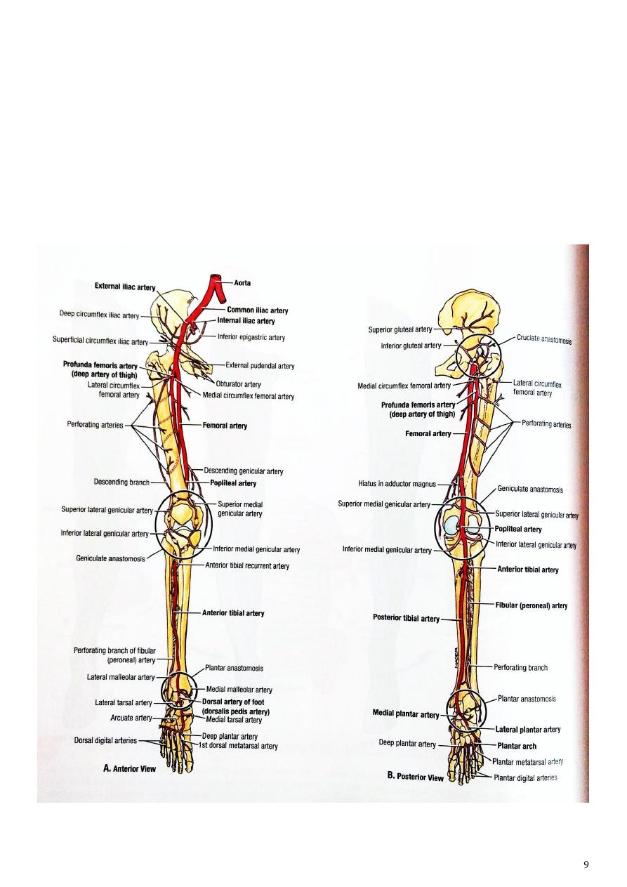
Branches:
1- Superficial arteries
2- Deep external pudendal artery.
3- Profunda femoris artery
4- Muscular arteries. To m. of ant. Comparetment
5- Descending genicular artery. Arise from the femoral artery short distance above the
adductor hiatus and share in the anastomose around the knee joint.
Profunda femoris artery
1- It is the principle artery of the thigh
2- arise from the posterolateral part of the femoral artery 5cm below the inguinal ligament
3- it descend deeply in the thigh between adductor longus and brevis m, behind the
femoral artery and vein on the medial side of the femur
4- In the lower third of the thigh the artery ends as the fourth perforating artery which
pierces the adductor magnus and distributes to the muscles of the posterior
compartment (hamstring muscles) at the back of the thigh
5- 4cm below the inguinal ligament in the femoral triangle it gives following branches :
a.
Lateral circumflex artery. Which is the largest branch of the profunda artery it runs
laterally among the branches of the femoral nerve then deep to rectus femoris m.
b.
Medial circumflex artery, either arise from the profunda or directly from the femoral
artery passes backwards between pectinus and iliopsoas muscles and continue
backward under the neck of the femur.
c.
3 perforating branches to muscles of the back of the thigh
d.
Finally the profunda femoris terminates as 4
th
perforating branch.
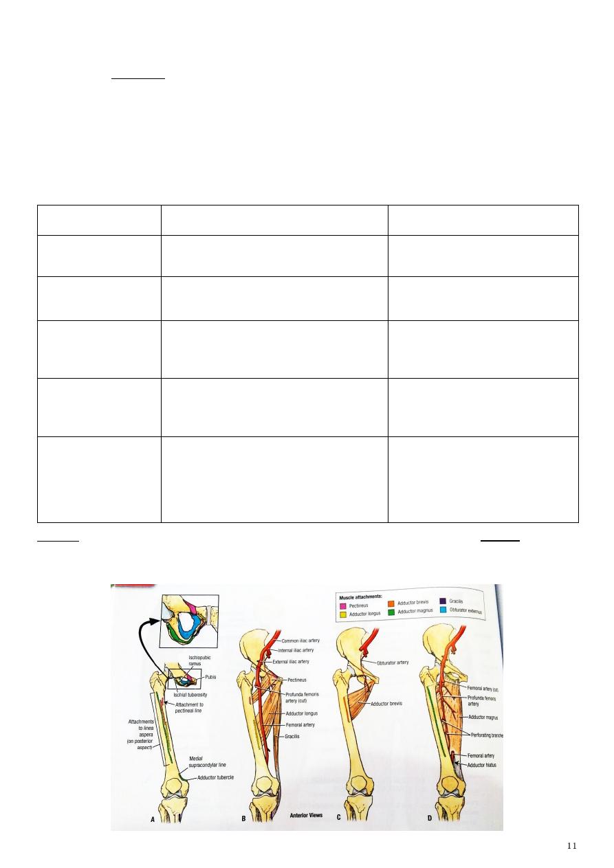
The medial side (adductor) of the thigh
includes the adductor muscles which arise from the external surfaces of the pubic rami and
the ramus of the ischium. they concerned with adduction at the hip joint, the muscles are:
1- Pectineus
2- adductor longus
3- adductor brevis
4- adductor magnus
5- gracilis.
Name of m.
origin
insertion
gracilis
From medial margin of pupic arch
Upper medial side of tibia
pectineus
Superior ramus of pupis
Spiral line between lesser
trochanter and linea aspera
Adductor longus
Pupic bone just below and medial to
pupic tubercle
Linea asperaa
Adductor brevis
From body and inferior ramus of
pupis
Linea aspera
Adductor magnus
a- Adductor part from inferior pupic
ramus and ischial tuberosity
b- hamsting part from ischial
tuberosity
To Linea aspera
Adductor tubercle
Note :
The nerve supply of these muscles is the obturator nerve (L2, 3, 4) . except
1- the hamstring portion of adductor magnus from sciatic N.
2- and pectineus m. receive nerve supply from both femoral and obturator N.
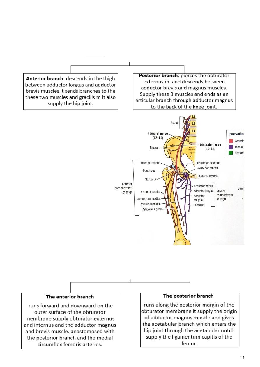
The obturator nerve:
1- arises from the lumbar plexus in the abdomen
2- it descends medial to the psoas m.
3- at the lateral wall of the pelvis here it join the obturator vessels and enters the
obturator canal where it divides into anterior and posterior branches:
The obturator artery
-
arises from the internal iliac artery it accompany the obturator nerve through the
obturator canal divides into anterior and posterior branches which forms an arterial
circle on the obturator membrane.

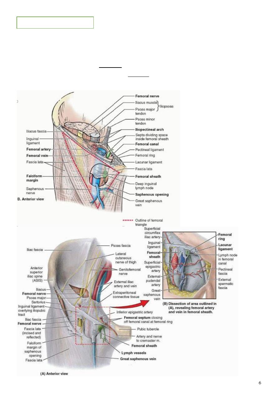
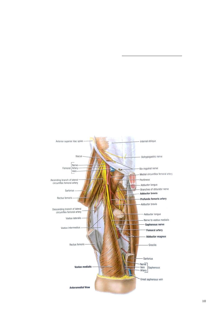
FEMORAL TRIANGLE
Occupy the upper third of the front of thigh.
Bounderies:
a. Superiorly (base): the inguinal ligament.
b. Medially: the medial border of the adductor longus muscle.
c. Laterally: medial border of the sartorious muscle.
d. Inferiorly (apex): is formed as Sartorius crosses over the lower part of adductor longus
m. continuous with the adductor canal.
e. The anterior wall of the triangle: composed of the skin and the fascia. In the superficial
fascia there are the following structures:
1- The upper part of the great saphenous vein.
2- Superficial inguinal lymph nodes and vessels.
3- Femoral branch of the genitofemoral nerve.
4- Superficial branches of the femoral vessels.
5- Branches of the ilioinguinal nerve.
f. The posterior wall (the floor): composed of muscles, from medial to lateral:, adductor
longus, pectineus, psoas major and iliacus muscles ( iliopsoas )
Contents of the triangle
1. Femoral sheath
- It is an extension of the transveralis fascia of the abdominal cavity which surrounds the
upper 2-3 cm of the femoral vessels below the inguinal ligament.
- The sheath is divided into 3 compartments :
1- the femoral artery occupy the lateral part of the sheath
2- while the vein is intermediate
3- medial to the femoral vein is the tubular femoral canal, through which femoral
hernia may pass.
Femoral canal
- It is a short fascial tube about 0.5 inch occupy the medial compartment of the femoral
sheath
- inferiorly it is rapidly decreased in width and closed by fusion of its walls.
- The wide upper end called the femoral ring which is separated from the abdominal
cavity only by peritoneum. It contains :
1- fatty connective tissues
2- efferent lymph vessels from the deep inguinal lymph nodes
3- and one or two of the deep inguinal lymph node.
Boundaries of the femoral ring:
1- Inguinal ligament anteriorly
2- The sharp edge of the lacunar ligament medially
3- The pectin pubis posteriorly
4- The femoral vein laterally

2. The femoral vessels
- enters the triangle behind the
midpoint of the inguinal ligament
traverse the triangle from the base to
the apex.
- The vein is medial to the artery at the
base, but it lies behind the artery at
the apex
3. Profunda femoris artery.
- It is the main artery of the thigh arise
from the posterolateral side of the
femoral artery, curves behind it and
passes posterior to the adductor
longus muscle.
- The profunda vein lies anterior to the
artery and ends in the femoral vein.
4. The lateral and medial circumflex
arteries
- arise from the profunda artery near
its origin.
- The lateral circumflex passes among
the branches of the femoral nerve and
leaves the triangle posterior to the
sartorious muscle.
- The medial one passes backwards
between psoas and pectineus muscle.
- The circumflex veins end in the
femoral vein.
5. Deep external pudendal artery
- it is small branch arise from the medial
side of the femoral artery runs
medially to the scrotum in male and
the labium majus in female.
6. 3-4 deep inguinal lymph nodes
- lie along the medial side of the
femoral vein
- receive afferent vessels from the
superficial inguinal lymph nodes and
popliteal lymph nodes and from the
deep structures of the limb.
- Efferent vessels pass from the deep
inguinal lymph nodes to the external
iliac nodes.
7. The femoral branch of the
genitofemoral nerve supply skin
over the triangle.
8. Lateral cutaneous nerve of the
thigh L2 L3.
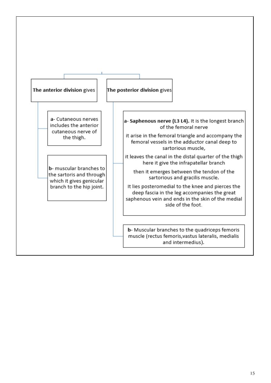
9. Femoral nerve (L2,3,4)
- arise from the lumber plexus in the abdomen descend in groove between psoas and
iliacus muscles and give branch to iliacs .
- it enters the thigh posterior to the inguinal ligament and lateral to the femoral
sheath. 2cm below the inguinal ligament it ends by dividing into anterior and
posterior branches.
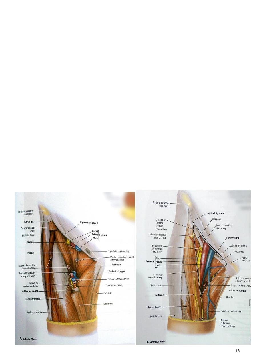
The adductor canal
- It is an intermuscular canal situated on the medial aspect of the middle of the thigh
beneath the sartorius m.
- it conducts the femoral vessels through the middle 1/3 of the thigh
- it begins about 15cm below the inguinal ligament at the apex of the femoral triangle and
ends at the upper limit of the adductor hiatus (a separation in the tendinous insertion of
the adductor magnus muscle allows the femoral vessels to pass to the back of the knee).
- it is triangular in section
- .
Boundaries of the canal are:
1- Sartorius muscle anteromedially.
2- Vastus medialis anterolaterally.
3- Adductor longus and magnus posteromedially.
The contents of the adductor canal are:
1- The femoral vessels.
2- Saphenous nerve.
3- Nerve to vastus medialis.
4- Branches of obturator nerve
5- Profunda femoris artery
Part2
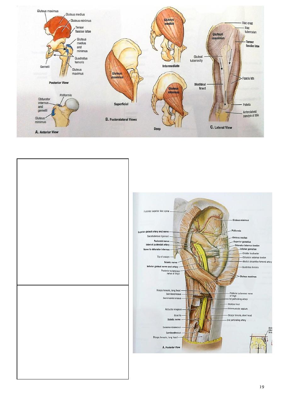
:
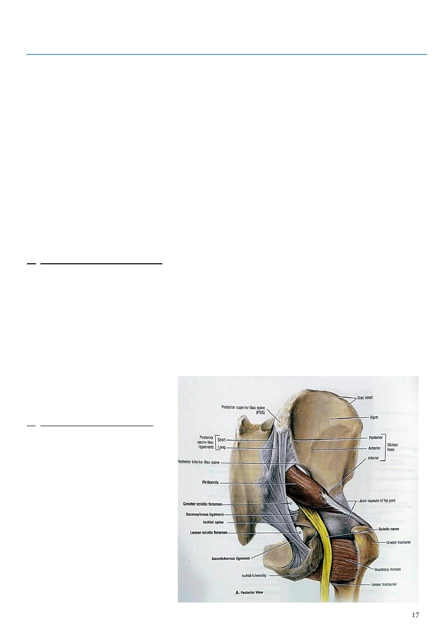
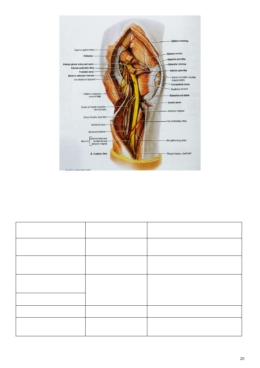
Gluteal region
- Extends from the iliac crest above to the gluteal fold below.
- The superficial fascia is thick dense and fatty, the deep fascia is thick It continuous
below with the fascia lata.
Sacrotuberous ligament:
- It is a strong band passes upwards from the medial side of the ischial tuberosity to the
margins of the sacrum and coccyx and to both posterior iliac spines.
Sacrospinous ligament:
- This is a thick triangular band, it passes from the the ischial spine to the margin of coccyx
and last piece of the sacrum deep to the sacrotuberous ligament.
These two ligaments share together to conver greater and lesser Sciatic notcges into
foramina
A. The greater sciatic foramen:
Transmits Structures which enter the gluteal region from the pelvis which include
1- Superior gluteal vessels and nerves.
2- the piriformis muscle
3- .inferior gluteal vessels and nerves
4- sciatic nerve,
5- the posterior cutanous of the thigh
6- pudendal nerve
7- nerve to quadrates femoris
B. The lesser sciatic foramen:
transmits structures between the
gluteal region and the perineum
these include :
1- the internal pudendal vessels
2- pudendal nerve
3- and nerve to obturator
internus muscle.

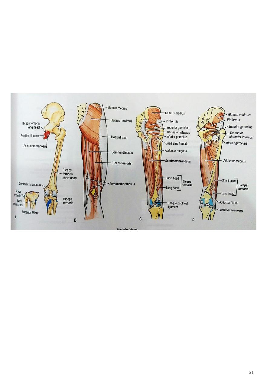
Muscles of the gluteal region
The muscles of the gluteal region can be divided into two groups:
Superficial group
Deep lateral rotators
-
a group of large muscles that abduct and
extend the femur
-
It includes
:
1- the gluteus maximus
2- gluteus medius
3- gluteus minimus
4- tensor fascia lata.
- A group of smaller muscles, that mainly
act to laterally rotate the femur.
- It includes:
1- the quadratus femoris
2- piriformis
3- gemellus superior
4- gemellus inferior
5- obturator internus
6- obturator externus.
a) action
1- Gl. Maximus is main extensor at hip joint assist in raising from sitting position and a
powerful muscle on climbing
2- Gl. Medius and minimus and tensor fasia lata are abductors and medial rotation of
thigh at hip
b) Nerve supply
1- Inferior gluteal nerve (L5 S1 S2): it is a branch from the sacral plexus enter the
gluteal region with the posterior cutanous nerve of the thigh inferior to the
piriformis m. supply gluteus maximus m.
2- Superior gluteal nerve (L4 L5 S1): it is a branch from the sacral plexus enters the
gluteal region above the piriformis m. divided into numbers of branches supply the
glueus medius , minimus and tensor fasciae lata m.
Names of m.
origin
insertion
Tensor Fascia Lata
anterior iliac crest, attaching to
the anterior superior iliac spine
iliotibial tract,
Gluteus maximus
a. Area behind the posterior
gluteal line
b. back of sacrum and coccyx
c. back of sacrotuberous lig.
a. 3/4 inserted into ilio-tibial
tract
b. 1/4to gluteal tuberosity
Gluteus minimus
Area between middle and inferior
gluteal line
Front of greater trochanter
Gluteus medius
Area bounded by iliac
crest,posterior and middle gluteal
lines
Greater trochanter
Gluteal vessels
Include:
1- Inferior gluteal artery:
- it is a branch of the internal iliac
artery emerges from the pelvis
below piriformis muscle
accompany the inferior gluteal
nerve supply the gluteus maximus
and gives branches to the back of
the thigh
- it also give a slender companion
artery to the sciatic nerve.
- The inferior gluteal artery
anastomosed with the medial
circumflex artery.
2- Superior gluteal artery:
- arise from the internal iliac artery
accompany the superior gluteal
nerve
- it enters the gluteal region above
the piriformis muscle.
- It follows the superior gluteal
nerve supply the gluteus medius,
minimus and the hip joint.
In addition gluteal region has the following small and short muscles which are located
deeply
Names of m.
Origin
insertion
Piriformis
Middle three peices of
the front of sacrum
Upper border of greater trochanter
and trochanteric fossa
Obturator internus
Pelvic surface of
obtur. Membrane
trochanteric fossa just inferior to
insertion of piriformis
Superior gemellus
Ischial spine and upper
border of ischial
tuberosity
Both insertion together with that of
obturator internus m. into
trochanteric fossa
Inferior gemellus
Quadrates femoris
Ischial tuberosity
Guadrate tubercle
Obturator externus
Outer surface of
obtur. Membrane
trochanteric fossa
a) action
All these short muscles are lateral rotaters of the thigh at the hip joint.
b) Nerve supply
1- sup.gemellus+obtu. Intr.by nerve to obtu. Int.
2- inferior gemellus+ quadrates femoris by nerve to quadratus femoris
3- piriformis by branches from S1andS2.
4- obtu. Exter. By obturater nerve

Part3
:
The back of the thigh
The muscles of the back of the thigh are the hamstring muscles which are
1- extensors of the hip joint and flexors of the knee joint
2- all arise from the ischial tuberosity except the short head of the biceps m.
3- and all are inserted in the bones of the leg.
4- These muscles include:
1-
biceps femoris
2-
semitendinosus
3-
semimembranosus.
5- All supplied by the sciatic nerve.
Names of m.
origin
insertion
Biceps femoris
a- long head has common origin
with semitendinossus from
ischial tuberosity
b- short head from linea aspira
By common tendone to the
head of fibula
semitendinosus
has common origin with long
head of biceps from ischial
tuberosity
Upper part of medial sureface of
tibia
semimembranousus from ischial tuberosity
Posteromedial part of medial
condyle of tibia
Ischial part of
adductor magnus
from ischial tuberosity
Adductor tubercle of femor
a) Action
1- hamstring muscles are extensors of the hip joint and flexors of the knee joint.
2- In addition both semiten.and semimem.act as medial rotaters of leg when knee joint
semiflexed.
3- biceps femoris act as lateral rotater of leg when knee joint semiflexed.
b) Sciatic nerve (L4 L5 S1 S2 S3):
-
it is the thickest nerve in the body arise from the sacral plexus, pass inferior to the
piriformis m through the greater sciatic foramen,
-
deep to the gluteus maximus m. in the upper part of its course it descends over:
1- ischial wall of the acetabulum.
2- Obturator internus m. and the 2 gemelli ms.
3 -Quadratus femoris m.
-
It leaves the buttock by passing deep to the long head of the biceps femoris
-
it supply the hamstring ms from the tibial side of the nerve except the short head of
biceps muscle receive its nerve supply from the common peroneal side
-
it also gives articular branch to the hip joint.
-
the sciatic nerve then descends on the posterior surface of the adductor magnus m.
-
at the lower third of the thigh it divided into:
1- medial branch (tibial nerve)
2- and lateral branch (common peroneal nerve).
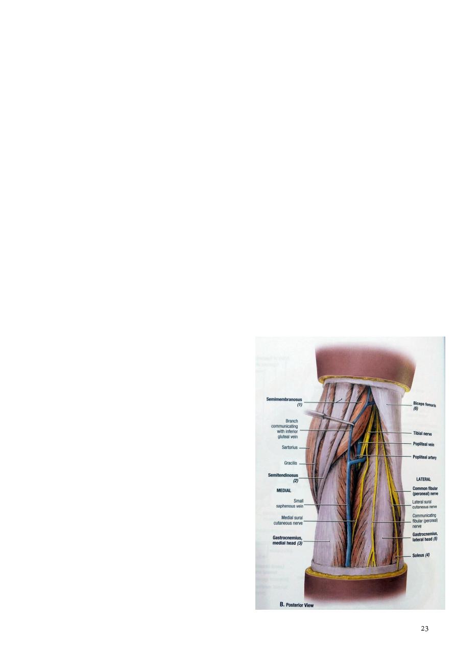
The popliteal fossa
1- It is a diamond shaped space ( depression) lies behind the knee, the lower 1/3 of the
femur and the upper part of the tibia.
2- The superficial fascia of the fossa contain little fat, while the deep fascia is thin and
strong
3- Bounderies:
a- superolaterally biceps femoris m.
b- superomedially semimembranosus and semitendinosus ms.
c- Inferolaterally lateral head of the gastrocnemius m.
d- Inferomedially Medial head of the gastrocnemius m.
e- The anterior wall ( floor) : from above
downward is the popliteal surface of the
femur, popliteus m the posterior capsule of
the knee joint and oblique popliteal ligament
f- The posterior wall ( roof) is the skin and
deep fascia of the fossa
Contents of the fossa
These include:
1- The popliteal vessels. The popliteal art. is most
anteriorly , it gives 5 genicular branches in the
fossa and bifurcates at lower border of
popliteus m. into anterior and posterior tibial
arteries
2- Branches of the sciatic nerve the tibial and
common peroneal nerves.
3- Popliteal lymph nodes.
4- Posterior cutaneous nerve of the thigh.
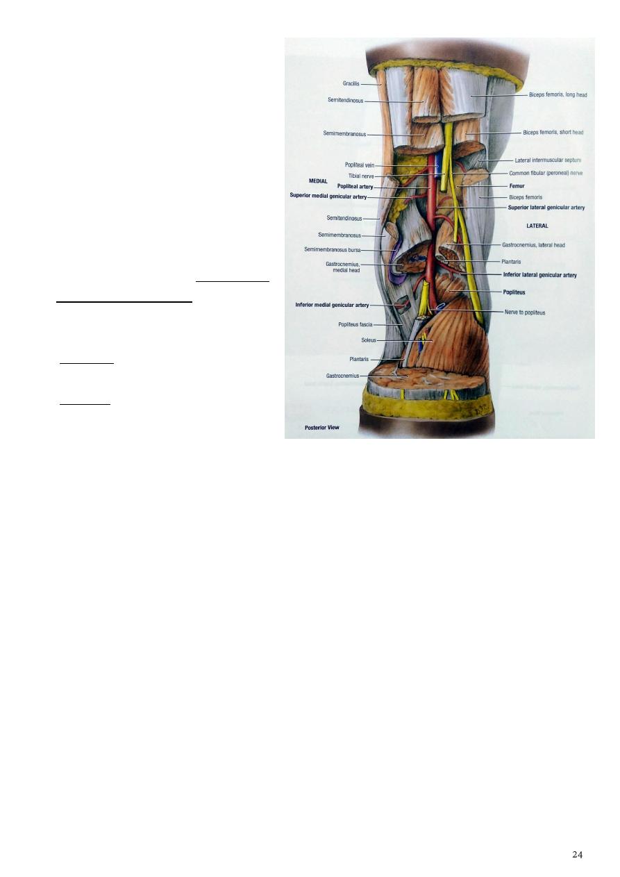
A- The popliteal artery
1- These are the direct continuation of
the femoral art. enter the fossa
through the adductor hiatus.
2- They lie anterior to the tibial nerve,.
3- it lies aganist the posterior part of
the capsule of the knee joint,
4- then it lies posterior to popliteus
muscle in the upper part of the leg.
5- The popliteal artery ends at the
lower border of the popliteus
muscle by dividing into anterior and
posterior tibial arteries.
Branches of popliteal artery:
1- muscular branches to the hamstring
ms. And to the muscles of the calf.
2- Articular branches these are :
a- the lateral and medial superior
and inferior genicular and middle
genicular arteries to the knee joint correspond to the genicular branches from the
tibial and common peroneal nerves.
b- they anastomosed with the branches from the lateral circumflex femoral,
descending genicular arteries, and the recurrent branches of the anterior tibial
artery.
B- The popliteal vein:
1- formed by the union of the anterior tibial, the posterior tibial and the peroneal veins at
the lower border of the popliteus muscle
2- it lies superficial to the artery and between it and the tibial nerve.
3- it receive tributaries correspond to the branches of the popliteal artery and the lesser
saphenous vein.
4- it become the femoral vein at the adductor hiatus.
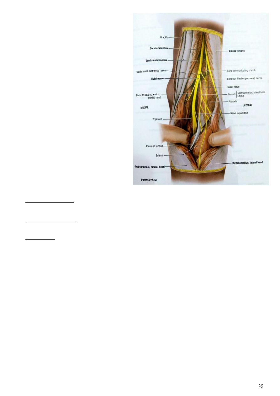
C- Tibial nerve (L4 L5 S1 S2 S3).
1- It is the largest of the two terminal
branches of the sciatic nerve
2- it begins above the popliteal fossa
descends vertically in the fossa, Lying
first on the lateral side of the
popliteal artery then posterior to it
and finally medial to it.
3- it pass between the two heads of the
gastrocnemius muscle and under the
soleus muscle.
4- It supply the muscles of the back of
the thigh and leg, the sole of the
foot, the skin of the lateral and lower
half of the back of the leg and sole of
the foot.
Branches in the popliteal fossa:
1- Articular branches, it gives superomedial, inferomedial and middle genicular branches to
the knee joint, accompanied the corresponding branches from the popliteal artery
2- Muscular branches to the muscles of the back of the thigh and to the gastrocnemius,
plantaris, soleus and popliteus ms.
3- sural nerve:
a- it is a cutaneous branch descend in the groove between the two heads of the
gastrocnemius m.
b- it pierce the deep fascia about the middle of the back of the leg, supply the skin of
the lower posterior part of the leg and the skin of the lateral side of the dorsum of
the foot.
c- It accompany the small saphenous vein.
D- Common peroneal nerve (L4 L5 S1 S2)
1- It is smaller than tibial nerve follow the tendon of biceps femoris m. along the upper
lateral border of the popliteal fossa to the back of the head of the fibula,
2- then curves forwards along the neck of the fibula deep to the peroneus longus m.
here it divides into deep and superficial branches.

Branches in the popliteal fossa:
1- cutaneous branches, these include
a-
the peroneal communicating branch which arise in the upper part of the popliteal
fossa descend on the posterolateral side of the calf , it supply the proximal 2/3 of
the posterolateral part of the leg.
b-
Lateral cutanous nerve of the calf arise on the lateral head of the gastrocnemius m.
supply the lateral side of the leg.
2- articular branches, these include:
a-
the superior and the inferior lateral genicular branches they are small branches
accompany the corresponding arteries.
b-
Recurrent genicular branch arise where the common peroneal nerve divides into
superficial and deep branches, it ascends to the knee joint.
3- muscular branch to the short head of the biceps femoris m. arise high up in the fossa.

Part4
:
Hip joint
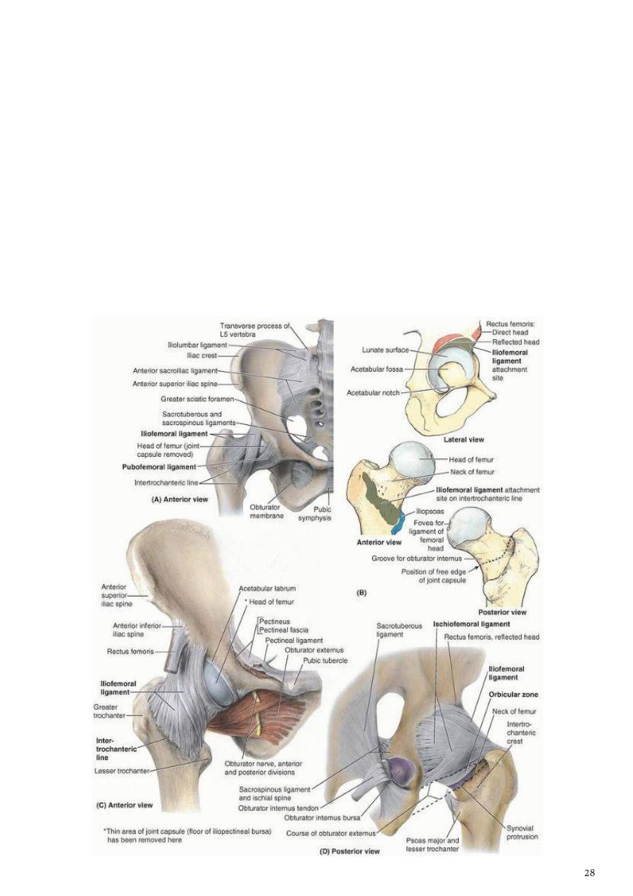
1- it is a synovial joint of ball and socket type, the joint formed between the head of the
femur and the acetabulum
2- the articular surface of which is horseshoe shaped and is deficient inferiorly at the
acetabular notch.
3- The cavity of the acetabulum is deepened by the presence of a fibrocartilaginous rim
called the acetabular labrum
4- the labrum is connected across the acetabular notch by the transverse acetabular
ligament.
5- The strength and stability of the joint depend on :
1- depth of the acetabulum which increased by the labrum acetabulae.
2- The strong ligaments and muscles surrounding the joint.
The fibrous capsule
1- which surrounds the joint attached to the margin of the acetabulum and transverse
ligament proximally.
2- Distally attached to the intertrochantric line
3- and greater trochanter anteriorly
4- and intertrochantric crest posteriorly.
The fibrous capsule is lined by the synovial membrane.
Ligaments of the joint:
1- iliofemoral ligament is a strong ligament lie in the front of the joint. it is inverted Y
shaped.it prevents hyperextens of the hip joint.
2- Pubofemoral ligament it is triangular ligament lie in the lower anterior part of the
capsule
.) it limits abduction)
3- Ischiofemoral ligament it is lie posteriorlyl
.) limit extension)
4- The transverse acetabular ligament it converts the notch into a tunnel through which
the blood vessels and nerves enter the joint.
5- Ligaments of the head of the femur it is attached to the pit on the head of the femur
and by its base to the transverse acetabular ligament
Blood supply of the joint
1- branch from lateral and medial circumflex femoral artery.
2- Acetabular branches of the obturator.
3- Branches of the superior and inferior gluteal artery.
Nerves of the joint
1- nerve to quadratus femoris.
2- The femoral nerve through nerve to rectus femoris.
3- Anterior division of the obturator nerve.
Movement at hip joint:
1- flexion which is very free it produced mainly by iliopsoas, assisted by rectus femoris and
sartorius.
2- Extension (restricted by the iliofemoral ligament.(it produced mainly by glut. Maximus
m. assited by hamstring m.
3- Abduction (restricted by the pubofemoral ligament.) it produced mainly glut. Medius
and minimus assited by Sartorius and tensor fascia latae.
4- Adduction is (restricted by the lateral portion of the iliofemoral ligament). it produced
mainly by 3 adductor m. assisted by pectineus and gracilis
5- Medial rotation (tightens the ischiofemorall ligament). it produced mainly by glut.
Medius and minimus assited by tensor fascia latae.
6- Lateral rotation is( limited by the pubofemoral ligament and the lateral part of the
iliofemoral ligament.) it produced mainly by deep glut. M.
Part5
: Front of the leg and dorsum of the foot
The deep fascia (crural fascia)
- The fascia lata of the thigh continuous onto the leg and called the crural fascia.
- It is connected to the bones by intermuscular septa, and forms thickened bands at the
ankle called retinacula which act as a pulley around the tendons of ms.
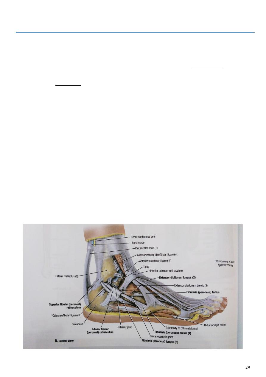
The Retinacula:
1- Superior extensor retinacula it is broad extends between the fibula and the medial
surface of the tibia.
2- Inferior extensor retinacula is Y shaped.
3- Superior peroneal retinaculum extends from the lateral malleolus downwards and
backwards attached to the lateral surface of the calcaneum
4- Inferior peroneal retinaculum attached to the lateral surface of the calcaneum above
and below the peroneal muscles.
5- Flexor retinacula extends from the medial malleolus downwards and backwards to be
attached to the medial tubercle of calcaneum
- The dorsum of the foot contains the structures which extend from the anterior
compartment of the leg.
- The fascia of the dorsum of the foot is thin, it is continuous with the extensor retinacula
curves over the margins of the foot and becomes the fascia of the sole.
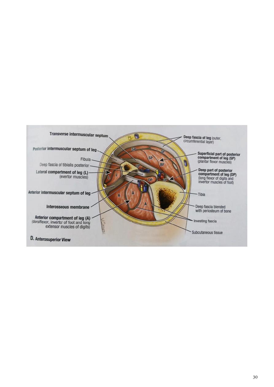
Intermuscular septa:
- These are extensions from the deep fascia of the leg to the tibia and fibula so that it
separate the leg into 3 compartments. These septa are:
1- The interosseous membrane between the tibia and fibula separate the anterior and
posterior compartments.
2- Anterior intermuscular septa attached to the anterior border of the fibula separate the
anterior and lateral compartments.
3-
4- The posterior septa attached to the posterior border of the fibula separate the posterior
and lateral compartments. From the posterior septa a broad transverse intermuscular
septa separating the superficial and deep groups of calf muscles.
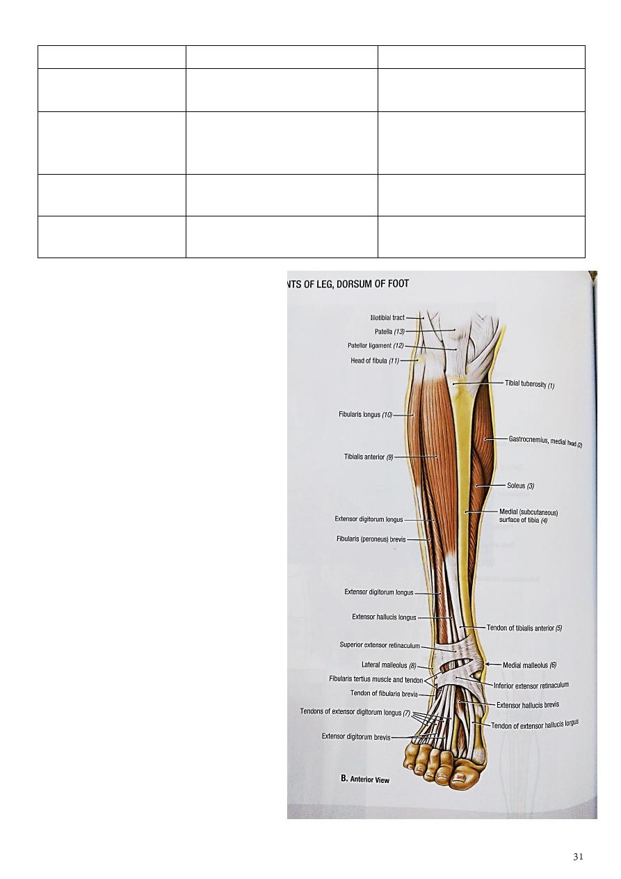
Anterior compartment of the leg
- The anterior compartment ( dorsiflexion) lies in front of interosseous membrane and the
fibula,
- it contains the following muscles:
1- Tibialis anterior m
2- extensor hallucis longus m
3- extensor digitorum longus m
4- peroneus tertius m
- Vessels of this compartment are the anterior tibial vessels and the nerve is the deep
peroneal nerve.
Action
1- TA: dorsiflexion of foot (at ankle)+
inversion of foot.
2- EDL:extension of toes +dorsiflexion
of foot.
3- EHL:extension of big
toe+dorsiflexion.
4- PT:eversion of foot
muscle
origin
insertion
Tibialis anterior( TA)
Upper 2/3of lateral surface of
tibia +interosseous membr.
Medial cuneiform +adjacent
part of first metatarsal.
Extensor digitorum
longus(EDL)
Upper 3/4of anterior surface
of fibula +interosseous
membr
Via 4 tendon into the lateral toes
,for extensor expantion
Extensor halucis
longus (EHL)
Middle 1/3of ant. surface of
fibula +interosseous membr
Base of distal phalanx of big toe
Peroneus tertius (PT)
lower 1/4of antrior surface of
fibula +interosseous membr
base of 5 metatarsal
Extensor expansions
- It is formed on the dorsum of the
proximal phalanx by the union of 5
tendons:
1- tendon of the extensor digitorum
longus
2- tendon of the extensor digitorum brevis
3- tendon of one lumbrical muscle.
4- tendon of two interossel m.
- the expantion divides into 3 parts .
1- The thick central part inserted in the
base of the middle phalanx.
2- The lateral and medial parts of the
expansion continue distally fused
together and inserted in the base of
the distal phalanx.
- The big toe has no ext.expantion.
- the ext. expantion of little toe is formed
by the union of 3 tendon
1- tendon of extensor digit. Longus
2- tendon of one lumbrical muscle
3- tendon of one interossel m.
Anterior tibial artery
1- Arises from the popliteal artery at the lower border of the popliteus m.
2- it passes forwards above the upper border of the interosseous membrane close to the
neck of the fibula
3- then descends forward on the membrane with the deep peroneal nerve passing behind
the superior extensor retinaculum.
4- The tendon of extensor hallucis m. lies on its medial side and the deep peroneal nerve
and tendons of extensor digitorum m. on its lateral side.
5- It ends in the front of the ankle joint by becoming the dorsalis pedis artery midway
between the malleoli.
Branches:
1- muscular branches to the muscles of the anterior compartment.
2- Anterior tibial recurrent artery passes upwards to the knee joint.
3- Medial and lateral malleolar arteries to the lateral and medial malleoli,
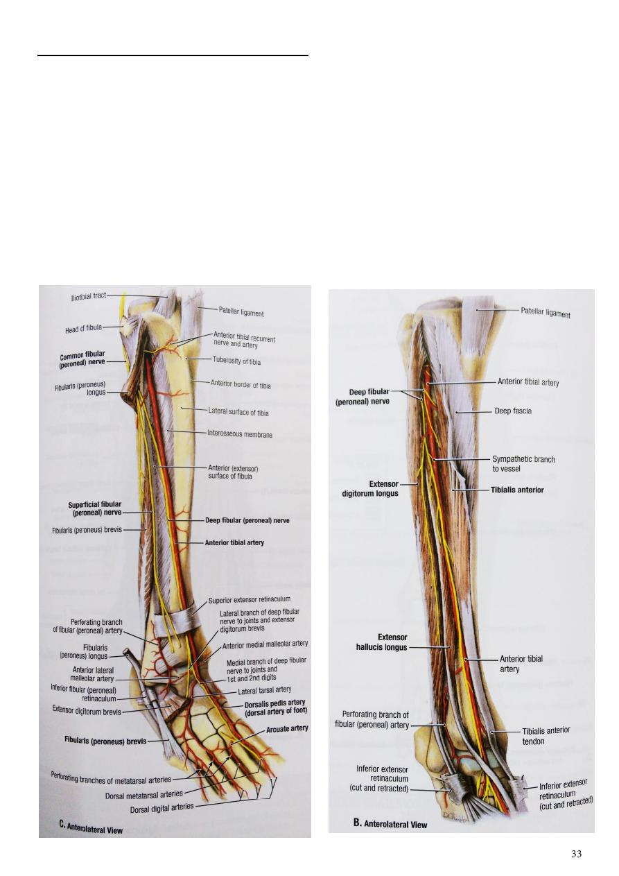
Deep peroneal nerve( ant. Tibial nerve)
1- Arises from the common peroneal nerve lateral to the neck of the fibula.
2- it pierces the peroneus longus m and descends in the anterior compartment between
EDL and TA in upper part then between EHL and TA.
3- It pass lateral to the anterior tibial vessels
4- near the ankle joint it crossed by the extensor hallucis longus m.
5- it enters the dorsum of the foot midway between the malleoli with the dorsalis pedis
artery.
It gives :
1- articular branch to ankle joint
2- Muscular branches to all muscles of the anterior compartment.
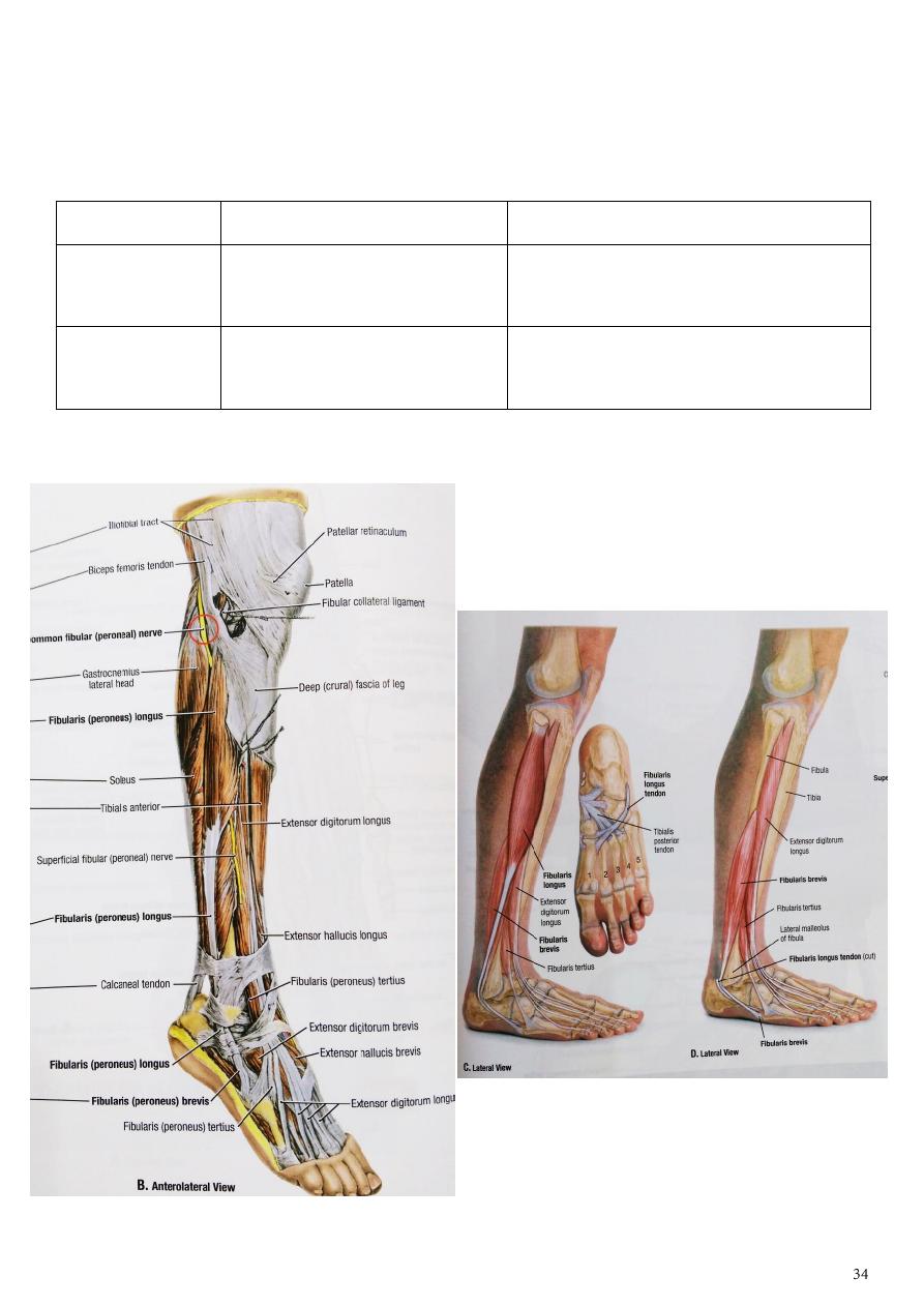
Lateral side of the leg
- Composed of the muscles which cover the lateral surface of the leg. These are peroneus
longus and brevis ms. supplied by the superficial peroneal nerve.
muscle
origin
insertion
Peroneus
longus
Upper 2/3 of lateral surface of
fibula
In the base of 1
st
metatarsal and
medial cuneiform on their lateral sides
Peroneus brevis Lower 2/3 of lateral surface of
fibula
On the medial aspect of base of 5
th
metatarsal bone
Action : both act as everter of foot (mainly) . Assist in planter flexion.

Superficial peroneal nerve:
1- Descend in the peroneus longus m. to reach the peroneus brevis m. supply both
muscles
2- then it descend between it and extensor digitorum longus m.
3- pierce the deep fascia in the distal 1/3 of the leg and divides into medial and
intermediate cutaneous nerves.
4- It supply the skin of the lower third of the front of the leg, the greater part of the
dorsum of the foot and most of the dorsal surface of the toes expect the first
interdigital cleft and the lateral side of the little toe.
The back of the leg
- The transverse intermuscular septa divide the back of the leg into superficial
posterior compartment and the deep posterior compartment
- they supplied by the tibial nerve.
The superficial layer consist of the muscles which inserted in the heel by the
tendocalcaneus , these muscles are the powerful planter flexors of the ankle joint, include :
6- gasterocnemius m.
7- soleus m.
8- plantaris m.
The deep layer consist of long flexors muscles of the toes these are:
1- flexor hallucis
2- flexor digitorum longus
3- popliteus
4- tibialis posterior.
muscle
origin
insertion
gastrocnemius Arises by 2 heads lateral and medial from
lateral and medial condyle of femur
By tendocalcaneus
tendon into the dorsum
of calcaneum bone
soleus
1-Post. Surface of head of fibula +post .
surf. Of upper 1/3 of fibula 2-Solial line of
tibia
plantaris
Lower part of lateral supracondylar ridge of
femur
Back of calcaneum
Action : raises the heel of the foot on the ground on propulsive movement during walking
in addition they share in planter flexion of foot.
muscle
origin
insertion
popliteus
Lateral surface of lateral
condyle of femur
Posterior surface of tibia above the
soleal line
Flexor digitorum
longus
Posterior surface of tibia Via its 4 tendon to the bases of the
distal phalanges of lateral 4 toes
Flexor halucis longus Lower 2/3of Posterior
surface of fibula
Distal phalanx of big toe
Tibialis posterior
Posterior surface of tibia
and fibula
Mainly into tuberosity of navicular
bone and to the all tarsal bone
except the talus
Action:
1- fl. Hallucis :flexion of big toe +planter flexion
2- Fl.digit. flexion of toes+planter flexion
3- Tibialis post. Inversion of foot +planter flexion
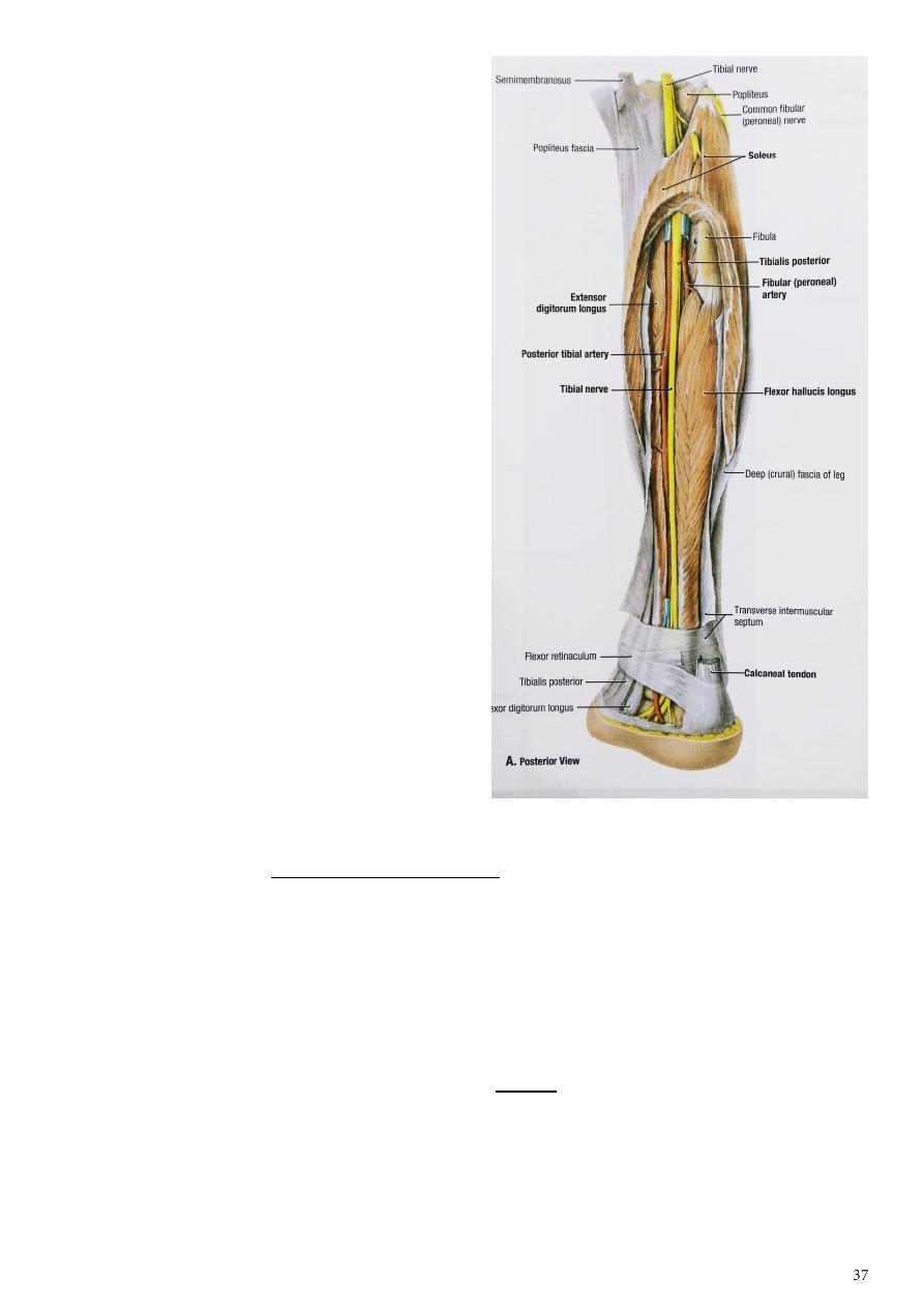
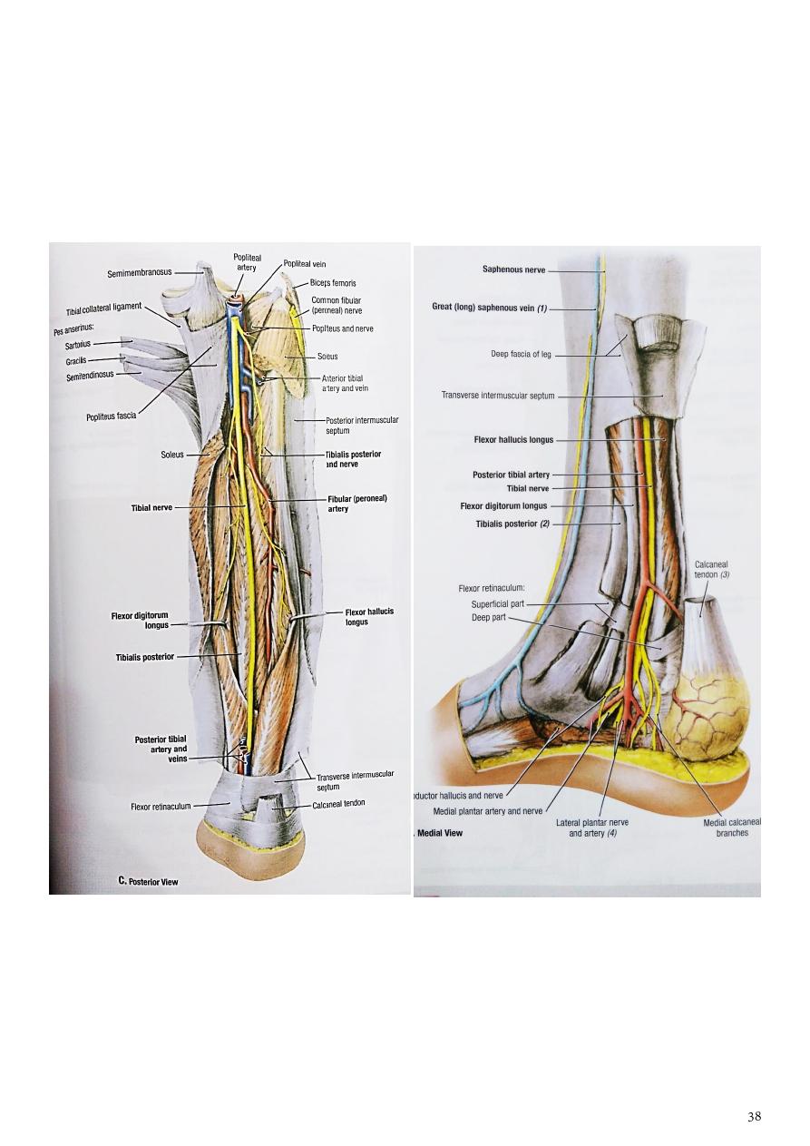
The princible nerve of the back of the leg is the Tibial nerve
(L4,5,S1,S2,S3)
1- passes under the tendinous arch of the soleus muscle and descend under the transverse
intermuscular septum superficial to the posterior tibial vessels.
2- in the upper part of the leg it lies on the popliteus m. then posterior to the tibialis
posterior m.
3- in the lower third of the leg it lies between tendons of flexor digitorum longus and flexor
hallucis longus ms.
4- the tibial nerve divides deep to the flexor retinaculum into medial and lateral planter
nerves.
Branches in the leg:
1- muscular branches to the tibialis posterior,
flexor digitorum longus, flexor hallucis
longus and deep part of soleus m.
2- cutaneous branches include medial
calcanean nerve (S1) arise in the ankle
pierce the flexor retinaculum supply the
skin on the posterior and lower part of the
heel.
3- Small articular branch to the capsule of the
ankle joint.
The posterior tibial artery
1- It is the direct continuation of the popliteal
artery, supply the muscles of the back
2- and it is the main artery of the foot begin at
the lower border of the popliteus m.
3- then descend with the tibial nerve and two
venae commitants deep to the
gastrocnemius, soleus and the transverse
intermuscular septum of the leg.
4- it runs first laterally to give the peroneal
artery
5- then it inclines medially passes behind the
medial malleolus
6- ends by dividing into medial and lateral planter arteries deep to the flexor retinaculum.
Branches in the leg:
1- peroneal artery
- it is the largest branch
- arises from the posterior tibial artery 2-3 cm below the lower border of the popliteus
muscle
- descend obliquely along the back of fibula deep to the flexor hallucis m.
- and ends in branches to the ankle and heel. it gives:
a- muscular branches to the muscles of the lateral compartment of the leg.
b- Nutrient branch to the fibula.
c- Perforating artery which pierces the distal end of the interosseous membrane
- The peroneal artery ends by giving post. lateral malleollar branch to the lateral side
of the back of the heel
2- circumflex fibular artery runs around the neck of the fibula
3- Nutrient artery to the tibia.
4- Muscular branches to the deep muscles of the back of the leg
5- Posterior medial malleolar to the posterior part of the medial malleolus.
6- medial and lateral planter art.
Part6
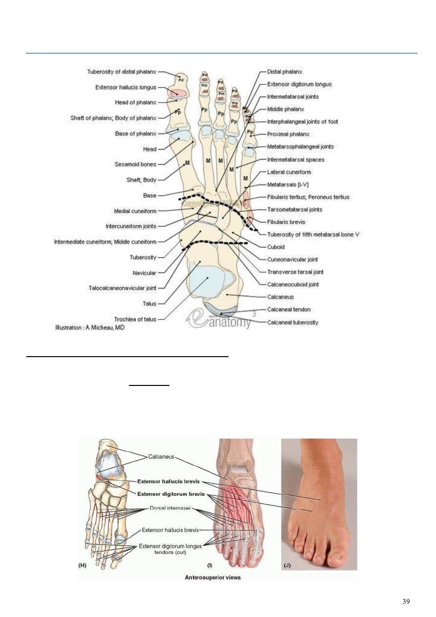
: Dorsum of the foot
There are two muscles at the dorsum of the foot
• Extensor digitorum brevis muscle
- originates at the calcaneus and divides into three muscle bellies whose tendons
insert at the dorsal aponeurosis and the middle phalanges of the second to fourth
toes.
• Extensor hallucis brevis muscle
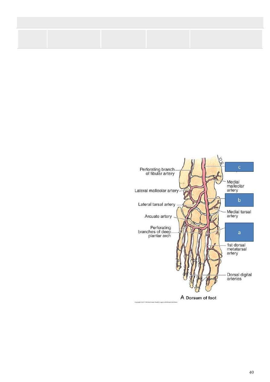
TABLE 5.14.III. MUSCLES OF FOOT: DORSUM OF FOOT
Muscle
Proximal
Attachment
Distal
Attachment
Innervationa
Main Action
Extensor
digitorum
brevis
Calcaneus (floor of
tarsal sinus);
interosseous
talocalcaneal
ligament; stem of
inferior extensor
retinaculum
Long extensor
tendons of four
medial digits
(toes 2-4)
Deep fibular
nerve (L5 or S1,
or both)
Aids the extensor digitorum
longus in extending the
four medial toes at the
metatarsophalangeal and
interphalangeal joints
Extensor
hallucis
brevis
In common with
extensor digitorum
brevis (above)
Dorsal aspect of
base of proximal
phalanx of great
toe (digit 1)
Aids the extensor hallucis
longus in extending the
great toe at the
metatarsophalangeal joint
( Artery of the dorsum of the foot
)
Dorsalis pedis artery
1- It is the continuation of anterior tibial
artery
2- Starts on the front of the ankle joint at a
point midway between the medial and
lateral melleolli
3- Descends anteromedially to 1
st
interosseous space and divides into
deep plantar and arcuate arteries
artery Branches:
1- Lateral tarsal branch.
2- Medial tarsal branch
3- Arcuate artery
4- The first dorsal metatarsal artery.
5- Deep planter art.
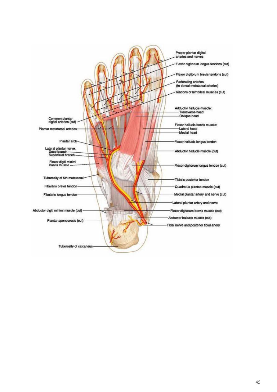
Sole of the foot
The skin of the sole is thickened over the heel and the heads of the metatarsal bones while
it is thin on the toes.
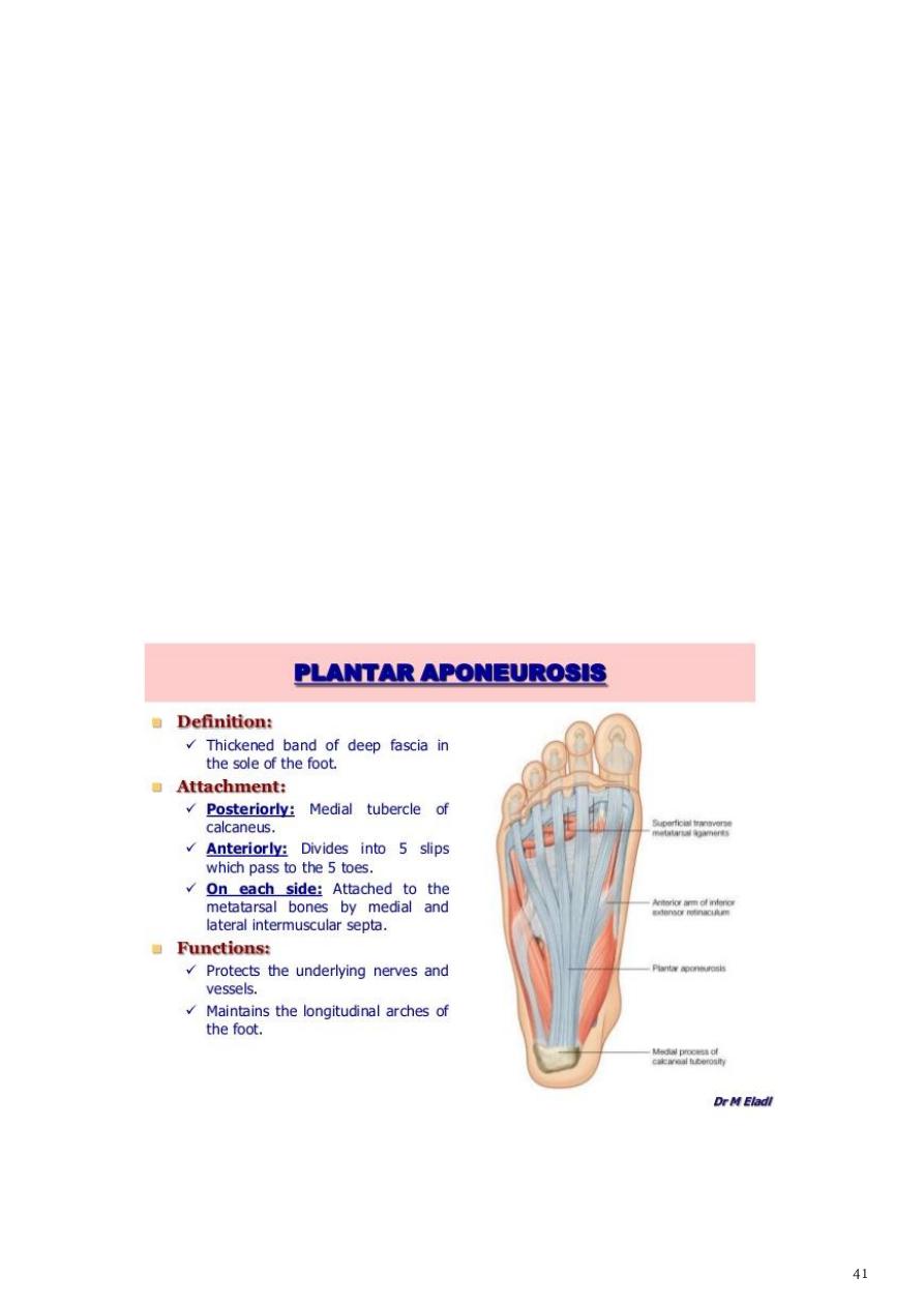
• Deep fascia (planter fascia)
1- it is continuous with the fascia of the dorsum of the foot
2- it is extremely thick in the intermediate region forming the planter aponeurosis but it
is thin medially and laterally where it covers the abductors of the big and little toe .
3- The thinner medial planter fascia covers the intrinsic muscles of the great toe.
4- The lateral planter fascia is thick near the heel and thin toward the little toe covers
the intrinsic muscles of the little toe.
• Planter aponeurosis
1- it consists of longitudinally arranged bands of white fibrous connective tissues which
diverge toward the toes from the medial process of the tuberosity of the calcaneus.
2- it is triangular in shape occupy the central part of the sole.
3- Anteriorly it widens and split into 5 slips near the heads of the metatarsal bones,
each slip pass to one toe bound to the proximal phalanx.
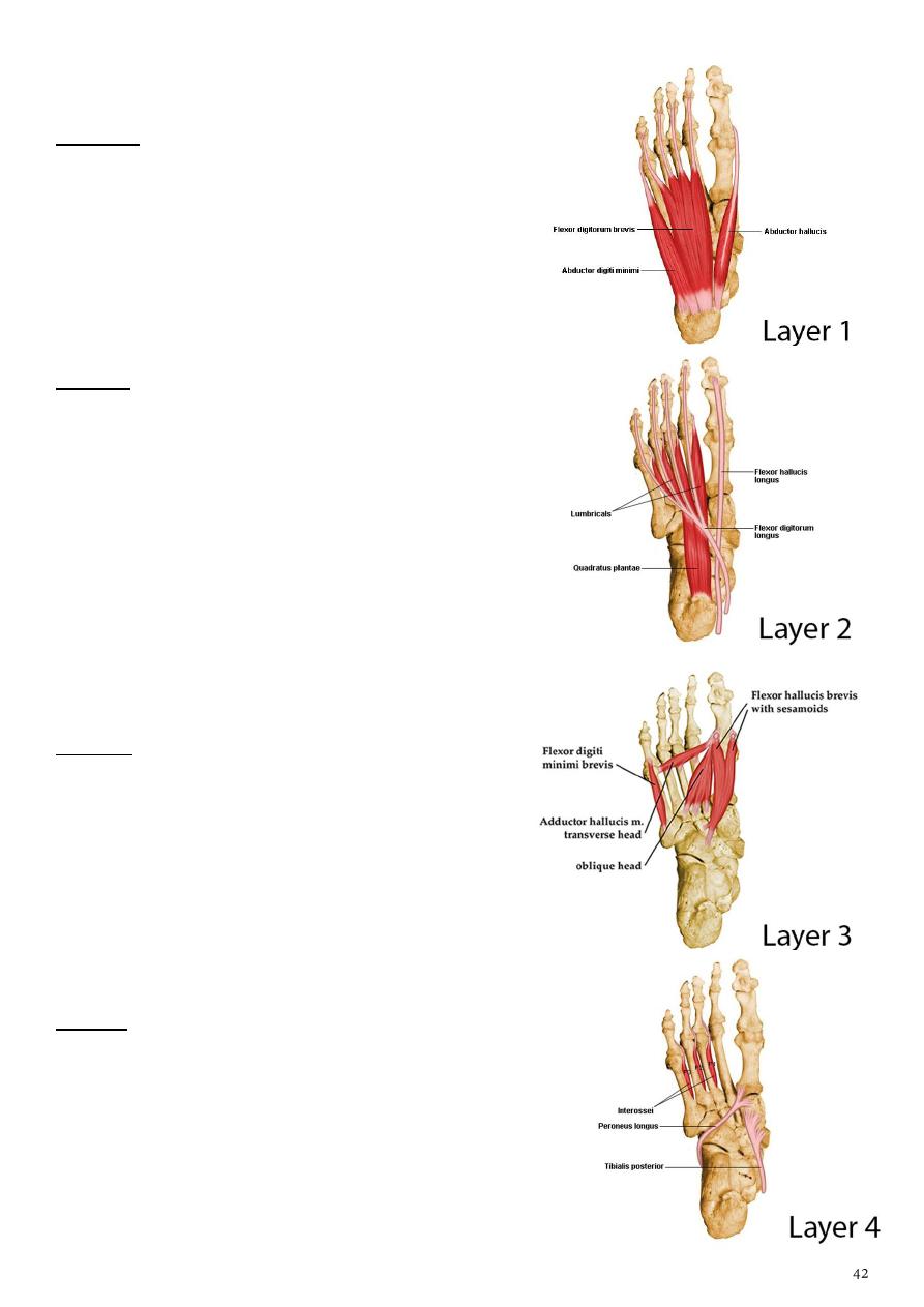
compartments of the sole
first layer ( most superficial layers)
contains 3 muscles:
1-
2-
3-
bductor digiti minimi
2
nd
layer
contains :
1-
2 Muscles (Quadratus plantae , 4 lumbricals )
2-
2 tendon ( of flexor halluces longus and flexor
digitorum longus)
3-
Neurovascular structures (medial planter nerve
and lateral planter nerve)
4-
medial and lateral plantar arteries
3
rd
layer
contains 3 muscles
1-
flexor hallucis brevis
2-
adductor hallucis
3-
flexor digiti minimi
4
th
layer
Contains:
1-
2 muscles (planter interossei – 3 muscles- , dorsal
interossei – 4 muscles-)
2-
2 Tendons ( of tibialis posterior and peroneus
longus )
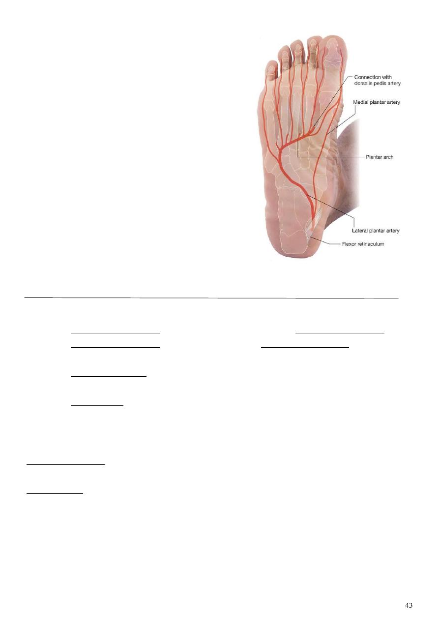
Arteries of the sole of the foot
• The medial planter artery
• Lateral planter artery
• The planter arch
A- The medial planter artery
The smaller branch of the posterior
tibial artery, it does not form an arch
it accompanied the medial planter
nerve and lies first under the
abductor hallucis and then between it
and the flexor digitorum brevis
muscle, supplying these muscles
It gives small branches to the skin, the
muscles and the joints, the artery
divided into three digital branches
which anastomosed with the three planter metatarsal arteries of the planter arch
at the base of the first three interdigital cleft
・Course:
1- The Posterior tibial artery divides into the medial and the Lateral plantar artery.
2- The Medial plantar artery runs forward along the Medial plantar nerve on the medial
side of the foot, and divides into a superficial and a deep branch.
3- The Superficial branch runs forward and medialward to supply the medial side of the
first toe.
4- The deep branch runs forward deep to the adductor hallucis muscle, lateral to the
lateral head of the flexor hallucis brevis muscle to join the first plantar metatarsal
artery.
・Supply:
Superficial branch:
Medial side of the first toe
Deep branch:
Adductor hallucis muscle
Flexor hallucis brevis muscle

B-
It is the larger of the two terminal branches of the posterior tibial artery. Arise deep to
the flexor retinaculum then pass deep to the abductor hallucis and flexor digitorum
brevis ms. runs lateral to the corresponding nerve. At the medial side of the fifth
metatarsal bone the artery sinks deeply, on reaching the base of the 5th metatarsal
bone the artery curve medially across the proximal ends of the second, third and fourth
metatarsal bones to form the planter arch.
1- is a branch of the Posterior tibial artery.
2- It is much larger than the Medial plantar artery.
・Course:
1- The Posterior tibial artery divides into the medial and lateral plantar arteries.
2- The Lateral plantar artery runs forward and lateralward toward the base of the Fifth
metatarsal, and turns medially toward the base of the First metatarsal bone, forming
the plantar arch with the deep plantar branch of the dorsalis pedis artery.
・Supply:
The sole
C- The planter arch
It is formed from the lateral planter artery, the arch completed medially by its union with
the deep planter branch of the dorsalis pedis artery which reaches the sole through the
proximal end of the first intermetatarsal space. The arch lies across the bases of the central
metatarsal bones and deep to the adductor hallucis muscle. The arch gives:
four planter metatarsal arteries run between the metatarsal bones, each artery divided into
pairs of proper digital arteries supply the adjacent sides of the toes. The proper digital
artery to the lateral side of the little toe arise from the lateral planter artery opposite the
base of the fifth metatarsal bone. Each planter metatarsal artery gives an anterior
perforating branch which passes through the interosseous space anastomosed with the
corresponding branch of the dorsal metatarsal artery.
The perforating branches arise from the arch, passes through the proximal ends of the
lateral three intermetatarsal spaces and between the heads of the dorsal interosseous
muscles to join the dorsal metatarsal arteries.
• It is formed from the lateral planter artery, the arch completed medially by its union
with the deep planter branch of the dorsalis pedis artery. The arch gives:
• four planter metatarsal s.
• The proper digital artery to the lateral side of the little.
• Each planter metatarsal artery gives perforating branches which passes through the
interosseous space anastomosed with the corresponding branch of the dorsal
• gives off numerous muscular
The medial planter nerve
It is the larger of the two terminal branches of the tibial nerve. It arise deep to the posterior
part of the abductor hallucis and passes forward accompanied by the small medial planter
artery It gives:
1- muscular branches to the abductor hallucis and flexor digitorum brevis muscles.
2- articular branches supply the joint and tarsal and metatarsal bones.
3- planter cutaneous branches supply the skin of the medial part of the sole.
The medial planter nerve become cutaneous at the middle of the sole divided into proper
digital branch to the medial side of the great toe which supply the flexor hallucis brevis
muscle. Three common digital branches supply the medial three and half toes. The first
common digital branch supply the first lumbrical muscle.

The lateral planter nerve
The smaller of the two planters nerves arising from the tibial nerve under the abductor
hallucis muscle has a distribution like the ulnar nerve in the hand. The nerve pass between
flexor digitorum brevis and quadratus plantae muscles. it gives:
• muscular branches to the abductor digiti minimi and quadratus plantae muscles.
• articular branches.
At the lateral margin of the quadratus plantae muscle the nerve divided into superficial and
deep branches.
The deep branch
• Sinks into the interosseous –adductor compartment with the lateral planter artery and
passes medially across the bases of the metatarsal bones posterior to the planter arch. It
gives:
1- muscular branches to the lateral 3 lumbrical muscles, the adductor hallucis m. The
interosseous muscles.
2- articular branches to the intertarsal and tarsometatarsal joints.
The superficial branch divided into proper digital branch to the lateral side of the little toe
supply the flexor digiti minimi muscle, and a common digital branch communicates with
the third common branch of the medial planter nerve and divides into two proper digital
branches to the adjacent sides of the fourth and fifth toes.
Part7
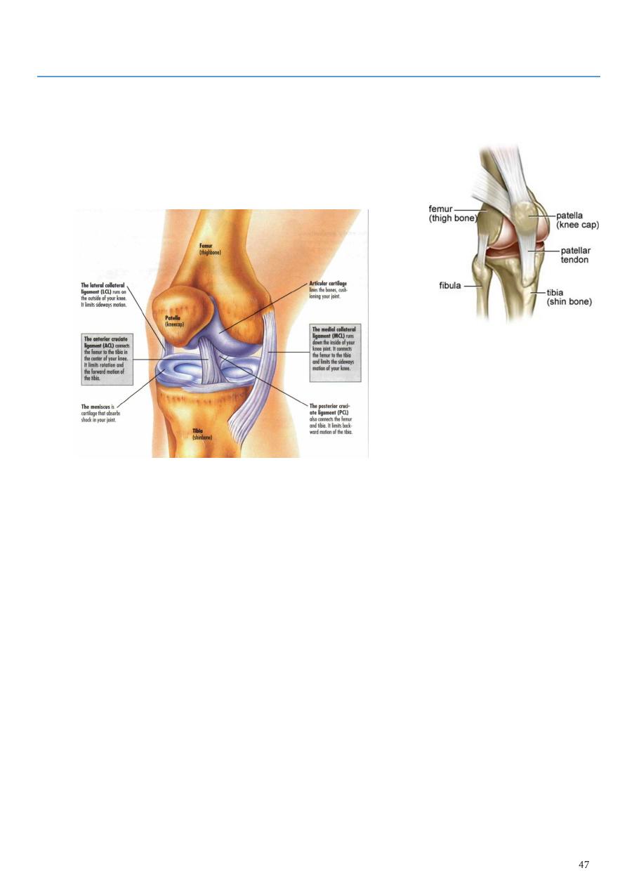
: Knee Joint
• It is a synovial joint of the hinge type, it is unstable joint but this overcome by certain
mechanism:
1- expansion of the upper end of the tibia and lower end of the femur.
2- Presence of the strong collateral ligament and tendons.
3- Strong capsule.
4- Presence of the intra-articular ligaments .
• The articular surface of the femur is the condyles while the articular surface of the
tibia is the tibial condyles which is deepen by the mensci. On the front of the joint
the capsule is absent permitting the synovial membrane to pouch upward beneath
the quadriceps tendon forming the suprapatellar bursa.
• The capsule of the joint attached to the condyles of the femur superiorly and to the
tibial condyles and the margin of the mensci inferiorly.
Ligaments of the joint
1- The ligamentum patellae which is a continuation of the quadriceps femoris tendon
run on the patella to reach the tibial tuberosity
2- Collateral ligament they are tibial and fibular collateral ligaments. They are very
strong ligaments.
3- cruciate ligaments : these are two ligaments lie inside the joint cross each other.
Anterior cruciate ligament extends from in front of condylar eminence of tibia to
the posterior part of the lateral condyle of the femur it passes upward and
backwards.
Posterior cruciate ligament passes upwards and forwards from the posterior part
of the tibial intercondylar area to the lateral surface of the medial condyle of the
femur. It prevents anterior displacement of the femur on the tibia
.

Synovial membrane
It lines all the structures which forms the wall of the cavity of the knee joint except the
articular surfaces of the bones, mensci and the posterior part of the fibrous capsule where
the synovial membrane turns forwards to enclose the cruciate ligaments.
Anastomosis around the knee joint
Formed by 8 arteries these are:
1- 2 lateral and 2 medial genicular arteries from the popliteal artery.
2- Descending genicular artery from the femoral artery.
3- Anterior and posterior tibilal recurrent arteries.
4- Genicular artery from the lateral circumflex artery.
The middle genicular artery play a little part since it supply the structures within the capsule
of the joint.
Nerves of the joint
1- femoral nerve through nerve of vasti muscles.
2- Common peroneal nerve through superior and inferior lateral genicular nerves.
3- Tibial nerve through superior and inferior medial genicular nerves.
4- Obturator nerve.
Movement of the joint
1- Flexion through biceps, semitendinosus and semimembranosus; assisted by the
sartorius, gracilis and popliteus.
2- Extension by quadriceps femoris m.
3- Rotation: medial rotation by sartorius, gracilis and semitendinosus. Lateral rotation by
biceps femoris m.

Part 8
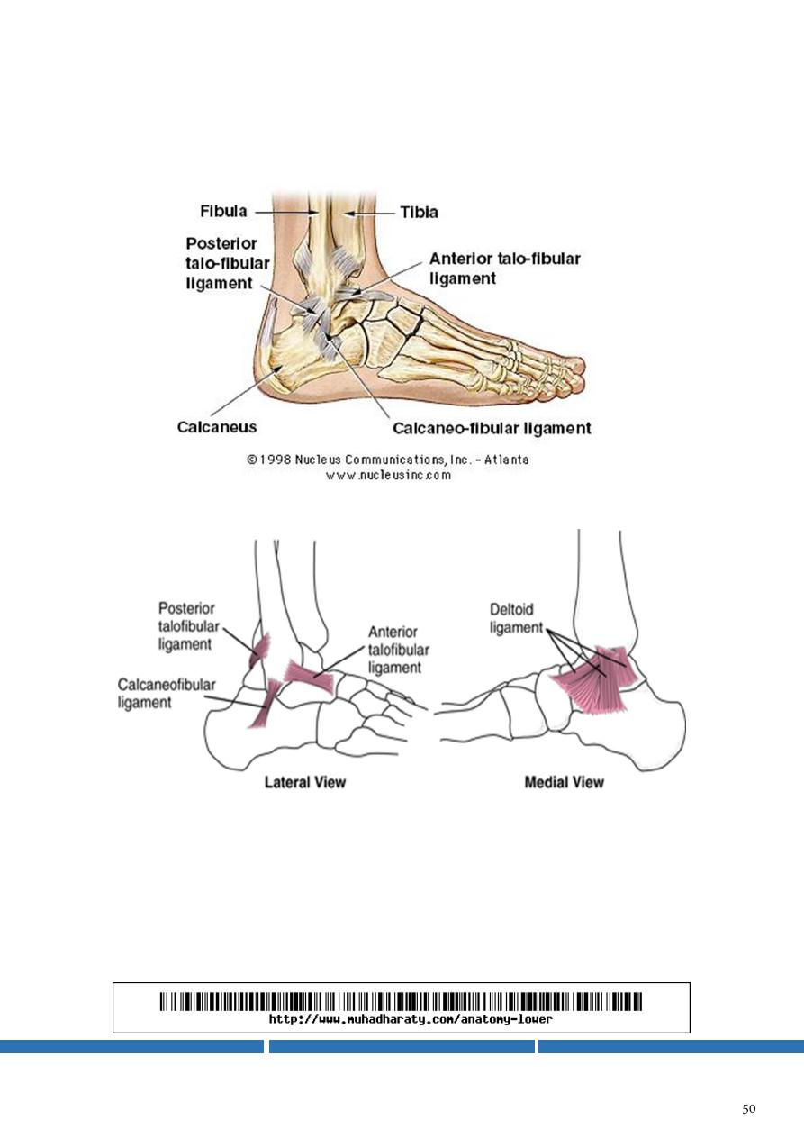
: Ankle joint
• This is a hinge type of joint between the trochlea of the talus with the distal end of the
tibia and medial malleolus medially and the lateral surface of the body of the talus with
the lateral malleolus laterally
It is strong and stable joint by:
1- The powerful ligament and tendons.
2- The insertion o the trochlea into the deep socket between medial and lateral malleoli.
Ligaments of the joint
1- medial (Deltoid) ligament. It is a very strong ligament radiates from the
distal border of the medial malleolus to the medial side of the talus, to the
medial surface of the calcaneus, to the navicular bone and to the neck of
the talus.
2- Lateral ligament consists of 3 bands, the anterior and posterior are
thickenings of the fibrous capsule, the anterior one is the anterior
talofibular ligament and the posterior is the posterior talofibular ligament.
And the calcaneofibular ligament extend from the distal end of the lateral
malleolus to the lateral surface of the calcaneus.
Anastomois around the ankle joint
1- on the lateral side the lateral malleolar branch of the anterior tibial artery
and the lateral tarsal branch of the dorsalis pedis artery anastomosed with
the perforating branch and terminal branches of the peroneal artery.
2- On the medial side the medial malleolar artery anastomosed with the
medial calcanean branch of the posterior tibial artery. The posterior tibial
artery itself also anastomosed with the peroneal artery posterior to the
ankle joint.
- Nerve supply of the joint from the tibial nerve and the lateral branch of the
deep peroneal nerve
- Movements of the joint are the dorsiflexion and planterflexion.
- Dorxiflexion is through the muscles of the anterior compartment of the leg;
while the planteflexion through the muscles of the superficial compartment
of the back of the leg.
- The maximum stability of the joint is achieved in dorxiflexion.