The Human
Digestive SystemOverview of Digestion
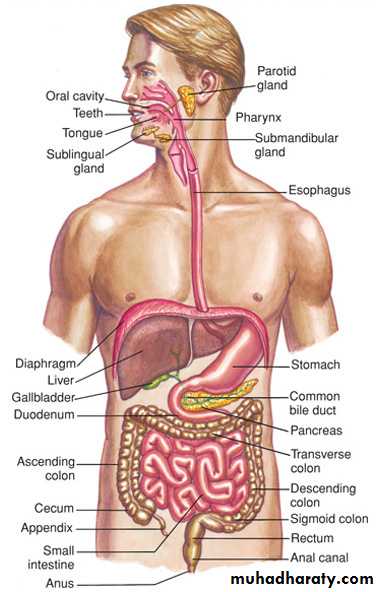
2 main groups of organs in the digestive system.1. Alimentary Canal (nutrition)
a. Mouth
b. Pharynx
c. Esophagus
d. Stomach
e. Small Intestine
f. Large Intestine
• Accessory Digestive Organs
• a. Teeth• b. Tongue
• c. Gall bladder
• d. Salivary glands
• e. Liver
• f. pancreas
The chemical Foundation of Digestion
All organisms need food, and all foods contain nutrients. Nutrients are the substances that provide the energy and the materials needed for growth, repair, regulation, and maintenance of the cells.
Therefore, food is what the organism consumes, and nutrients are substances within food that are needed by the cells to sustain life.
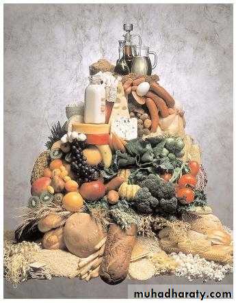
The 6 Essential Nutrients
• Carbohydrates• Source
Plants
• Function: Major source of energy in the body
Such as : sugar from candy bars or fruits and vegetables
• Lipids (Fats)
• Sources
Ingestion of animal and plant fats conversion of carbohydrates into fats
• Functions
Storage of energy, component of cell membranes, cushion for delicate organs, carriers for certain vitamins, raw materials for important chemicals
The 6 Essential Nutrients
• Protein• Sources
Meat, Fish, Poultry, milk, cheese, legumes, eggs, whole grains
• Function
Broken down into amino acids which are used in the construction of human proteins
Proteins are essential for the building, repair, and maintenance of cell structure.
• The predominant part of muscles, nerves, skin, and hair is protein.
• Functional Molecules in the blood and in the organ system such as enzymes , antibodies and some types of hormones are specialized proteins.
The 6 Essential Nutrients
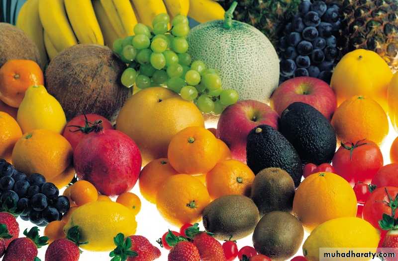
• VitaminsSources
Various foods contain different types of vitamins
Functions
Required in small amounts for various metabolic functions including enzymatic activity
Some are fat soluble and stored in the body, while others are water soluble and need to be replenished on a daily basis
The 6 Essential Nutrients
• Minerals• Sources
Various foods we eat contain different minerals
Ie. Milk contains calcium, salt contains sodium, cereals often contain iron, bananas contain potassium
Function
Used throughout the body for many functions
Calcium – tooth and bone formation
Iron – haemoglobin
Sodium / potassium – nervous system
The 6 Essential Nutrients
• Water• Sources
Various foods and drink – ie. The tap
• Function
Used mostly as a solvent throughout the body, but also responsible for maintaining cell structure
Carbohydrates, lipids, and proteins require digestion.
Vitamins, water, and minerals do not require digestionThe Human Digestive System
Foods taken into the body consist of large complex organic compounds.Digestion must occur in order to release the nutrients contained within the food.
Digestion will break down the large complex organic compounds into smaller, simpler units that can be absorbed and used by the cells of the organism.
Two Types of Digestion
• Mechanical DigestionPhysical breaking up of food into smaller pieces by the teeth.
The tongue manipulates the food into a mass called the bolus
The squishing action in the esophagus and intestines further break up the food mass
The Churning action of the stomach muscles contracting to mix food with the digestive juices in the stomach
Functions of Gastro Intestinal Tract
18-5
Is movement of food through GI tract by means of:
Ingestion--taking food into mouthMastication--chewing food and mixing it with saliva
Deglutition--swallowing food
Peristalsis--rhythmic wave-like contractions that move food through GI tract
Motility
18-6
Includes release of exocrine and endocrine products into Gastro Intestinal tract
Exocrine secretions include: HCl, H2O, HCO3-, bile, lipase, pepsin, amylase, trypsin, elastase, and histamineEndocrine includes hormones secreted into stomach and small intestine to help regulate GI system
e.g. gastrin, secretin, Cholecystokinin(CCK), GIP, GLP-1, guanylin, VIP, and somatostatin
Secretion
18-7
Is passage of digested end products into blood or lymph
Absorption18-8
Digestion
Refers to breakdown of food molecules into smaller subunits
Includes temporary storage and subsequent elimination of indigestible components of food
Storage and Elimination
18-9
Immune Barrier
Includes physical barrier formed by tight junctions between cells of small intestineAnd cells of the immune system that reside in connective tissue just below epithelium
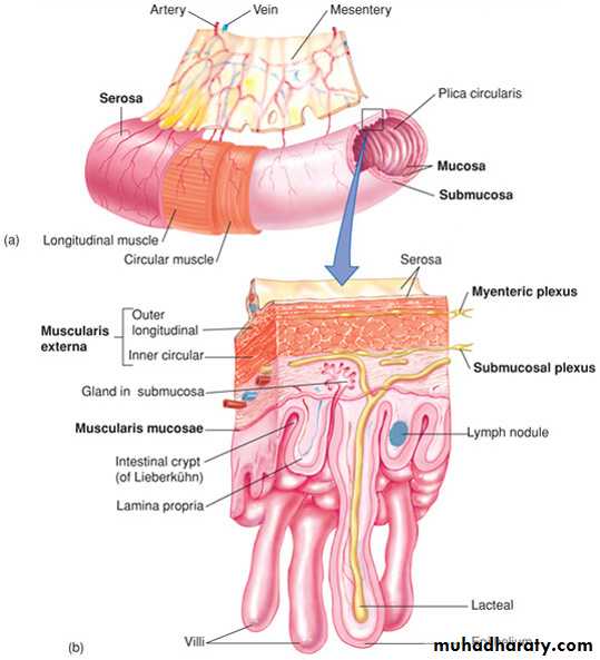
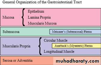
Layers of the Gastro Intestinal Tract
• Wall of GI tract from lower esophagus to anal canal has same basic 4 layers• Mucosa – inner lining
• Epithelium protection, secretion, absorption
• Lamina propria – connective tissue with blood and lymphatic vessels and mucosa-associated lymphatic tissue (MALT)
• Muscularis mucosae – thin layer of smooth muscle making folds to increase surface area
• Submucosa
• Connective tissue binding mucosa to muscularis
• Contains many blood and lymphatic vessels
• Submucosal plexus
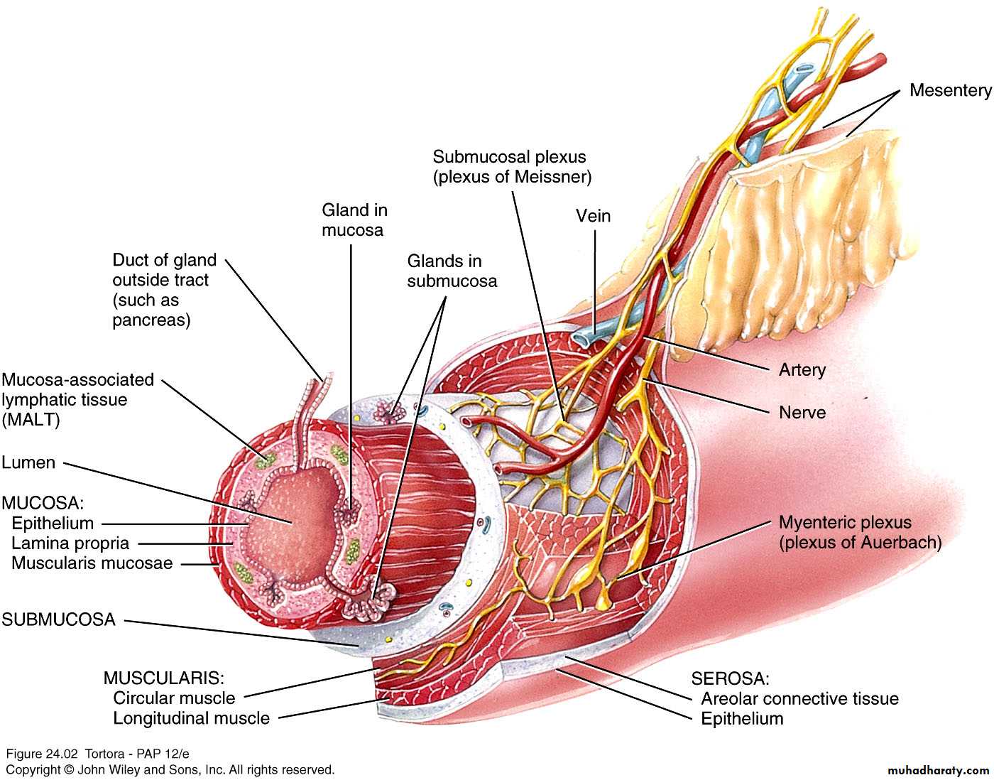
Layers of the GI tract
• Muscularis• Voluntary skeletal muscle found in mouth, pharynx, upper 2/3 of esophagus, and anal sphincter
• Involuntary smooth muscle elsewhere
• Arranged in inner circular fibers and outer longitudinal fibers
• Myenteric plexus between muscle layers
• Serosa
• Outermost covering of organs suspended in abdomino pelvic cavity
• Also called visceral peritoneum
• Esophagus lacks serosa – has adventitia
Peritoneum
Largest serous membrane of the bodyDivided into
Parietal peritoneum – lines wall of cavity
Visceral peritoneum – covers some organs
Also called serosa
Space between is peritoneal cavity
5 major peritoneal folds
Greater omentum, falciform ligament, lesser omentum, mesentery, and mesocolon
Weave between viscera binding organs together
Copyright 2009, John Wiley & Sons, Inc.
Layers of the gastrointestinal tract
The Tongue:
• is accessory digestive organand composed of skeletal muscle covered by mucous membrane .
the most important articulator for speech production.
• Maneuvers food for chewing, shapes mass, forces food back for swallowing, during speaking the tongue can make amazing range of movements
The primary function of the tongue is to provide a mechanism for taste. Taste buds are located on different areas of the tongue, but are generally found around the edges. They are sensitive to four main tastes: Bitter, Sour , Salty & Sweet
Digestion in the mouth
Mechanical digestion in the mouthChewing or mastication
Food manipulated by tongue, ground by teeth, and mixed with saliva
Forms bolus
Chemical digestion in the mouth
Salivary amylase secreted by salivary glands acts on starches
Only monosaccharides can be absorbed
Continues to act until inactivated by stomach acid
Lingual lipase secreted by lingual glands of tongue acts on triglycerides
Becomes activated in acidic environment of stomach
Pharynx
Passes from mouth into pharynx
3 parts
Nasopharynx
Functions only in respiration
Oropharynx
Digestive and respiratory functions
Laryngopharynx
Digestive and respiratory functions
Esophagus
Secretes mucous, transports food – no enzymes produced, no absorptionMucosa – protection against wear and tear
Submucosa
Muscularis divided in thirds
Superior 1/3 skeletal muscle
Middle 1/3 skeletal and smooth muscle
Inferior 1/3 smooth muscle
2 sphincters – upper esophageal sphincter (UES) regulates movement into esophagus, lower esophageal sphincter (LES) regulates movement into stomach
Adventitia – no serosa – attaches to surroundings
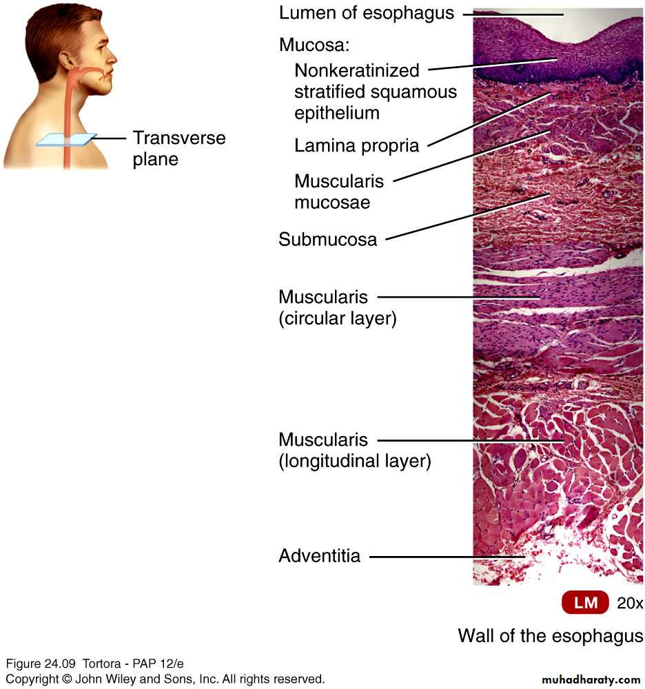
Histology of the esophagus
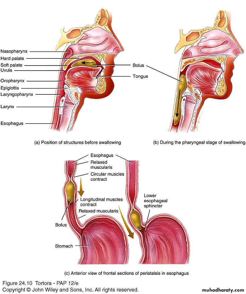
DeglutitionAct of swallowing
Facilitated by secretions of saliva and mucus
Involves mouth, pharynx, and esophagus
3 stages
Voluntary – bolus passed to oropharynx
To swallow, larynx is raised so that epiglottis covers entrance to respiratory tract
Pharyngeal – involuntary passage through pharynx into esophagus
Esophageal – involuntary passage through esophagus to stomach
Peristalsis pushes bolus forward
After food passes into stomach, the gastroesophageal sphincter constricts, preventing reflux
Deglutition (swallowing)
Is most distensible part of GI tract
Empties into the duodenumFunctions in: storage of food; initial digestion of proteins; killing bacteria with high acidity; moving soupy food mixture (chyme) into intestine
• Stomach
Stomach
Serves as mixing chamber and holding reservoir4 main regions
Cardia, fundus, body, pylorus
Same 4 layers
Mucosa – gastric glands open into gastric pits
3 types of exocrine gland cells – mucous neck cells (mucus), parietal cells (intrinsic factor and HCl), and chief cells (pepsinogen and gastric lipase)
G cell – endocrine cell – secretes gastrin
Submucosa
Muscularis – additional 3rd inner oblique layer
Serosa – part of visceral peritoneum
Stomach
Is enclosed by gastroesophageal sphincter on top and pyloric sphincter on bottomIs divided into 4 regions:
Cardia
Fundus
Body
Pylorus
18-26
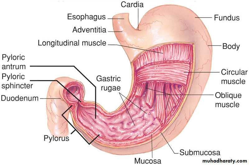
Inner surface of stomach is highly folded into rugae
Contractions of stomach churn chyme, mixing it with gastric secretionsEventually these will propel food into small intestine
Stomach continued
18-27Stomach continued
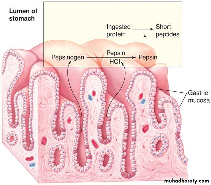
Gastric mucosa has gastric pits in its foldsCells that line folds deeper in the mucosa, are exocrine gastric glands
18-28
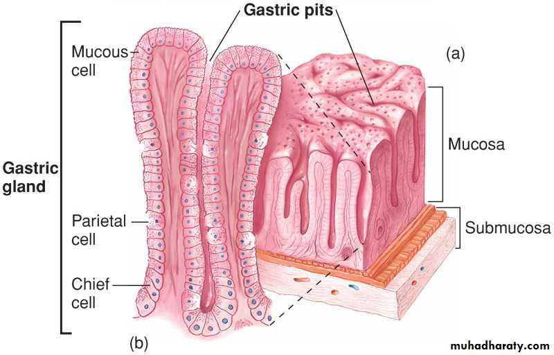
Gastric glands contain cells that secrete different products that form gastric juice
Goblet cells secrete mucusParietal cells secrete HCl and intrinsic factor (necessary for B12 absorption in intestine)
Chief cells secrete pepsinogen (precursor for pepsin)
Stomach continued
18-29
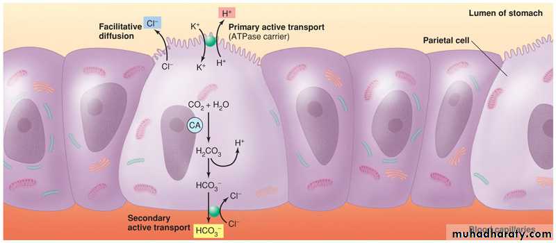
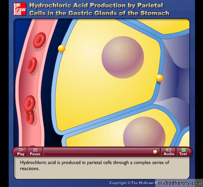
HCl in Stomach
Is produced by parietal cells which pump H+ into lumen via an H+/ K+ pump (pH ~1)Cl- is secreted by facilitated diffusion
H+ comes from dissociation of H2CO3
Cl- comes from blood side of cell in exchange for HCO3-
18-31
Mechanical and Chemical Digestion
Mechanical digestionMixing waves – gentle, rippling peristaltic movements – creates chyme
Chemical digestion
Digestion by salivary amylase continues until inactivated by acidic gastric juice
Acidic gastric juice activates lingual lipase
Digest triglycerides into fatty acids and diglycerides
Parietal cells secrete H+ and Cl- separately but net effect is HCl
Kills many microbes, denatures proteins
Is secreted in response to the hormone gastrin; and Acetylcholine from vagus nervous
These are indirect effects since both stimulate release of histamine which causes parietal cells to secrete HClHCl in Stomach
18-32
Makes gastric juice very acidic which denatures proteins to make them more digestible
Converts pepsinogen into pepsinPepsin is more active at low pHs
HCl in Stomach
18-33
Both HCL and pepsin can damage lining and produce a peptic ulcer
1st line of defense is the adherent layer of mucus= a stable gel of mucus coating the gastric epithelium
Contains bicarbonate for neutralizing HCL
Is a barrier to actions of pepsin
Gastric epithelial cells contain tight junctions to prevent HCL and pepsin from penetrating the surface
Gastric epithelial cells are replaced every 3 days
Protection of Stomach Against HCL and Pepsin
18-34
Digestion and Absorption in Stomach
Proteins are partially digested by pepsinCarbohydrate digestion by salivary amylase is soon inactivated by acidity
Alcohol and aspirin are the only commonly ingested substances that are absorbed
18-35
Gastric and Peptic Ulcers
Peptic ulcers are erosions of mucous membranes of stomach or duodenum caused by action of HClIn Zollinger-Ellison syndrome, duodenal ulcers result from excessive gastric acid in response to high levels of gastrin
Helicobacter pylori infection is associated with ulcers
Antibiotics are useful in treating ulcers
And also proton pump inhibitors such as Prilosec
Acute gastritis is an inflammation that results in acid damage due to histamine released by inflammation
Is why histamine receptor blockers such as Tagamet and Zantac can treat gastritis
18-36
Zollinger-Ellison syndrome is a complex condition in which one or more tumors form in your pancreas or the upper part of your small intestine (duodenum). These tumors, called gastrinomas, secrete large amounts of the hormone gastrin, which causes your stomach to produce too much acid. The excess acid, in turn, leads to peptic ulcers.
Zollinger-Ellison syndrome (ZES) is rare. The disease may occur at any time in life, but people are usually diagnosed between ages 30 and 50. Medications to reduce stomach acid and heal the ulcers is the usual treatment for Zollinger-Ellison syndrome.
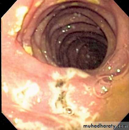
Gastritis describes a group of conditions with one thing in common: inflammation of the lining of the stomach. The inflammation of gastritis is often the result of infection with the same bacterium that causes most stomach ulcers. However, other factors — such as injury, regular use of certain pain relievers prolonged use of nonsteroidal anti-inflammatory drugs (also known as NSAIDs) such as aspirin or ibuprofen. Sometimes gastritis develops after major surgery, traumatic injury, burns, or severe infections. or drinking too much alcohol — also can contribute to gastritis. and stress; certain autoimmune disorders can cause gastritis as well. The most common symptom is abdominal upset or pain. Other symptoms are indigestion, abdominal bloating, nausea, and vomiting and pernicious anemia. Some may have a feeling of fullness or burning in the upper abdomen.
Gastritis may occur suddenly (acute gastritis) or it can occur slowly over time (chronic gastritis). In some cases, gastritis can lead to ulcers and an increased risk of stomach cancer. For most people, however, gastritis isn't serious and improves quickly with treatment.
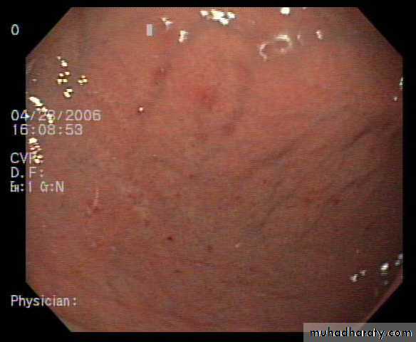
Acute gastritis with superficial erosions.
Small Intestine90% of absorption occurs in the small intestine
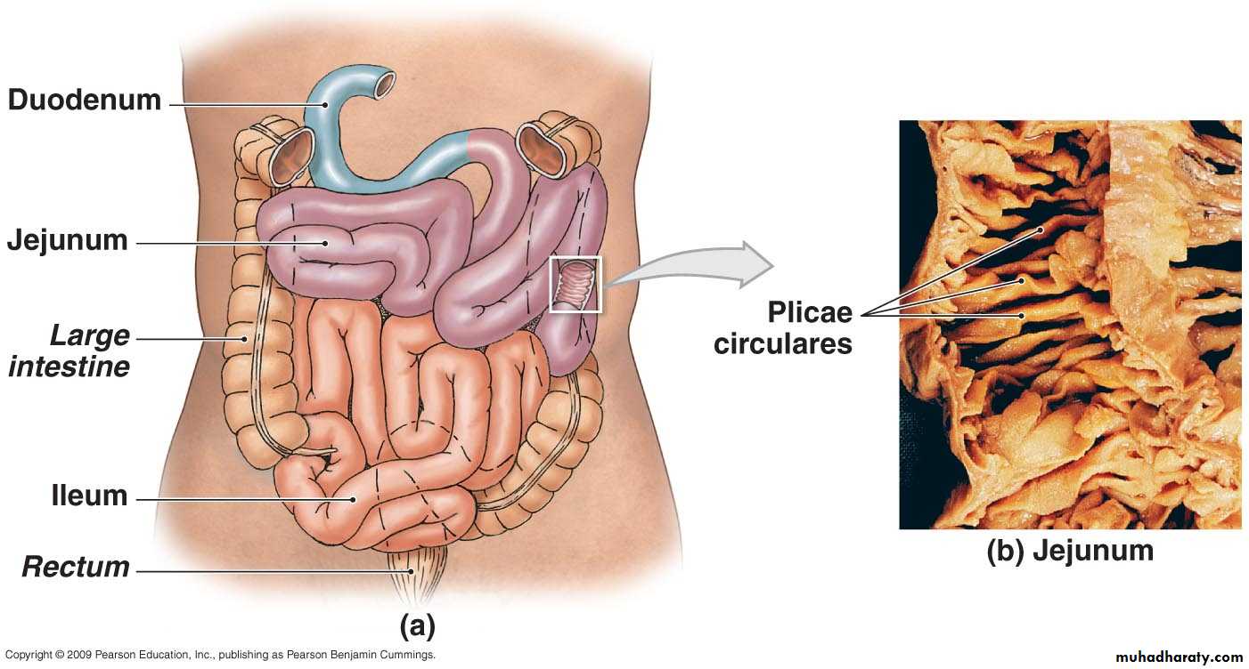
Small intestine
• Anatomically is 3 regions – duodenum, jejunum, and ileum• Histologically as the same of other portion of Gastro intestinal track is composed of 4 layers
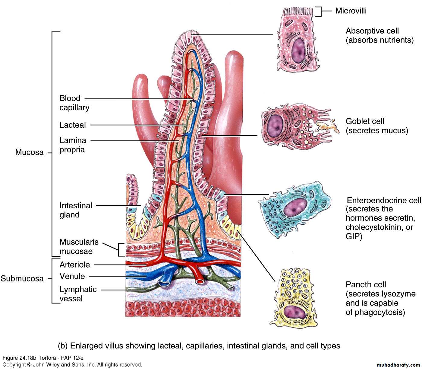
• Mucosa
• Absorptive cells (digest and absorb), goblet cells (mucus), intestinal glands (intestinal juice), Paneth cells (lysozyme), and enteroendocrine cells
• Abundance of Mucosa-Associated Lymphoid Tissue (MALT) responsible for immunity
• Submucosa
• Duodenal glands secrete alkaline mucus
• Muscularis
• Serosa
• Completely surrounds except for major portion of duodenum
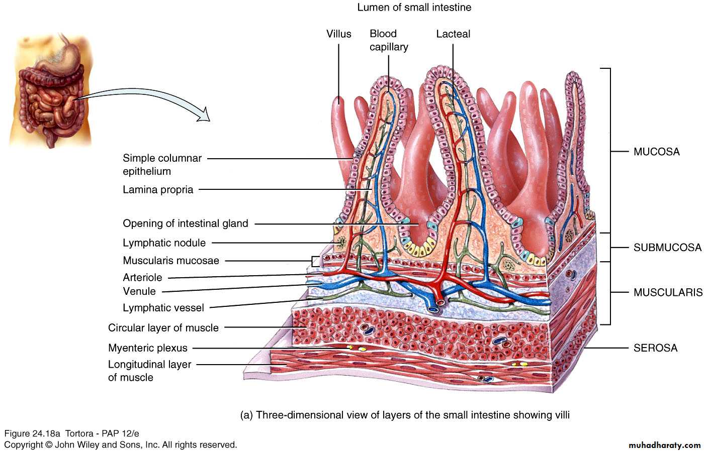
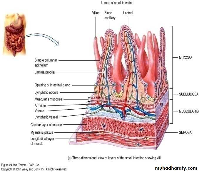
Special structural features increase surface area for digestion and absorption
• cause chyme to spiral and afford an increased surface for absorption. They are covered with small fingerlike projections called villi Each villus, in turn, is covered with microvilli.
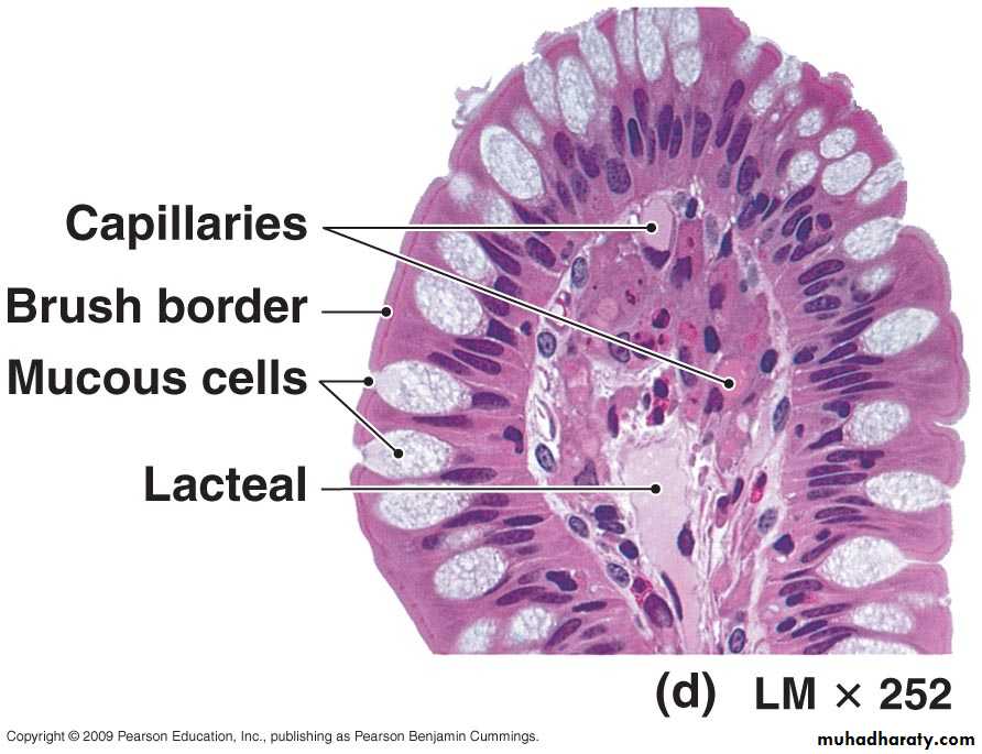
Villi: Fingerlike projections of mucosa
Contains arteriole, venule, blood capillary, and lacteal
Microvilli
Projects of apical membrane of absorptive cells
Brush border with brush border enzymes
Absorb fats and nutrients from the chyme.
Histology of the small intestine
Small Intestine
The DuodenumThe segment of small intestine closest to stomach
24-27 cm (10 -12 in.) long
“Mixing bowl” that receives chyme from stomach and digestive secretions from pancreas and liver
Functions of the duodenum
To receive chyme from stomach
To neutralize acids before they can damage the absorptive surfaces of the small intestine
Small Intestine
The Jejunum
Is the middle segment of small intestine
2.5 meters (8.2 ft) long
Is the location of most
Chemical digestion
Nutrient absorption
Has few plicae circulares
Small villi
Small Intestine
The IleumThe final segment of small intestine
3.5 meters (11.48 ft) long
Ends at the ileocecal valve, a sphincter that controls flow of material from the ileum into the large intestine
Small Intestine
Intestinal juice and brush-border enzymes
Intestinal juice1-2L daily
Contains water and mucus, slightly alkaline
Provide liquid medium aiding absorption
Brush border enzymes
Inserted into plasma membrane of absorptive cells
Some enzymatic digestion occurs at surface rather than just in lumen
α-dextrinase, maltase, sucrase, lactase, aminopetidase, dipeptidase, nucleosidases and phosphatases
Myenteric Plexus: are intricate layers of nervous tissue that control movements in the esophagus, stomach, and intestines.Two major nerve centres are involved: the myenteric plexus (Auerbach’s plexus) and the submucous plexus (Meissner’s plexus). The myenteric plexus is situated between the circular muscle layer and the longitudinal muscle layer
• The myenteric plexus receives its messages from the vagus nerve and responds by transmitting the message to muscle cells. the myenteric plexus stimulates the muscles to contract in peristaltic waves and promotes
• secretions of intestinal juices,
• and allows
• muscular constrictions (sphincters)
• to open, thus permitting food to pass
• from
• one part of the digestive system t
• o another.
Mechanical Digestion
Governed by myenteric plexusSegmentations
Localized, mixing contractions
Mix chyme and bring it in contact with mucosa for absorption
Migrating motility complexes (MMC)
Type of peristalsis
Begins in lower portion of stomach and pushes food forward
Chemical digestion
Carbohydrates
Pancreatic amylase
α-dextrinase, sucrase, lactase, maltase in brush border
Ends with monosaccharides which can be absorbed
Proteins
Trypsin, chymotrypsin, carboxypeptidase, and elastase from pancreas
Aminopeptidase and dipeptidase in brush border
Lipids and Nucleic Acids
LipidsPancreatic lipase most important in triglyceride digestion
Emulsification by bile salts increases surface area
Amphipathic – hydrophobic and hydrophilic regions
Nucleic acids
Ribonuclease and deoxyribonuclease in pancreatic juice
Nucleosidases and phosphatases in brush border
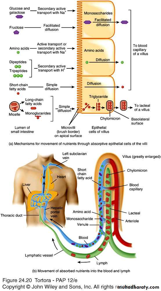
Absorption of:
MonosaccharidesAll dietary carbohydrates digested are absorbed
Only indigestible cellulose and fibers left in feces
Absorbed by facilitated diffusion or active transport into blood
Amino acids, dipetides and tripeptides
Most absorbed as amino acids via active transport into blood
½ of absorbed amino acids come from proteins in digestive juice and dead mucosal cells
Lipids
All dietary lipids absorbed by simple diffusion
Short-chain fatty acids go into blood for transport
Long-chain fatty acids and monoglycerides
Large and hydrophobic
Bile salts form micelles to ferry them to absorptive cell surface
Reform into triglycerides forming chylomicrons
Leave cell by exocytosis
Enter lacteals to eventually enter blood with protein coat of chylomicron keeping them suspended and separate
Absorption of digested nutrients in the small intestine
Absorption of:
ElectrolytesFrom GI secretions or food
Sodium ions (Na+) reclaimed by active transport
Other ions also absorbed by active transport
Vitamins
Fat-soluble vitamins A, D, E, and K absorbed by simple diffusion and transported with lipids in micelles
Most water-soluble vitamins also absorbed by simple diffusion
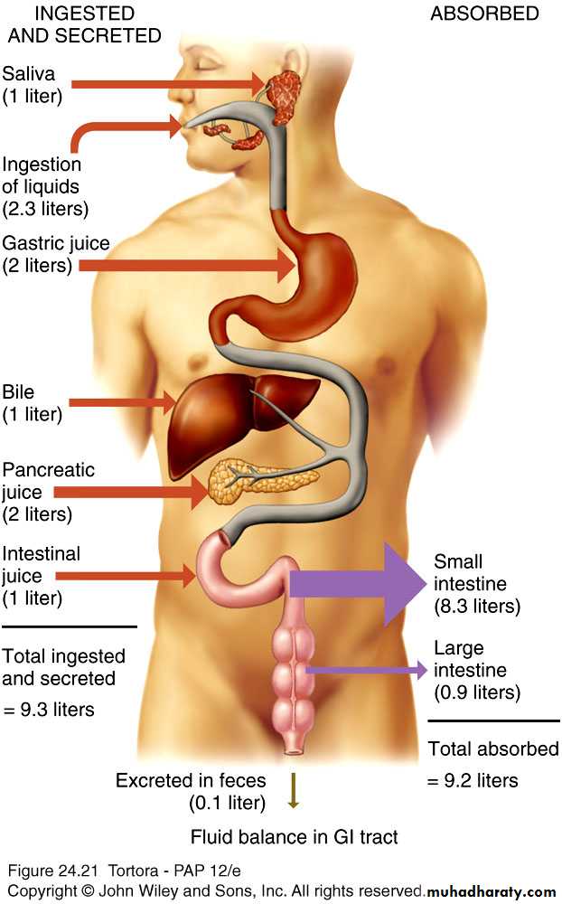
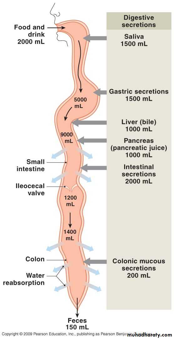
Water
9.3L comes from ingestion (2.3L) and GI secretions (7.0L)
Most absorbed in small intestine, some in large intestine
Only 100-150 ml excreted in feces
All water absorption by osmosis
Daily volumes of fluid ingested, secreted, absorbed, and excreted from the GI tract
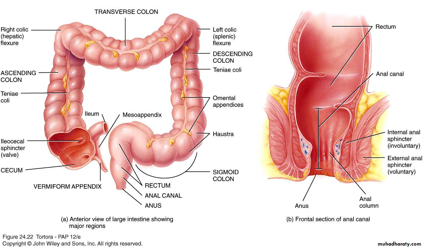
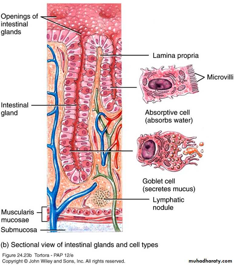
Large intestine
Overall function to complete absorption, produce certain vitamins, and form and expel feces4 major regions – cecum, colon, rectum, and anal canal
Ileocecal sphincter between small and large intestine
Colon divided into ascending, transverse, descending and sigmoid
Opening of anal canal (anus) guarded by internal anal sphincter of smooth muscle and external anal sphincter of skeletal muscle
Anatomy of the large intestine
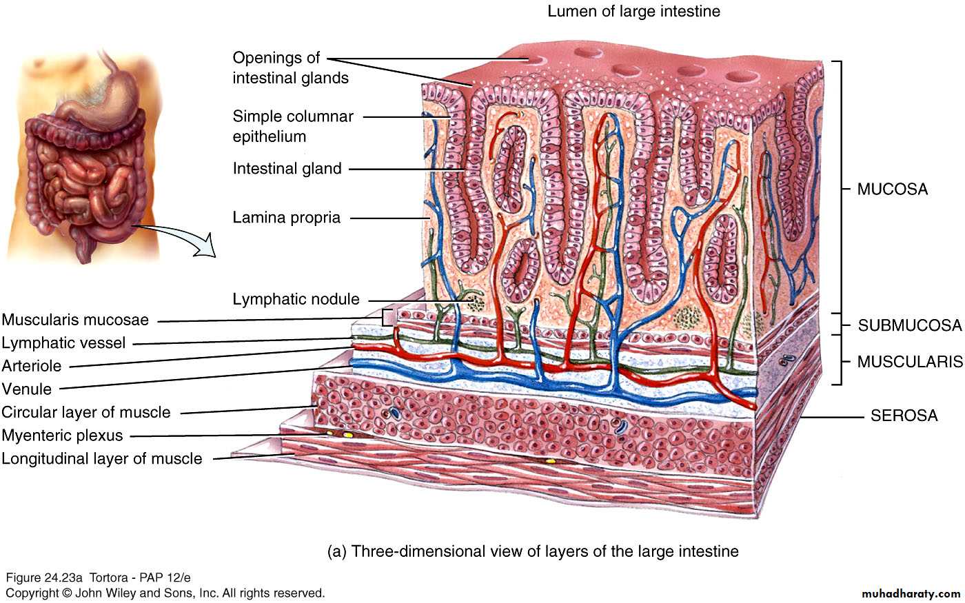
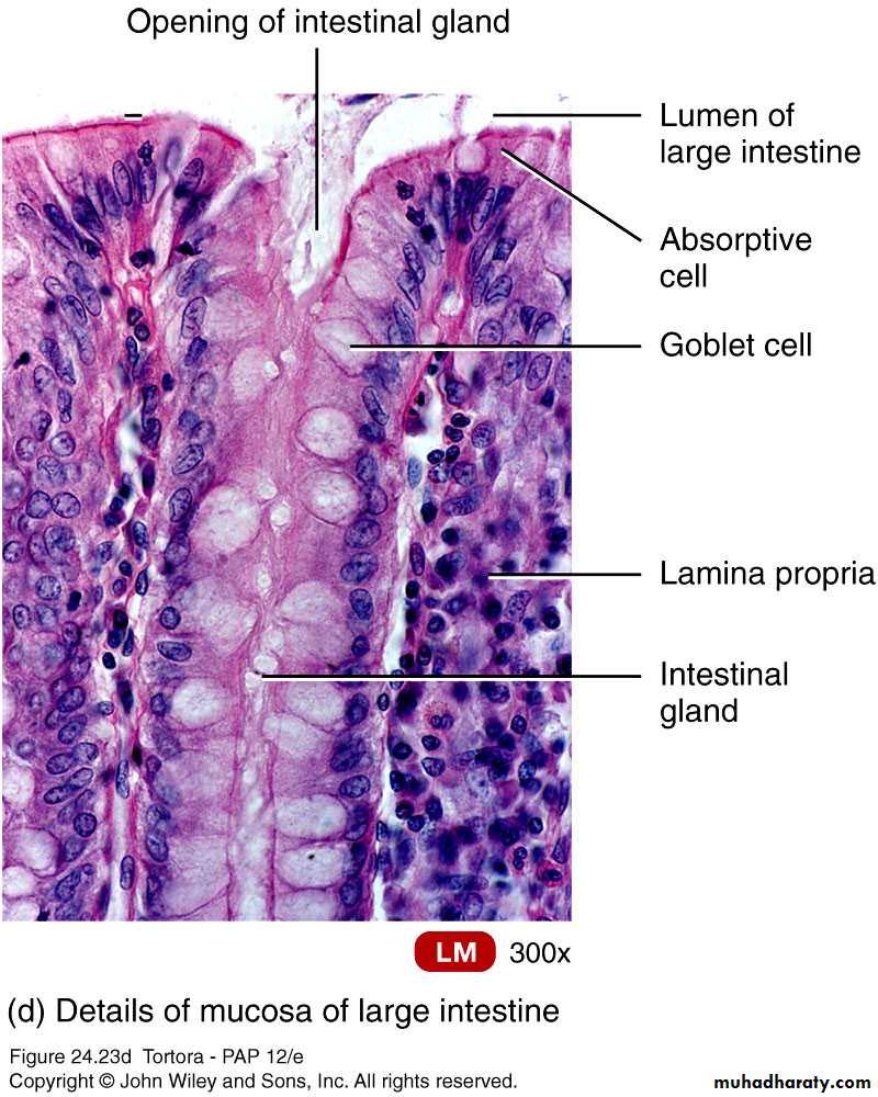
Large Intestine
Same 4 layersMucosa – mostly absorptive and goblet cells
No circular folds or villi
Does have microvilli
Submucosa
Muscularis
Longitudinal muscle modified to form teniae coli
Forms haustra – pouches
Serosa
Digestion of the Large Intestine
Mechanical digestion
Haustral churning
Peristalsis
Mass peristalsis – drives contents of colon toward rectum
Chemical digestion
Final stage of digestion through bacterial action
Ferment carbohydrates, produce some B vitamins and vitamin K
Mucus but no enzymes secreted
Remaining water absorbed along with ions and some vitamins
Secretions of Large Intestine
Mucus provides protectionParasympathetic stimulation increases rate of goblet cell secretion
Pumps
Exchange of bicarbonate ions for chloride ions
Exchange of sodium ions for hydrogen ions
Bacterial actions produce gases called flatus
Histology of the large intestine
Copyright 2009, John Wiley & Sons, Inc.
Histology of the large intestine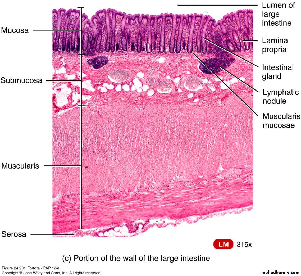
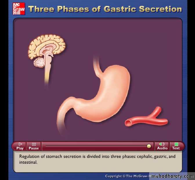
Phases of digestion
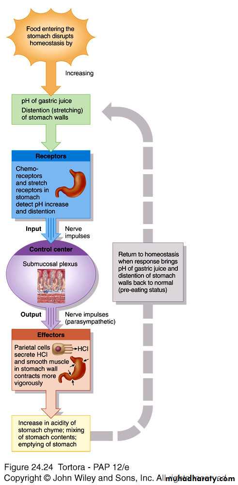
Cephalic phaseSmell, sight, thought or initial taste of food activates neural centers – prepares mouth and stomach for food to be eaten
Gastric phase
Neural and hormonal mechanisms promote gastric secretion and motility
Intestinal phase
Begins when food enter small intestine
Slows exit of chyme from stomach
Stimulates flow of bile and pancreatic juice
The gastric phase of digestion