
Connective tissue

Connective tissue
•
Its support and protect organs, consist of:-
•
Cells
•
Fibers
•
Ground substance: it's transparent, homogenous, random in
shape may be viscous, semisolid or solid.
Cells
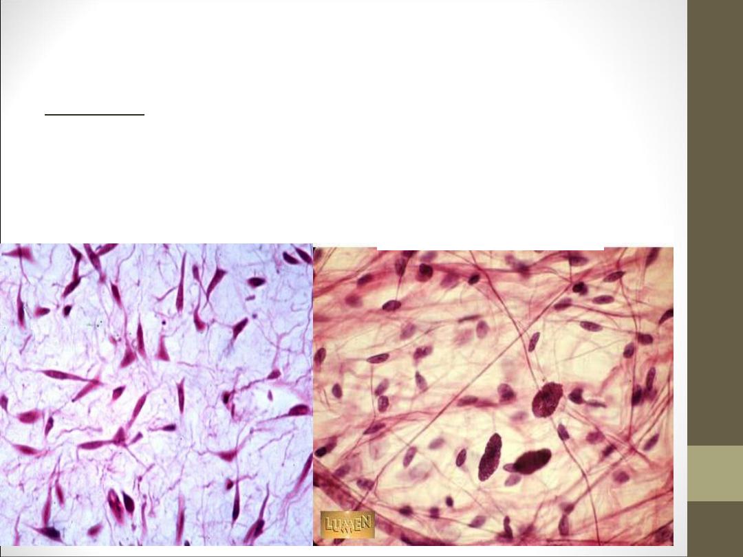
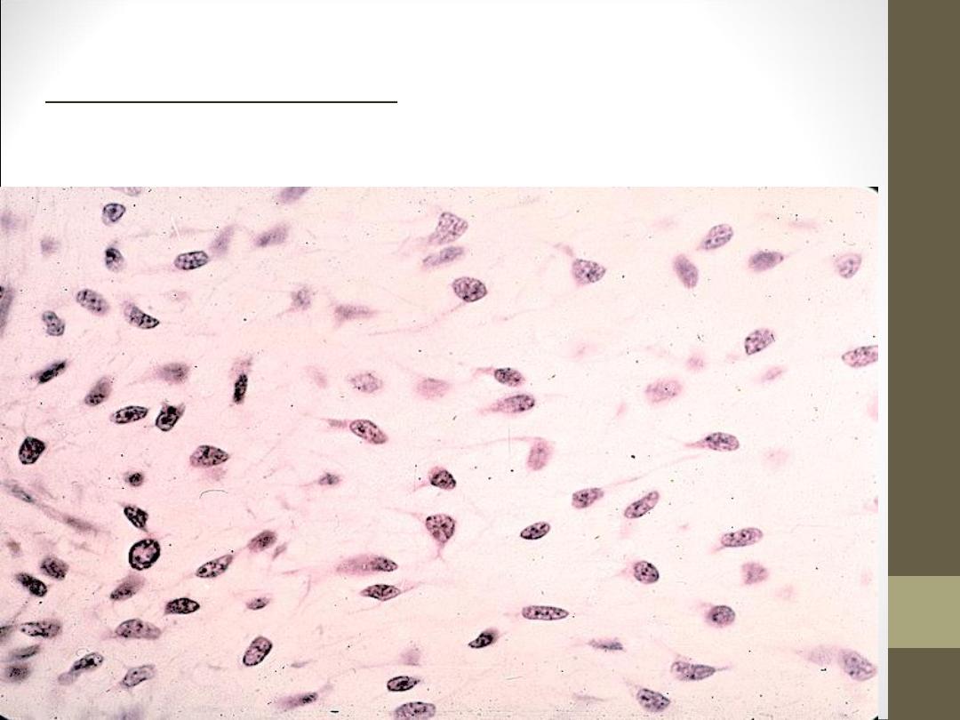
Fibroblast: they are large, flat, branching cell which appear
spindle shape in side view, the nucleus is oval and appears pale
and has one or two nucleoli .cytoplasm very pale so that the out
line of the cells are indistinct .Fibroblasts are responsible for
formation the fibers. We can see in Areolar connective tissue.
Cells
•
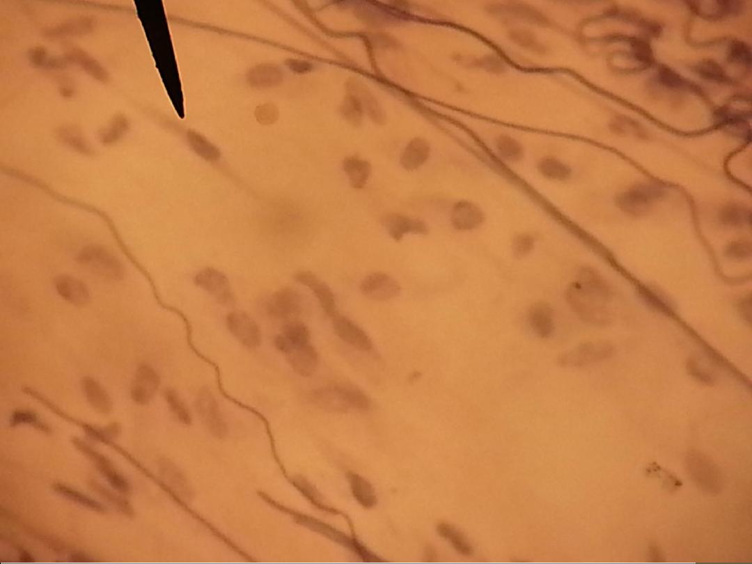
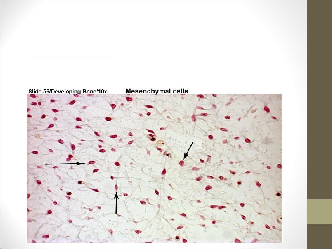
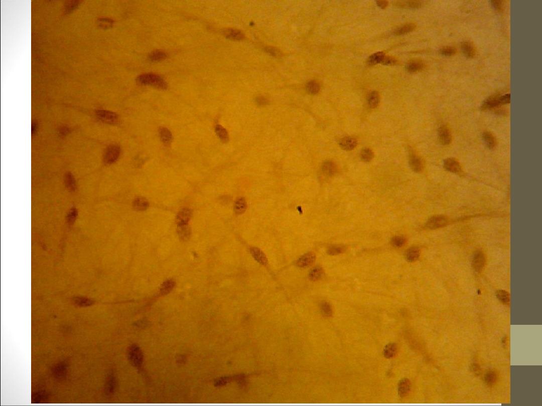
Mesenchymal cells: they are smaller than the fibroblast in the
size. We can see in the embryo sections.
•
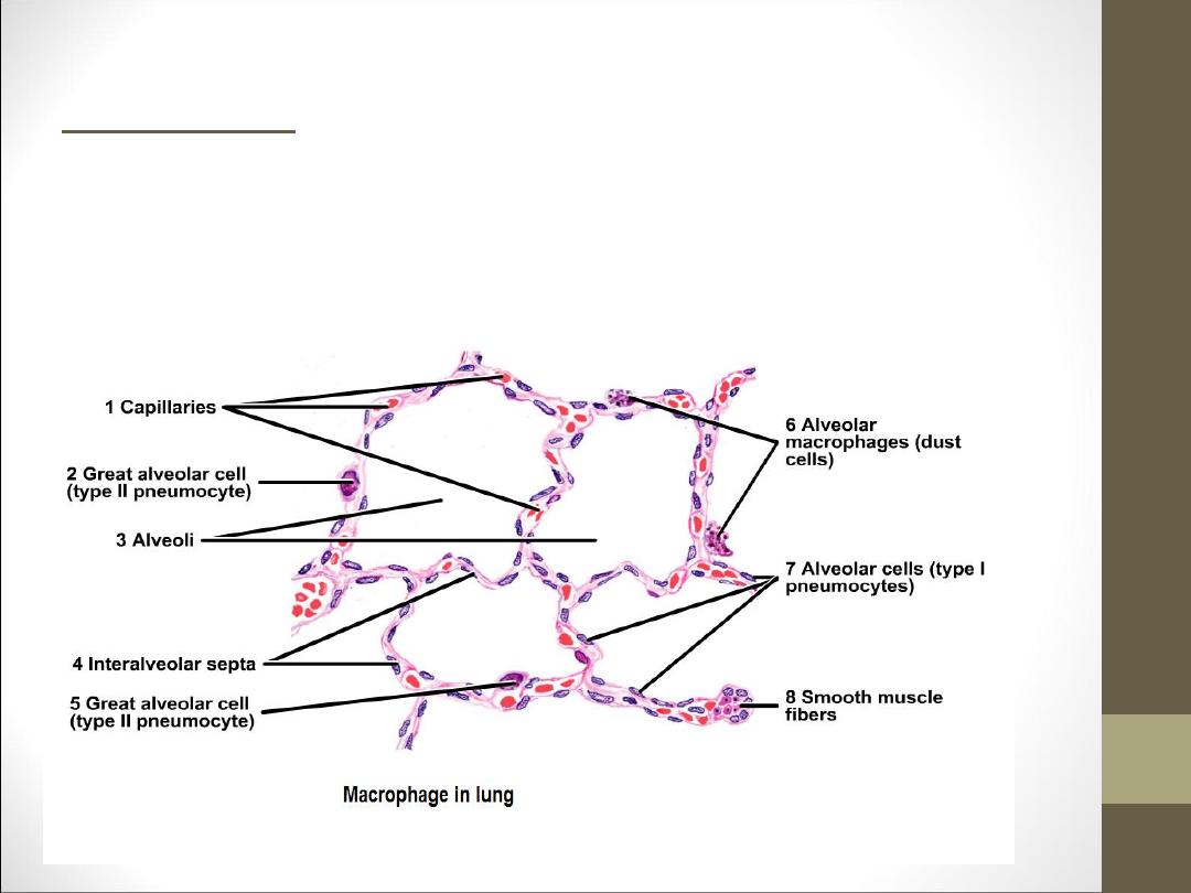
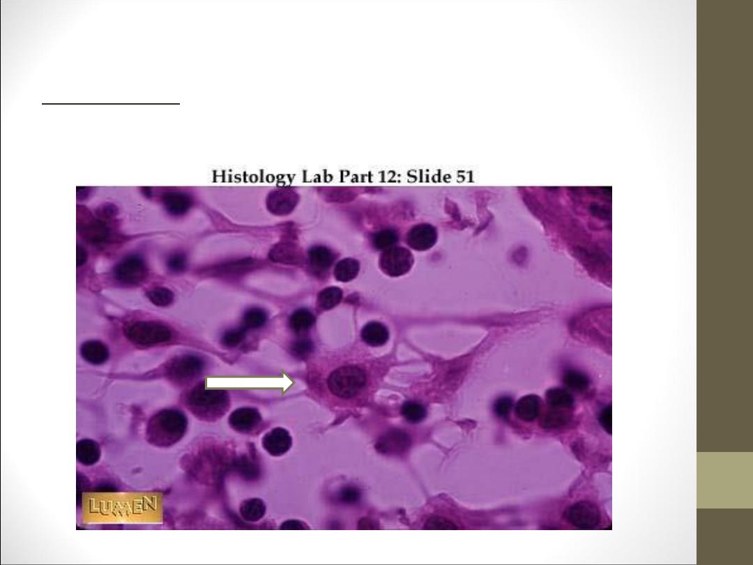
Macrophages: they are irregularly shape with processes
which usually are short. The nucleus is ovoid, small and more
heterochromatic. Cytoplasm fills with granules or vacuoles.
Macrophages are important agents of defense because of their
phagocytic activity. We can see in the section of the lung.
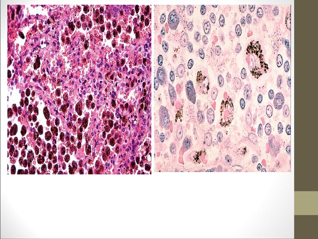
Macrophages
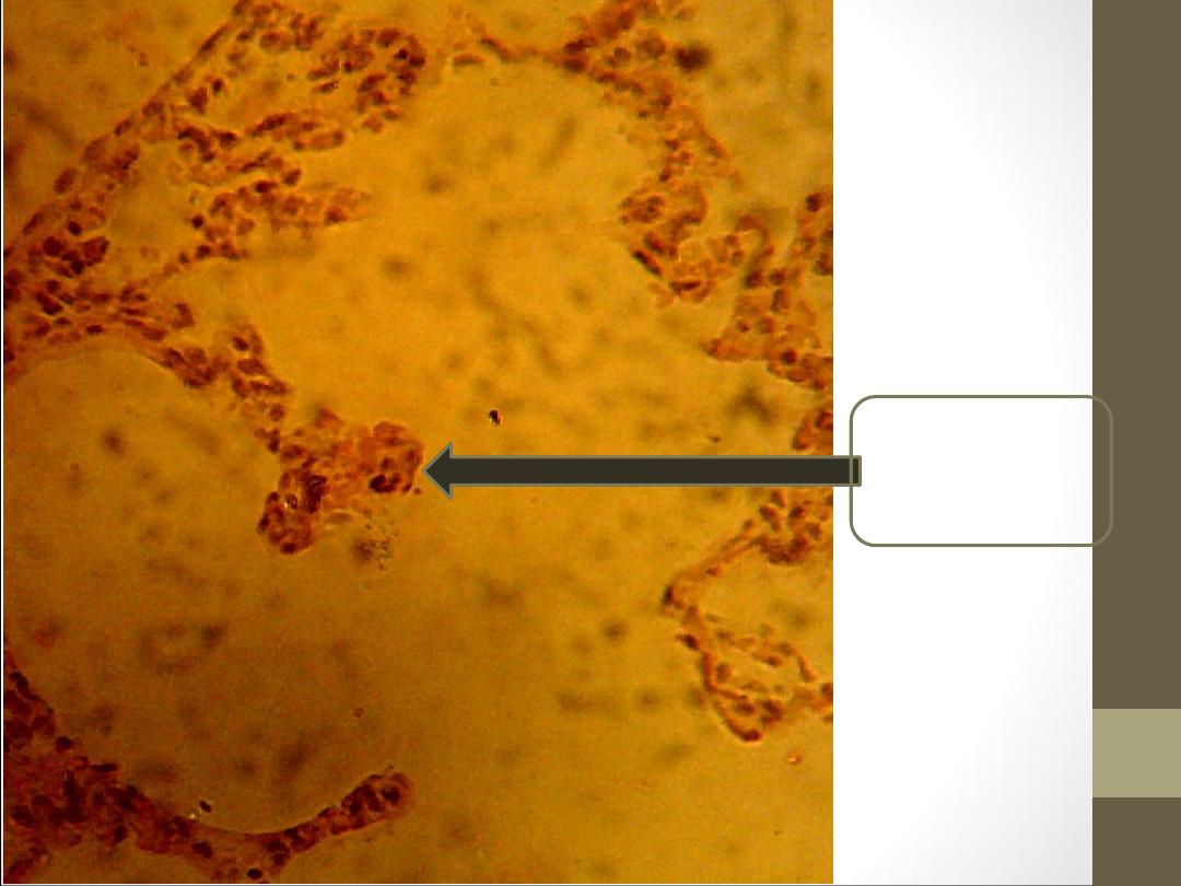
Macrophage
Cells
•
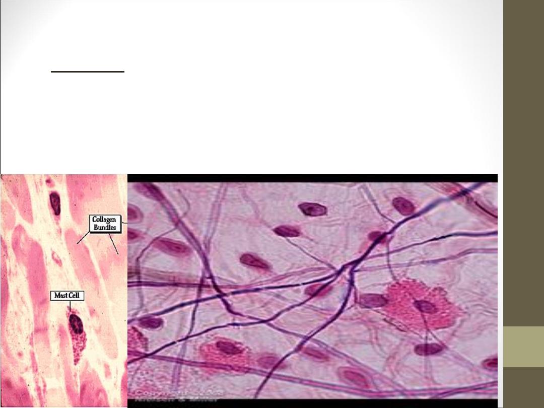
Mast cells: they are large in size irregularly oval in out line,
mast cells are identified easily by the content of cytoplasmic
granules .the nucleus is small spherical and central. Their
function is formation of heparin or anticoagulant and
histamine for vasodilatation. (. )موسع لالوعية الدمويةWe can see
in Areolar connective tissue.
Plasma cells:
are rare in connective tissue but are found
in lymphoid tissue .They are large, ovoid cells and the
nucleus is spherical and eccentrically placed, with
chromatin occur in course clumps peripherally and
arranged in pattern like wheel or clock face, plasma cell
responsible for antibodies production.
Plasma cell
Mast cell
Fibroblast
White fiber
Elastic fiber
Cells
•
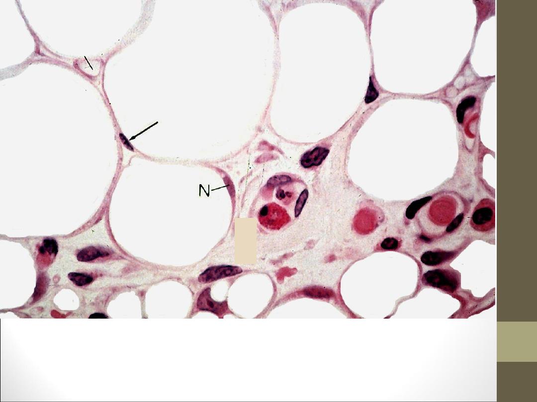
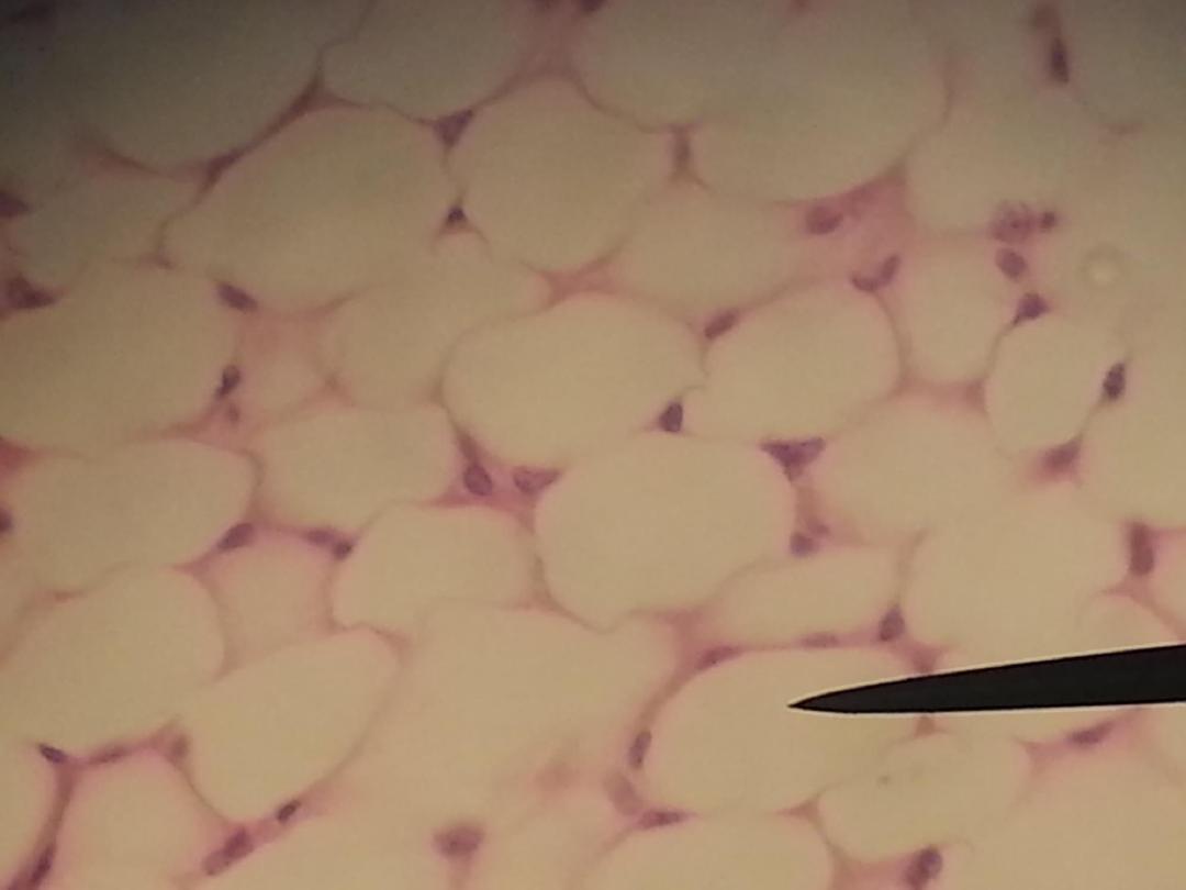
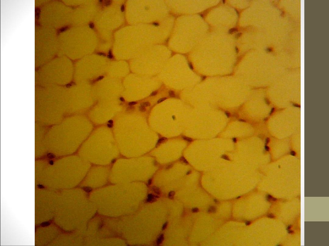
Adipose cells: their shape spherical to ovoid contains a single large
droplet of oil and thin rim cytoplasm contains in one area the
flattened nucleus.
Adipocytes (fat cells)
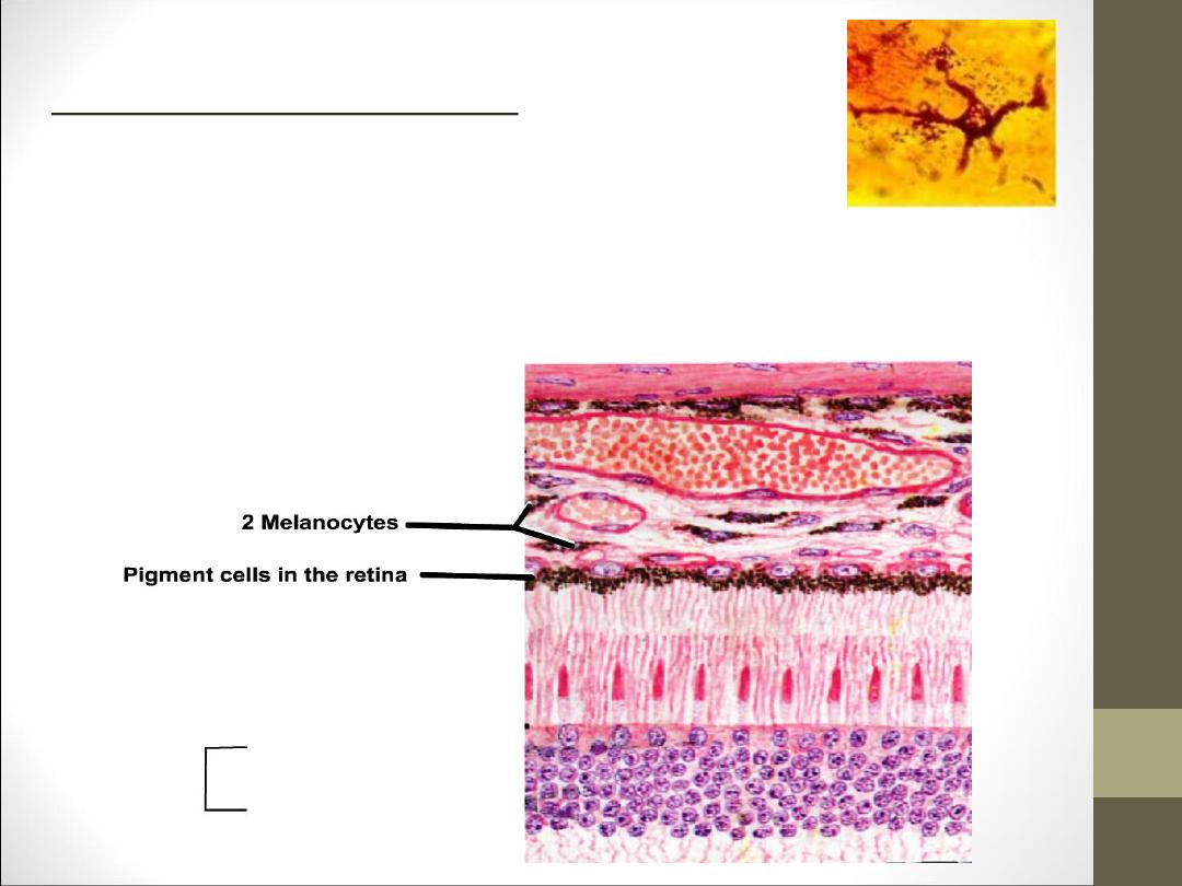
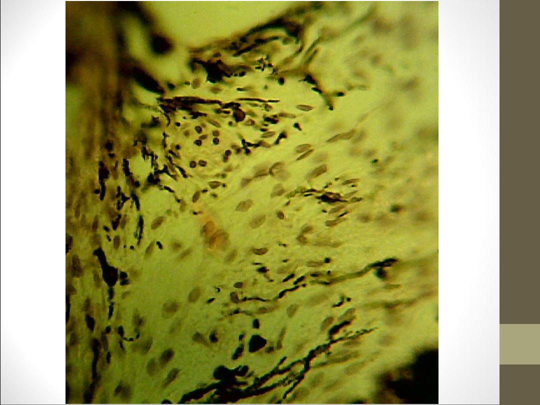
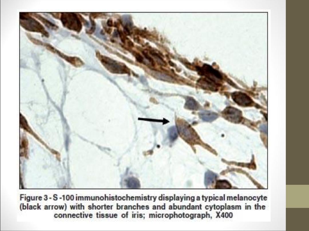
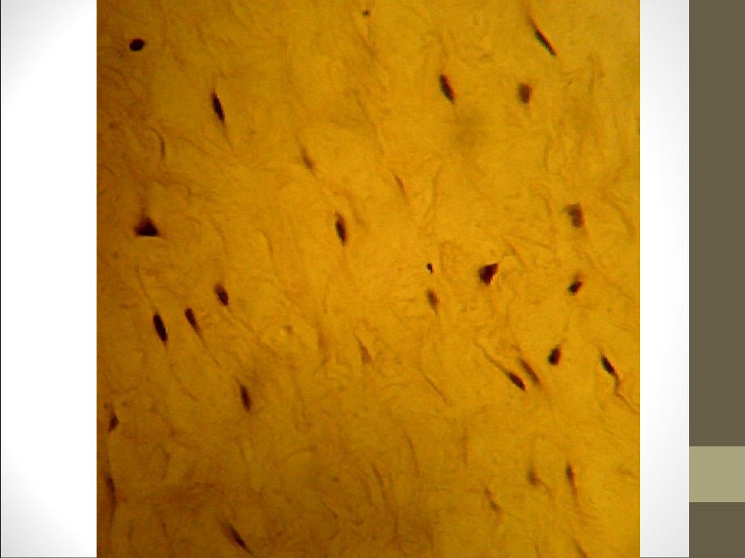
Melanocyte and pigment cells: cells have irregular
cytoplasmic processes like the genera cytoplasm
contain small granules of pigment called
melanosomes which contain melanin. It has a role in absorption
alight rays pigment cell found in dermis of skin.
Cells
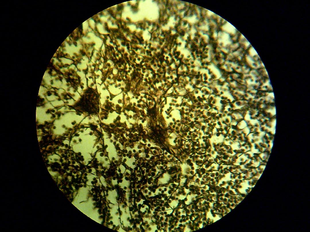
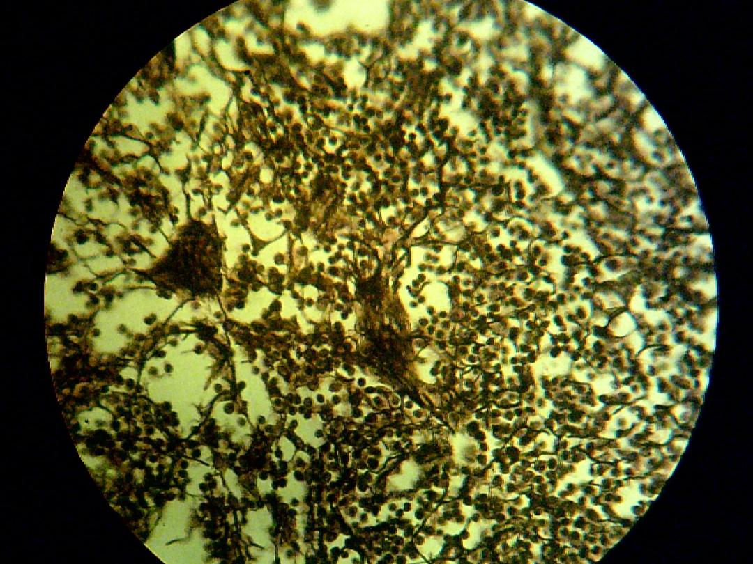
Reticular cells: are stellate they have long cytoplasmic extension,
which appear to join with other cell extensions. They have pale,
large nuclei, and basophile cytoplasm. It's found in lymph node.
Fibers
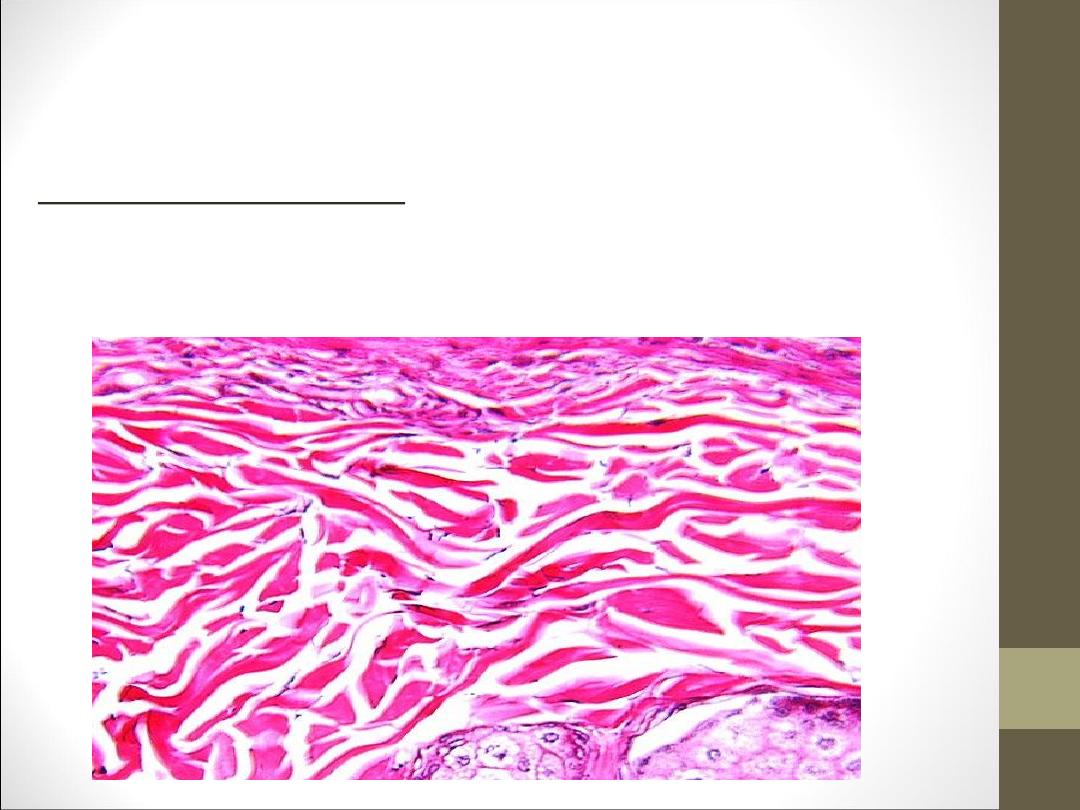
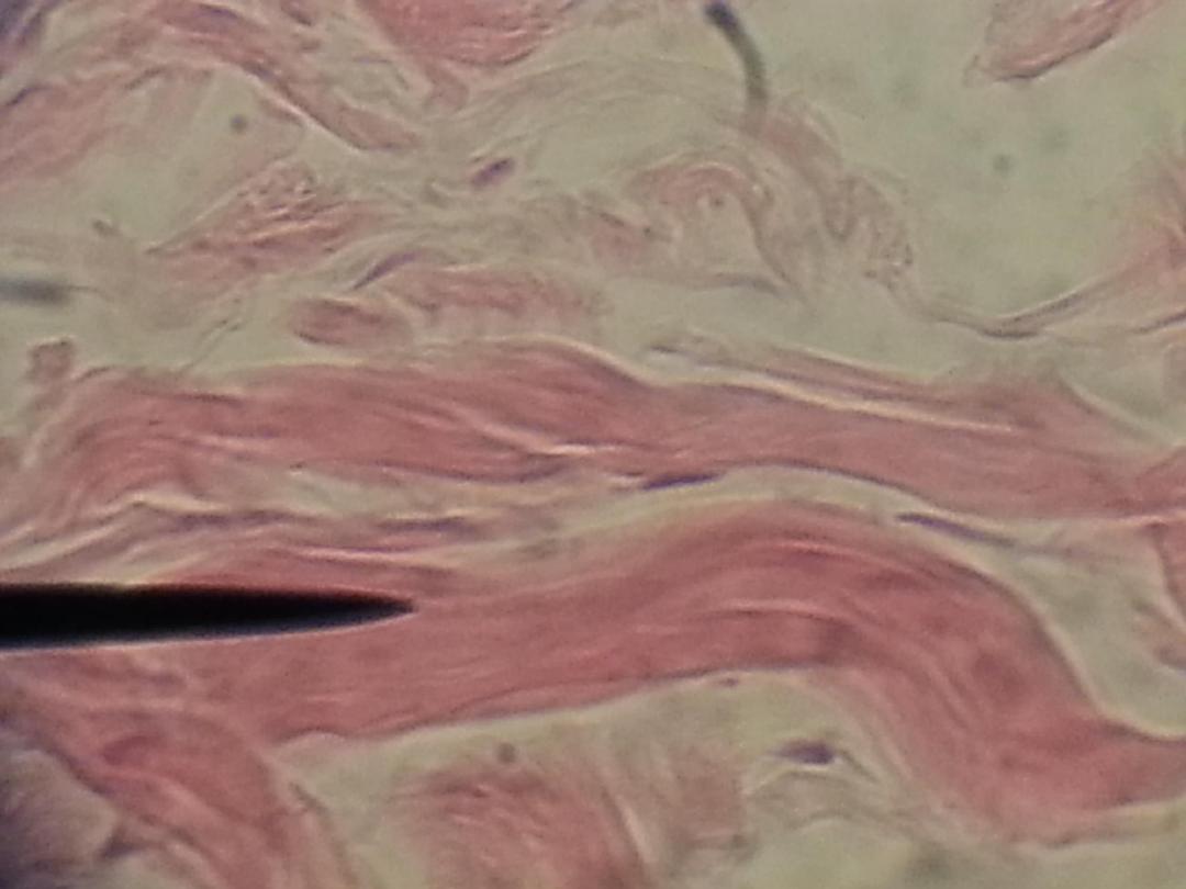
White (collagenous) fibers: are seen as straight wavy bundle, each
bundle consists of fibers, each fibers consist of fibrils, which appear
white in fresh state. White fiber is soft, flexible and inelastic that gives
the tissue strength. We can see in dermis.
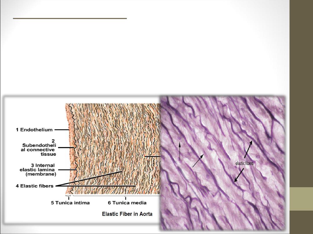
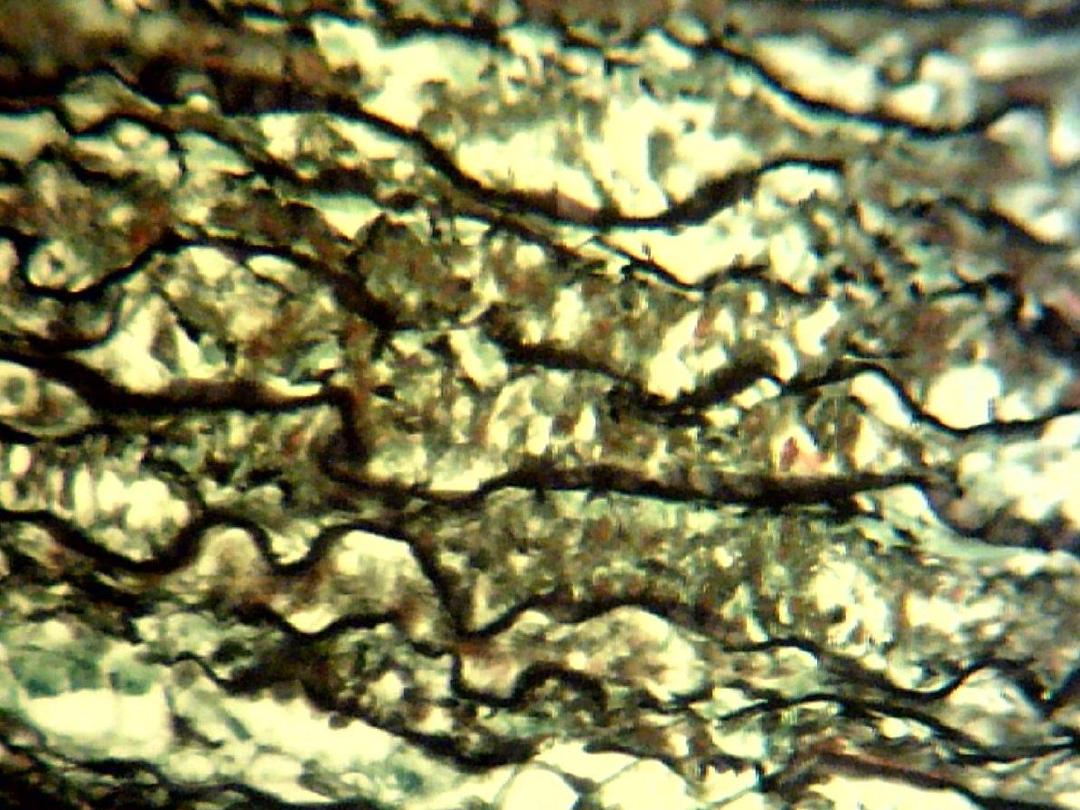
Yellow (elastic) fibers:
are seen as along, thin and single threads and un
branched bundle, in fresh state has a yellowish color.
Yellow fibers are elastic and easily to stretching .we
can see in cross section in aorta.
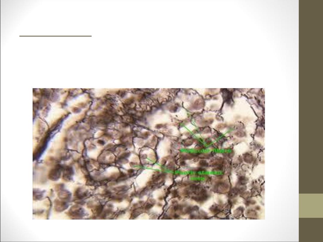
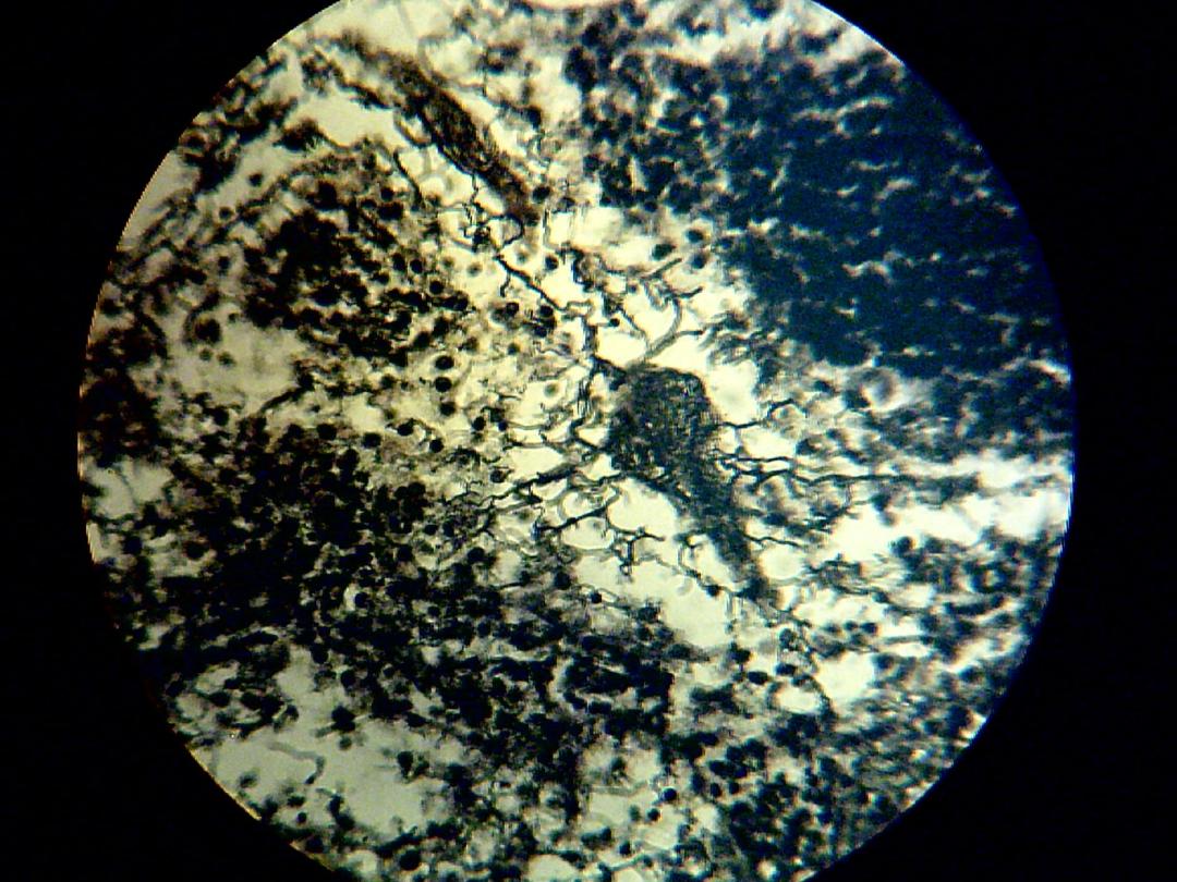
Reticular fibers: are very fine threads arrange to form a net
colored brown when staining them by silver impregnation.
They are found in lymphoid organs.

Types of connective tissue
•
Proper connective tissue
•
Special connective tissue

Proper connective tissue classify
according to fibers ratio to;
1.
Loose connective tissue
2.
Dense connective tissue
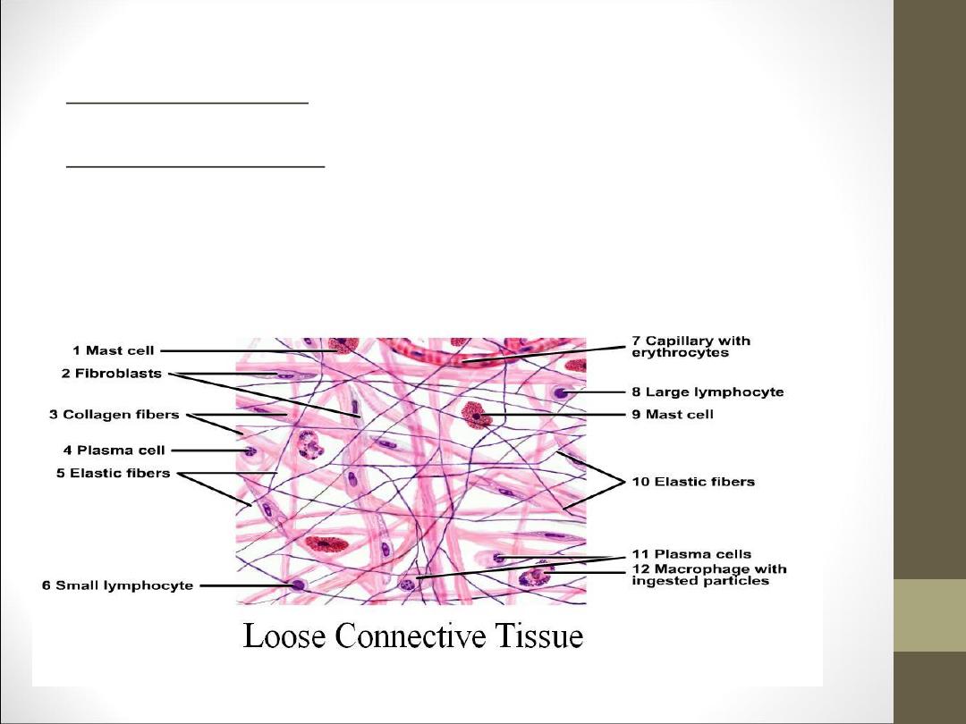
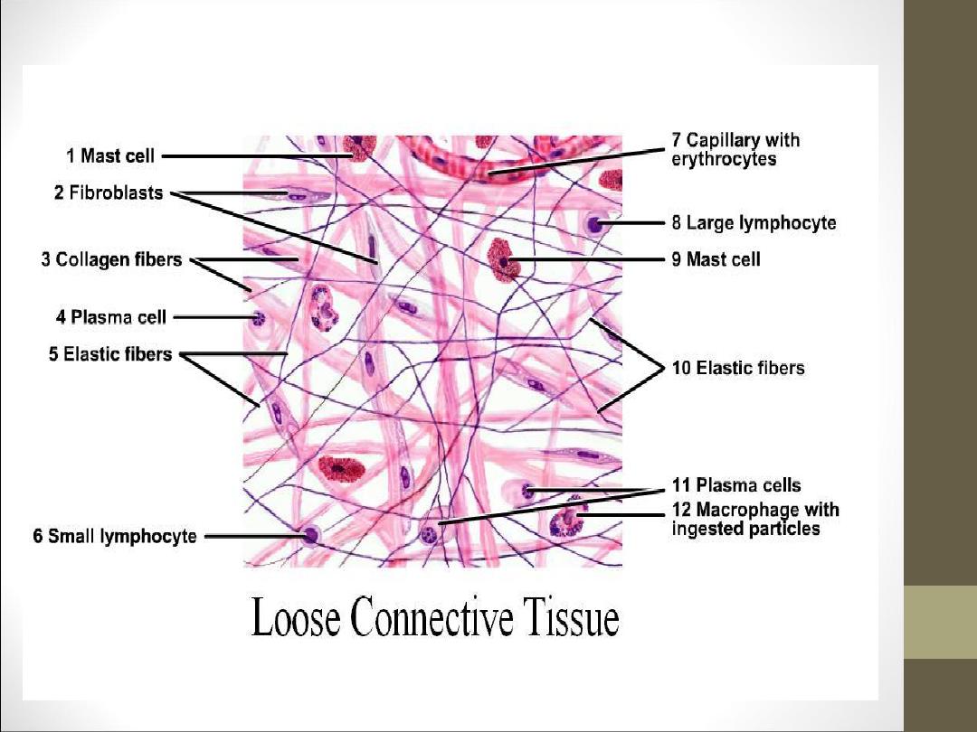
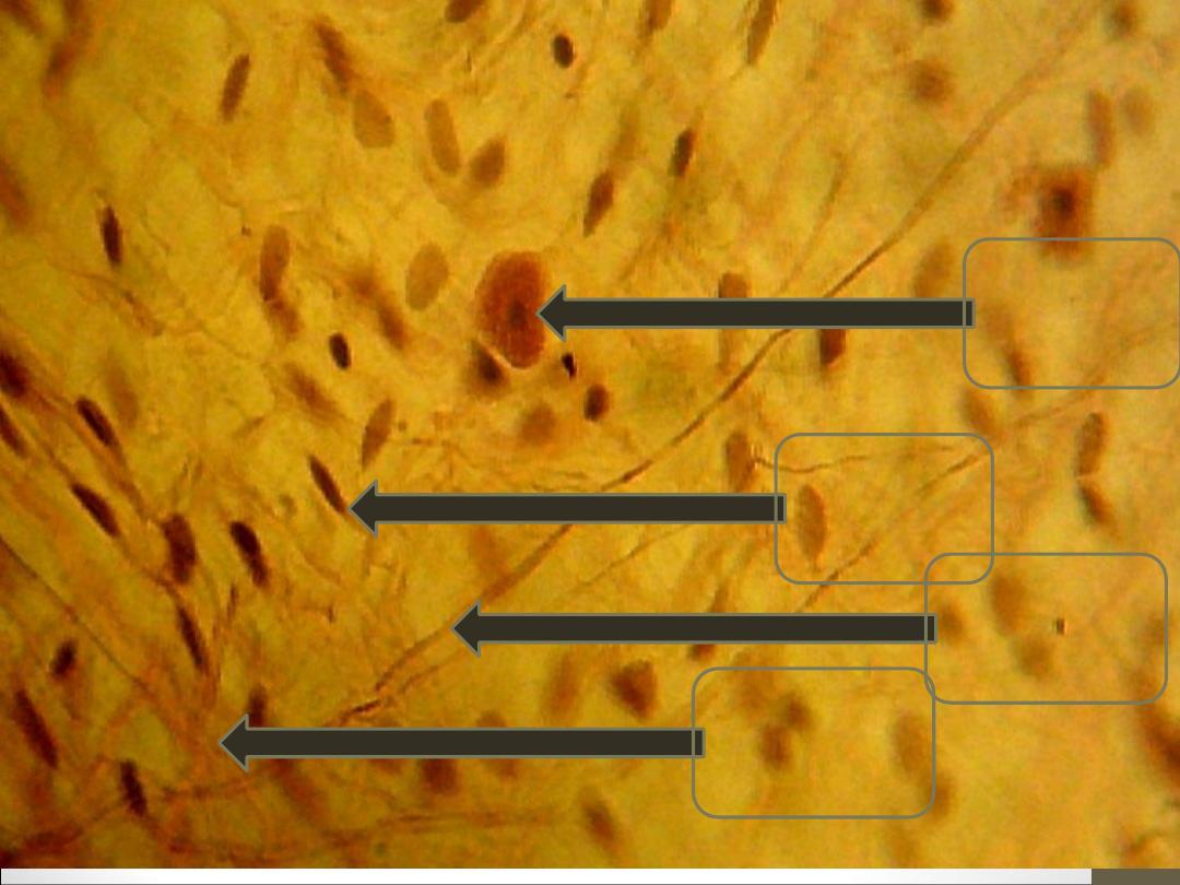
Loose connective tissue: Are characterized by loose arrangement of fibers,
with low concentration of fibers.
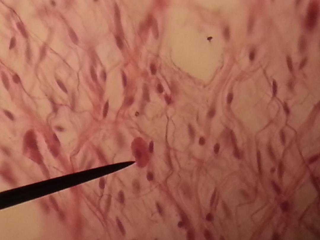
Areolar connective tissue: contain vacuoles or intercellular distance. It's found
in the substance of lung, heart and digestive trunk. The ground substance is
semisolid which contain yellow and white fibers and little of reticular fibers.
The cells which the most common in this tissue are: fibroblast, mast cell,
macrophage, and plasma cell.
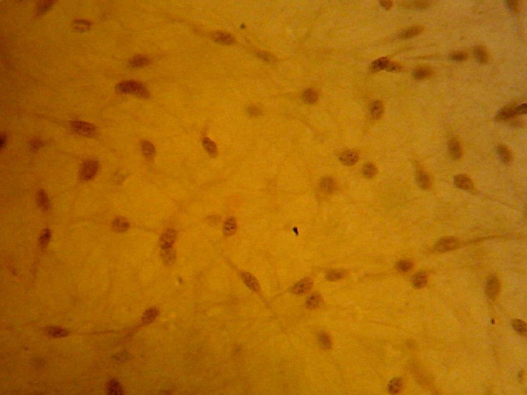
Mucoid connective tissue: contain fibroblast with ratio
of white fibers and fewer ratios of yellow and reticular
fibers. It's found in umbilical cord.
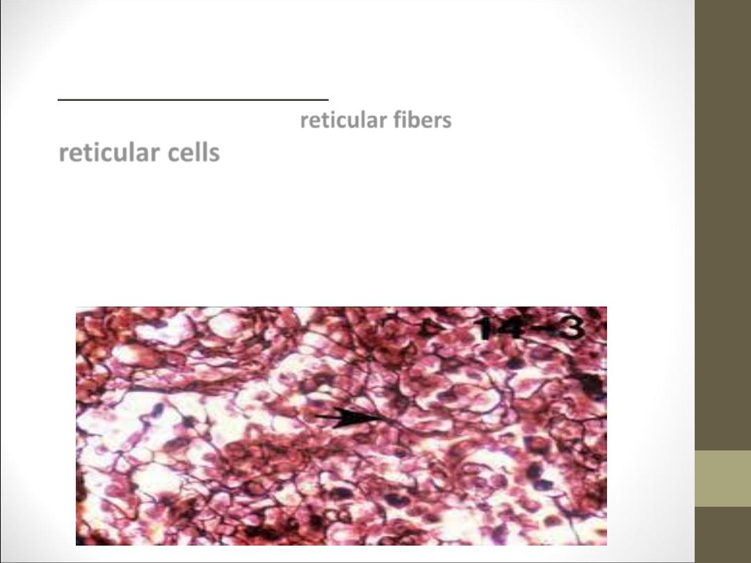
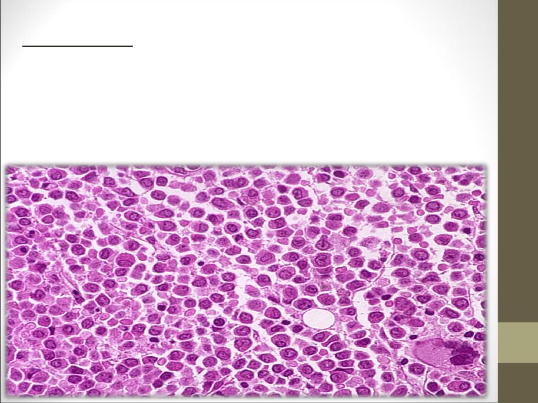
Reticular connective tissue: which is characterized by
presence of network of reticular fibers associated with
reticular cells
which are stellate and have long
cytoplasm extensions which appear to join with
extension of other cells. Also it is contain lymphocytes
which have darkly nucleus and occupied most of the
cell. It's found in lymph node.
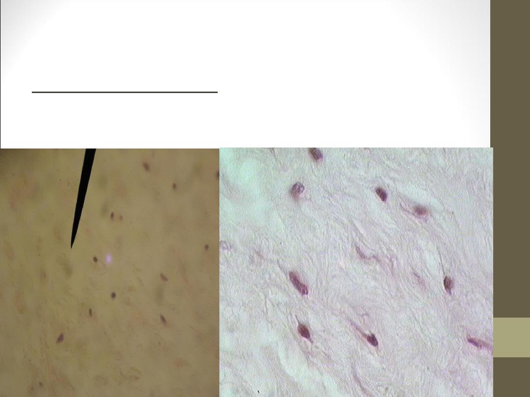
Mesenchymal connective tissue: its component of mesenchymal cell
whose branching process that swimming in ground substance
transparent with out any fibers.
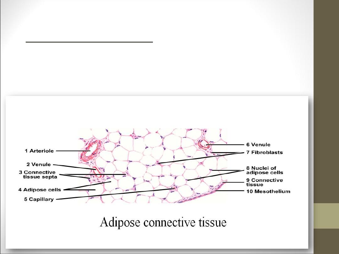
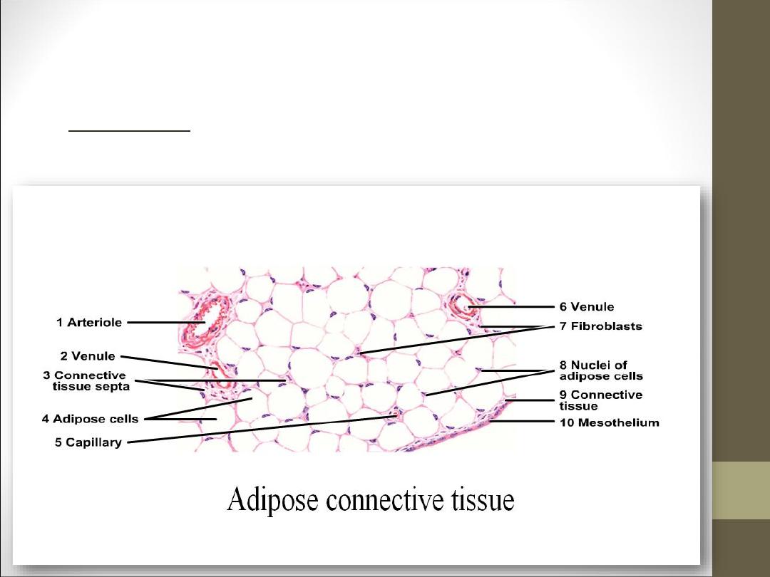
Adipose connective tissue: fat cells form large
aggregation .each fat cell is surrounded by a web of
different fibers and fibroblast. It found in the skin,
mesenteries and bone marrow.

•
THANKS