
ZOOLOGY
HIGHER SECONDARY - FIRST YEAR
Untouchability is a sin
Untouchability is a crime
Untouchability is inhuman
TAMIL NADU
TEXTBOOK CORPORATION
College Road, Chennai - 600 006.
A Publication under
Government of Tamilnadu
Distribution of Free Textbook Programme
(NOT FOR SALE)
Revised based on the recommendation of the
Textbook Development Committee

© Government of Tamilnadu
First Edition - 2005
Revised Edition - 2007
Chairperson
Prof. T. SARGUNAM STEPHEN
Dept. of Zoology
Govt. Arts College
Nandanam, Chennai - 600 035.
Reviewers
Dr. D. Mony
Dr. D. Sudarsanam
Reader in Zoology
Reader and H O D
R. M. Vivekananda College
Dept. of Zoology
Mylapore
Loyola College, Nungambakkam
Chennai - 600 004.
Chennai - 600 034.
Authors
Tmt. V. M. Gayathri Rani
P. G. T. in Zoology
Govt. Girls Higher Sec. School
Ashok Nagar,Chennai - 600 083.
Thiru. T. Sekar
P. G. T. in Zoology
Govt. Girls Higher Sec. School
Choolaimedu
Chennai - 600 094.
Price : Rs.
This book has been prepared by The Directorate of
School Education on behalf of the Government of Tamilnadu.
This book has been printed on 60 GSM Paper.
Tmt. Anne Freeda Chandran
P.G.T. in Zoology
Bentinck Hr. Sec. School for girls
Vepery, Chennai - 600 007.

Preface
This book has been prepared with an idea that on completion of
the course, the student can opt for any field related to the Biological
Sciences in his / her higher studies. A foundation for learning various
subjects had already been provided in the lower classes through
appropriate revisions in the syllabus. The revision of syllabus made,
has provided us with an opportunity to offer adequate information
related to several fields in biology to the students of Higher Secondary
classes. The 11
th
and 12
th
standard lessons are interlinked. A sound
knowledge of the material provided in this book will be essential for
pursuing the next level.
The lessons are written in such a way that the student is
encouraged to do further reference work. A list of books for such
reference work is provided. The students can also visit websites related
to each lesson.
Sample questions are provided at the end of each unit. The
teachers can frame more questions to test knowledge, understanding,
application and extension of this discipline.
As the scope of life sciences is widening due to increasing
demands and ultra developments in all fields, an active, interested
indulgence in the study of Biology will certainly be beneficial.
T. Sargunam Stephen
Chairperson
Biology (Zoology)
Text book writing committee.

Standard XI - Zoology Syllabus
Theory
Unit I : Bio - diversity
Taxonomic systems : Introduction to Taxa - Species concept - Methods
of Taxonomy - Phenetic methods - Identification keys - Cytotaxonomy -
Chemotaxonomy - Palaeotaxonomy - Nomenclature methods.
Animal groups : Methods of grouping animals - Major Phyla - General
characters with appropriate examples - Protozoa - Porifera - Coelenterata
- Platyhelminthes - Aschelminthes - Annelida - Arthropoda - Mollusca -
Echinodermata - Chordata.
Type Study : Plasmodium - Earthworm - Amphioxus-Pigeon.
Unit II : Cell Biology
Introduction : Microscopy and Cytological techniques.
Animal cell - Ultra structure : Plasma membrane - Nucleus -
Mitochondria - Ribosomes - Endoplasmic reticulum - Lysosomes -
Golgibodies -
Centrosomes - Chromosomes.
Cancer Biology : Cancer definition - Types of cancer - Management of
cancer-Radio therapy-Chemotherapy.
Unit III : Human Anatomy
Human systems : History - The integumentary - Skeletal - Muscular -
Digestive - Respiratory - Circulatory - Lymphatic - Nervous - Sense
organs - Endocrine - Excretory - Reproductive.

Unit IV : Genetics
Introduction - Multiple alleles - Quantitative inheritance - Sex
determination - Sex linked inheritance - Pleiotropy-Hardy Weinberg law-
Population genetics.
Uint V : Developmental Biology
Introduction - Types of eggs - Cleavage and types - Frog’s egg -
Gastrulation in frog embryo - Organogenesis in frog-Developmental stages
in eye and heart.
Unit VI : Economic Zoology
Beneficial animals : Corals - Earthworm - Vermiculture - Beneficial
insects - Prawns - Lobsters - Crabs - Pearl oysters - Fishes - Guano -
Aquarium - Vivarium-Planaria-Regeneration studies.
Harmful animals : Disease causing organisms - Vectors - Poisonous
organisms - Fouling organisms - Pests.
Unit VII : Origin of life
Theories - Geological time scale - Fossils - Extinct animals - Mass
extinction-Evidences for evolution-Comparative anatomy-Embryology-
Physiology-Vestigeal organs-Geographical distribution.

Standard XI - Zoology Syllabus
Practical
I
Earthworm - Mounting of Body setae - minimum 3 setae
II
Shark - Mounting of Placoid scales
III
Study of parts of a compound microscope and dissection
microscope.
IV
Prepared slides - observation - drawing and writing notes on
1. Plasmodium - any 2 stages
2. Paramoecium - entire, Paramoecium - conjugation
3. Hydra - entire
4. Tapeworm - Scolex
5. Earthworm - Body setae and Peneal setae - Cross section of body
6. Amphioxus - entire
7. Amphioxus - Cross section through different regions
8. Shark - Placoid scales
V
Museum specimens
Simple sponge, Corals, Tapeworm - entire, Ascaris - entire (male and
female), Earthworm - entire, Prawn - entire, Cockroach - Dorsal and ventral
views, Apple snail, Sepia, Star fish, Sea urchin, Amphioxus, Shark, A teleost
fish, Frog, Calotes, A snake, Pigeon, Quill feather, Rat
VI
Demonstration only
1. Earthworm - Viscera and Nervous system
2. Circulation of Blood in the wing of a live cockroach.
3. Frog - Buccal cavity, viscera and Digestive system.
VII
Human anatomy
1. Upper and lower jaw with dentition
2. Models / actual bones - humerus, radius ulna, femur, tibia, fibula, vertebrae,
pelvic girdle

CONTENTS
Page
1.
BIODIVERSITY
1
2.
CELL BIOLOGY
76
3.
HUMAN ANATOMY
104
4.
GENETICS
180
5.
DEVELOPMENTAL BIOLOGY
201
6.
ECONOMIC ZOOLOGY
223
7.
ORIGIN OF LIFE
268
vii

1
1. BIODIVERSITY
Our planet, earth, is occupied by diverse kinds of living organisms.
They live in various environments. The world is estimated to have 5 to 30
million species of living organisms. At present about 2.5 million species of
living organisms have been given scientific names. Over 1.5 million of them
are animal species and out of which 750,000 belong to insect species alone.
There are 350,000 species of plants including algae, fungi, mosses and higher
forms of plants. Thus the existence of different forms of a species or genus
and diverse adaptations for, varied surroundings are referred to as “biodiversity”.
The survival of such a vast range of living beings could be ensured
only when their habitats and environmental conditions remain without
alterations. The term ‘biosphere’ had been coined to highlight the interde-
pendence of living and non-living world. It represents a stable environment of
various physical and biological factors which have been operating since the
past. The organic continuity of the system rests on a delicate network of inter-
dependent relationships. The air, the water, the animals, the plants, the mi-
crobes and human beings are all interlinked in a life sustaining system, called
the environment.
Safeguarding the entire biosphere with all its intricacies is of prime
importance today. The nations of the world have convened
several conferences and adopted important resolutions for safeguarding the
sustainability of earth. In this background, the United Nation’s ‘Environmental
Agency’ organised the “International Conference on Human Environment” at
Stockholm in 1972. This conference adopted the motto ‘Only one earth’. In
1982, a UN conference on Environment was held at Nairobi. The UN again
convened “Earth summit ” at Rio de Janeiro highlighting “o ur
common future”, in 1992. Once again a world summit on sustainable
development was organised in Johannesberg in 2002. One of the agenda
commonly placed and accepted in all these meets was the significance of
biodiversity and its conservation to ensure sustainable earth.
Biodiversity in Indias
India’s immense biological diversity represents about 7% of world’s
flora and 6.5% of world’s fauna. About 62 % animals in India are endemic to
the country. India is one of the 12 countries identified as mega centres of
biological diversity.

2
As per the State Forest Report 1999, based on visual and satellite data
from IRS-1B, 1C and 1D, the total forest cover of India is 637,293 sq. km. It
is 19.39 % of the total geographic area of the country. It comprises about 64
million hectares.
Indian flora comprises about 15,000 flowering plants of which roughly
around 1,500 plant species are threatened. Mammalian fauna of India is 372
species with 63% in Assam. India’s 1228 bird species represent about 13% of
world’s total. Reptilian and amphibian fauna includes 446 and 204 species
respectively.
Since the world has a vast range of organisms, identifying the useful,
as well as harmful living beings is a need. Differentiating, grouping and
giving names to living things has been an ancient activity of every human
culture. Without proper classification it would be impossible to deal with enor-
mous diversity of life forms.
1.1 Taxonomic systems
The initiation for eolving taxonomic systems was provided by Aristotle
(384-322 BC). He emphasized that animals can be classified
according to their way of living, actions, habits and body parts. He observed
insects, fishes, birds and whales. The insect orders like Coleoptera, Diptera
were created by him. Due to his contributions, he is considered as the ‘father
of biological classification’.
For modern taxonomy, the first work was carried out by John Ray
(1627 - 1705) of England. His most interesting systematic work ‘Synopsis
Methodica Animalium Quadrupedum et Serpentini Generis’ was published
in 1693. He divided animals into those with blood and those without blood. He
also classified animals based on gills, lungs, claws, teeth and other
structures. He provided the first good definition of the species as ‘a
reproducing unit’.
The great Swedish naturalist Linnaeus (Caroli Linnaei) (1707 - 1778)
exerted an important influence on further advancement in taxonomy. Hence
he has been called the father of taxonomy. In 1758 he published his famous
book, systema naturae. He first introduced the hierarchic system, both in
animal and plant kingdoms. He followed four categories namely class, order,
genus, species for the animal world. His greatest contribution to taxonomy
was the use of binomial nomenclature for all species of animals and plants.

3
Michael Adamson (1727 - 1806), a French botanist, stressed that
classification should be based on as many characters as possible. His concept
helped to develop a new type of taxonomy called ‘Numerical Taxonomy’.
Lamarck (1744 - 1829) made the first attempt to improve Linnaen
system. He published seven volumes of his ‘Histoire Naturelle des Animaux
sans Vertebres’. He arranged animals according to evolution. He displayed
the groups of animals in the form of a branching tree. It was the beginning of
the use of phylogeny in systematics.
Cuvier (1769 - 1832) insisted that extinct fossil forms should be
included in the table of classification. He divided animals into four branches.
They are Vertebrata-fishes to mammals, Mollusca-mollusca and barnacles,
Articulata-annelids, crustaceans, insects and spiders and Radiata-
echinoderms, nematodes and coelenterates.
Charles Darwin in 1859, published his famous work ‘Origin of
species’. The new evolutionary concept of Darwin had an
immediate acceptance among biologists. Due to the influence of evolutionary
ideas, taxonomy was studied as an important evidence in favour of evolution.
The taxonomists were encouraged to learn that evolution theory of Darwin
gave meaning to their classifying activities. A large number of species were
discovered and described.
The development of modern taxonomy started during 1930s. During
this period taxonomy was based on population studies .
E. Mayr (1942) considered species as “groups of interbreeding natural popu-
lations”. His book ‘New Systematics’ became a landmark in the history of
taxonomy. The taxonomists were forced to accept species as a ‘population’.
Hence the taxonomist started moving from the laboratory to the field.
Morphological characters were studied along with other characters as behaviour,
sound, ecology, genetics, zoogeography, physiology and
biochemistry. Thus taxonomy was transformed into ‘biological taxonomy’.
1.1.1 Introduction to taxa and species
While grouping or arranging the organisms, a biologist faces three
scientific ideas, namely taxonomy, systematics and classification. These
disciplines though appear similar have slight deviations in their meaning.
The term taxonomy is a Greek word. Its components are taxis and
nomos. While taxis means arrangement, nomos means law. Thus taxonomy
is defined as the “theory and practice of classifying organisms” (E. Mayr
1966).

4
The term systematics originates from the Greek word systema. It
means ‘placing together’. Thus systematics means classification of living things
in accordance with their natural relationships. G.G Simpson (1961) defines
systematics as follows “Systematics is the scientific study of the kinds
and diversity of organisms and of any and all relationships among them”.
The term classification in meaning partly overlaps with taxonomy.
However it simply means the activity of classifying. Thus
according to Simpson “Zoological classification is the ordering of ani-
mals into groups on the basis of their relationships”.
A certain amount of overlap in meaning between the terms systamatics,
taxonomy and classification is unavoidable.
Taxon.
Based on specific charateristics, animals are grouped in various cat-
egories. These categories are otherwise called taxa (singular: taxon). “A taxon
is a taxonomic group of any rank that is sufficiently distinct to be
worthy of being assigned to a definite category”.
The several taxa in animal taxonomy are the Phylum, Class, Order,
Family, Genus and Species. This arrangement from Phylum to Species is
designated as the hierarchic system of classification. In this system each
taxon is based on specific characters of a group of organisms. Eventhough
such an arrangement appears to be man made, each taxon is a natural assem-
blage. However, human error in identification and grouping may happen.
The taxon, ‘Phylum’ is the largest group. There are several such Phyla
constituting the animal kingdom. Members of a Phylum are recognised by
certain distinctive features as shown below.
Characters
Phylum
Single celled animals
-
Protozoa
Pore bearers
-
Porifera
Common body cavity
and digestive cavity
-
Coelenterata
Flatworms
-
Platyhelminthes
Thread-like worms
-
Nematoda
Metamerically segmented animal
-
Annelida
Having jointed legs
-
Arthropoda
Soft bodied
-
Mollusca
Spiny skinned
-
Echinodermata
Having notochord
-
Chordata

5
Apart from one specific character, the members of the Phylum may
also show other common characters. Since a Phylum comprises enormous
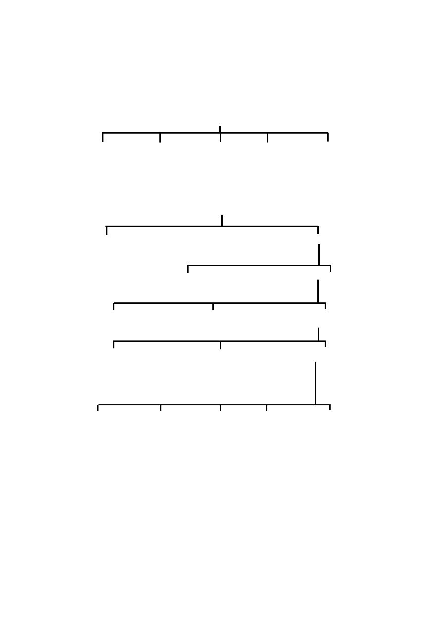
varieties of animals, it is further subdivided as given below
Phylum
Phylum
Phylum
Subphylum
Superclass
Classes
Classes
Classes
A Class is the next level in the hierarchy. There are only few Classes
in a Phylum. The members of each Class are identified by some specific char-
acter. Thus for example the Phylum : Protozoa comprises four Classes as
follows.
Class
Character
Rhizopoda
with root like pseudopodia
Ciliata
having cilia
Flagellata
having flagellum
Sporozoa
producing spores
Each Class may further be divided into Superorders or Orders
Class
Class
Subclass
Orders
Superorders
or
Orders
An Order is another level in the taxonomic hierarchy. It is marked by
some specific feature. A Class may have several Orders. For example, the
Class : Insecta is subdivided into nearly 29 Orders. Each Order is identified by
a specific character.
Order
Character
Example
Aptera
No wing
Lepisma
Coleoptera
Horny wings
Beetles
Lepidoptera
Scaly wings
Butterflies
Diptera
Two winged
Mosquitoes
Hymenoptera
Membranous wings
Wasps.
The Order is subdivided into Families
Order
Order
Superfamily
or
Families.
Families
or
or

6
Each Family will contain several Genera (singular : Genus). Each
Genus again is subdivided into Species.
In this hierarchy, the Species is considered as the most important taxon.
A Species represents a natural unit. All other taxa remain arbitrary and are
subjected to revision. A Species is considered a reality. It is the
fundamental unit in taxonomy. Evolution basically operates at the Species level
only. Hence the concept of Species has received much attention.
Concept of Species
Initially the Species was considered as a group of organisms showing
similar or specific characters. However modern workers have identified three
main concepts regarding Species.
1. Typological Species concept - This concept has its beginning from the
essentialism concept of Aristotle. According to this concept a Species is
recognised by its essential characters expressed in morphology.
2. Nominalistic Species concept - According to this concept Species are
man made ideas. Nature produces individuals and not Species. Thus a Spe-
cies is considered as a mental concept.
3. Biological Species concept - According to this concept, “Species are
groups of interbreeding natural populations that are reproductively isolated
from other such groups”. This concept is mostly accepted by present day
taxonomists.
1.1.2 Methods of taxonomy
Phenetic method or Numerical taxonomy
This method involves clustering or grouping of individuals of a taxon
or several taxa. Based on overall similarity, identifications are being made.
The desired size of the clusters or groupings is called the operational
taxonomic unit (OTU).
The identification method involves measurement of taxon to taxon simi-
larity or dissimilarity. It is measured using a scale of 0 to 1. ‘1’represents
perfect identity. ‘-1’designates dissimilarity between taxa. In this method
enormous amount of data are collected for related groups. Analyses are made,
using statistical tools and computers.
1.1.3 Cytotaxonomy
The characterization and identification of a cell’s complete
chromosome set is referred to as karyotyping. It is the first stage in the

7
process of using chromosomes in taxonomy.
Karyotypes within interbreeding populations of a species are usually
constant. Between species there may be variation in chromosome number
and size. Final stages of chromosomal aberrations such as inversions and
translocations can give clues regarding intermediary stages.
1.1.4 Chemotaxonomy
Chemotaxonomy refers to the use of information about small molecules
produced by the action of enzymes. Protein fractions in
electrophoretic techniques, identification of amino acids in chromatography,
prevalence of isoenzymes in tissue materials are all tools
employed in chemotaxonomy. The occurrence of specific pheromones, colour
pigments, toxins also help as keys in taxonomy.
1.1.5 Palaeotaxonomy
This method depends on identification and dating of fossils. Availabil-
ity of a good complete fossil provides better chance for
identification. In several fossils, their sections taken through laborious pro-
cesses have provided the identification features.
The fossils are normally studied along with other accompanying fos-
sils, its geographic location and other factors. Even though it is possible to
assign a fossil to a genus or other higher level, fixing the species is not always
possible.
1.1.6 Nomenclature methods
Nomenclature forms the basis by which scientists can name and cross
refer to organisms. It is an integral part of taxonomy. In fact, modern tax-
onomy started in 1753 with the publication of first part of Systema by Linnaeus.
According to Linnaeus a Species is specified by the combination of both its
specific and generic names. Since it requires two names, it is referred to as
the binomial system. This system is now firmly established in Biology.
In modern times International Commissions are responsible for nam-
ing each major group of organisms. There are several such commissions. These
commissions authorize the usage of scientific names in biology. Naming of
animals is monitored by International Code of Zoological Nomenclature
(ICZN) (International Commission of Zoological Nomenclature, 1985).
The rules are set out in the ‘codes’. The codes are modified by
occasional science congresses.

8
Basic principles of nomenclature
1. Providing stability in the naming and classification of organisms is
emphasized. Any taxon must have only one correct name.
2. If two or more names are already in use the correct name will be the one
that was published earlier. This system is referred to as the law of priority.
3. If two or more workers at one particular time describe the same organism
using different names, it results in synonyms. However only one name will be
held as a valid name. The validity is provided to the senior synonym.(law of
priority)
4. When names referring to two separate taxa of the same nomenclatural
level are spelt the same, the two names are called homonyms. This situation
arises when two separate authors used the same name to refer to two differ-
ent taxa. This condition is called homonymy. In this situation the junior name
is invalid and a new replacement has to be proposed.
5. A material on which an original description is based, gets a special status. It
will form the basis for any future identity of a taxon. This idea is called the
type concept. Thus the concept of a genus and species are fixed by their
type genus or type species.
6. Names that were used prior to those included by Linnaeus in the “Systema
Naturae”, tenth edition, 1758 are not recognised.
7. Scientific names must be either Latin or latinized. The name should be
mentioned in italics.
8. The genus name should be a single word beginning with a capital letter.
9. The species name sholud be a single or compound word beginning with a
smalll letter.
1.1.7 Identification keys
Identification of animals is an integral part of taxonomy. Identification
could be made thorugh literature, keys, pictures and comparison with type
specimens. Of these, the most commonly used method is, using of keys.
A key is essentially a printed information or a computer software pack-
age. The construction of the key is an important job of a systematist.
A good key is strictly dichotomous and not having more than two al-
ternatives at any point. The language of a key is telegraphic.

9
The key may be either bracketed or indented. In a bracketed key
alternative contrastive characters are used for identification. The number on
the right side indicates the next alternative character for consideration.
In an indented key a series of choices are provided for identifying a
taxon. The user should choose from among the choices.
The following examples provide the keys for identification four
species of frogs in Tamil Nadu, namely Rana hexadactyla, R. tigrina,
R. cyanophlictis and R. limnochoris.
The Bracketed key (Genus : Rana)
(1) Large size, snout - vent 100 - 200mm .......3
(1) Small size,snout to vent less than 100 mm ........2
(2) Pointed snout ................... .......R. limnochoris
(2) Obtusely pointed snout ......... ...R. hexadactyla
(3) 4
th
toe longer than others ...........R. tigrina
(3) 4
th
toe not longer ............... ...... R. cyanophlictis.
The Indented key (Genus : Rana)
Large sized body
skin smooth .................... R.hexadactyla
skin with folds ................. R. tigrina
Small size
blunt snout ...................... R. cyanophlictis
pointed or round snout ......... R. limnochoris
1.2 Animal groups
1.2.1 Methods of grouping animals
There are several ways of grouping animals. In all these methods the
basic Taxon remains without any change. However the taxa are rearranged in
different groups. All these groupings are mostly provided for the convenience
in identifying similar taxa.
I. One of the earliest method of grouping the animals could be dividing the
Animal kingdom into two assemblages called Invertebrata and Vertebrata.
10
This scheme was provided initially by Aristotle. This scheme does not have a
place for the Prochordates.
II. Animals can also be grouped as single celled and multicellular. The
single celled organisms are called the Protozoans. The multicellular could be
called the Metazoans. In this arrangement among the metazoans the unique
nature of the sponges in not having a tissue grade of body constuction is not
mentioned.
III. In yet another method the animals are grouped under following three
assemblages.
1. Protozoa - single celled animals
2. Parazoa - Multicellular without tissue grade (sponges).
3. Eumetazoa - Multicellular with tissue grade.
Eumetazoa is a large group including most of the multicellular
animals. Hence it is subdivided further into two groups.
1. Diploblastic animals - having ectoderm and entoderm as two layers in the
body wall. Ex : Coelenterata.
2. Triploblastic animals - having ectoderm, mesoderm and endoderm as three
layers in the body wall.
The Triploblastic animals are further divided into three groups based
on the presence or absence of an embryonic body cavity called coelom.
1. Acoelomata - no coelom Ex : Platyhelminthes
2. Pseudocoelomata - with a false coelom Ex : Nematoda
3. Coelomata - with a true coelom
IV. In a recent system, the entire living world is subdivided into 5 kingdoms.
This system is much more broader including algae, fungi, and plants. It is
known as the Five kingdom concept.
Fig. 1.2.1 Coelomic cavity
Acoelomate
Coelomate
coelomic cavity
endoderm
mesoderm
ectoderm
11
FIVE KINGDOMS
Kingdom 1
Kingdom 2
Kingdom 3
Kingdom 4
Kingdom 5
Monera
Protoctista
Fungi
Plantae
Animalia
Outline Classification of Animal Kingdom
ANIMALIA
PROTOZOA
METAZOA
Now kingdom : Protoctista
Eg : Ameoba
PARAZOA
EUMETAZOA
PHY : PORIFERA
Eg : Sponges
PHY : COELENTERATA
RADIATA
BILATERIA
Eg :
Hydra
DIPLOBLASTIC
TRIPLOBLASTIC
ACOELOMATA
PSEUDOCOELOMATA
EUCOELOMATA
PHY : PLATYHELMINTHES
PHY : ASCHELMINTHES
Eg : Tapeworm
(or)
NEMATODA
Eg : Ascaris
PHY : ANNELIDA PHY : ARTHROPODA PHY : MOLLUSCA PHY : ECHINODERMATA PHY : CHORDATA
Eg : Earthworm
Eg : Cockroach
Eg : Pila
Eg : Starfish
Eg : Rat

12
PHYLUM - CHORDATA
SUB-PHYLUM
SUB-PHYLUM
SUB-PHYLUM
SUB-PHYLUM
HEMICHORDATA CEPHALOCHORDATA UROCHORDATA VERTEBRATA
Eg : Balanoglossus Eg : Amphioxus
Eg : Ascidian
Eg : Fish
CLASS
1. PISCES
Eg : FISH - MUGIL
2. AMPHIBIA
Eg : FROG - RANA
3. REPTILIA
Eg : GARDEN LIZARD - CALOTES
4. AVES
Eg : PIGEON - COLUMBA
5. MAMMALIA
Eg : RAT - RATTUS
Sub class :
1. Monotremata (Prototheria)
Eg. Anteater
2. Marsupalia (Metatheria)
Eg. Kangaroo
3. Placentalia (Eutheria)
Eg. Elephant, tiger
Order :
Primates
Eg. Man
13
1. Kingdom : Monera - It includes all bacteria and the cyanobacteria. A
circular DNA occurs in the cytoplasm. The cell wall is a rigid structure.
a) Phylum :Cyanobacteria b) Phylum : Bacteria.
2. Kingdom : Protoctista or Protista - It includes single celled eukaryotes. It
has two subkingdoms, namely Protozoa and Algae.
3. Kingdom : Fungi
4. Kingdom : Plantae (green plants)
5. Kingdom : Animalia : multicellular, eukaryotic animals.
1.2.2 Major phyla
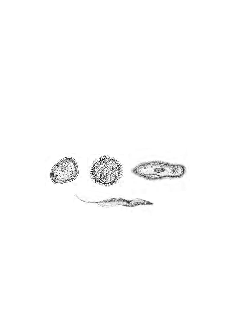
Phylum : Protozoa
This phylum includes a great diversity of small, microscopic organ-
isms. These are single celled eukaryotes. Their locomotion happens using
pseudopodia, cilia or flagella.
The nutrition is either autotrophic or heterotrophic. They reproduce
either asexually or by sexual methods. Ex : Amoeba, Paramoecium,
Plasmodium.
Phylum : Porifera.
These are multicellular, aquatic organisms. They have a cellular grade
of construction without the occurrence of tissues. The sponges belonging to
this phylum are characterised by the presence of a canal system in their
body. The body wall contains spicules. They can reproduce both by asexual
Fig. 1.2.2 Protozoans
Entamoeba
Volvox
Paramoecium
Trypanosoma
14
and sexual methods. Ex : Sponges.
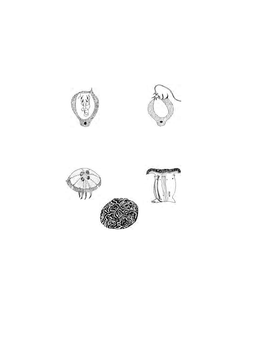
Phylum : Coelenterata or Cnidaria
All coelenterates are aquatic animals. They are mostly marine. The
body is radially symmetrical. The body wall is of two layers of cells. The outer
layer is called the ectoderm. The inner layer, entoderm is seperated from
the ectoderm by a non-cellular mesogloea. The mesogloea is a jelly-like sub-
stance. Due to the presence of two layers in the body wall, these are said to
be diploblastic animals.
Many coelenterates exhibit polymorphism. In this phylum, organisms
exist in two different body forms namely, a polyp, and a medusa.The
ectoderm contains stinging cells called nematocysts (cnidoblasts). These
cells when triggered can explosively penetrate prey and inject poison.
Fig. 1.2.3 Sponges
gastro- vascular cavity
Fig. 1.2.4 Coelenterate-body wall
Polyp
medusa
Body wall
ectoderm
entoderm
mesogloea
15
The layers in the body wall contain several cells and tissues such as
muscle cells epithelial tissues, gland-cells and sensory cells.
They reproduce both asexually and sexually. They are divided into
three classes, namely Hydrozoa, Scyphozoa and Anthozoa. In Hydrozoa,
the animal has a dominant polyp body form and a reduced medusa stage. (e.g)
Hydra, Obelia.
In Scyphozoa the medusa form is permanent. This group includes jelly
fishes such as Aurelia. They swim in the surface waters. They have a bell
shaped medusa stage.
The Anthozoans mostly remain as polyps. Their body cavity is
divided by large radial partitions called mesenteries.
(eg)
sea-anemone and corals.
All animals of subsequent phyla show the following general
characters.
Fig. 1.2.5 Nematocyst
in action
at rest
Fig. 1.2.6 Coelenterates
Brain coral
Sea anemone
Aurelia
16
Planaria
Tape worm
Liver fluke
1. All of them have three layers in the body wall. They are named
as outer ectoderm, middle mesoderm, and inner endoderm. Thus they are
called as Triploblastic animals.
2. The body is bilaterally symmetrical.
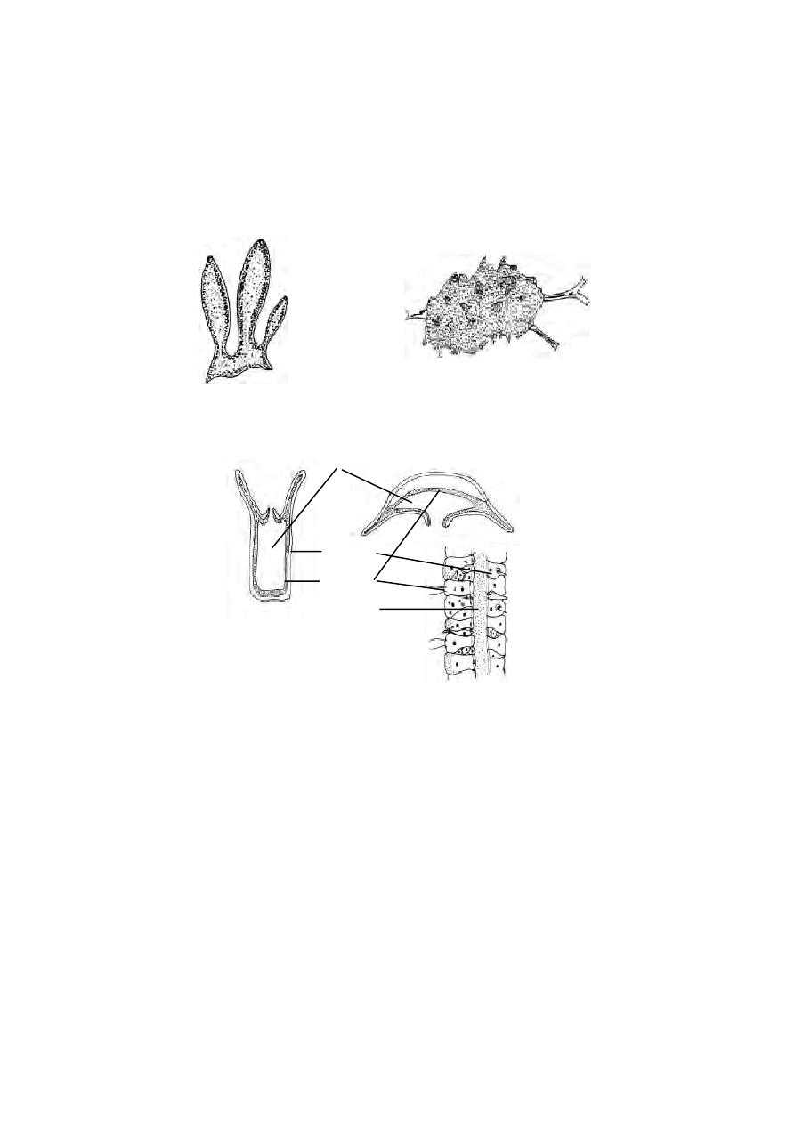
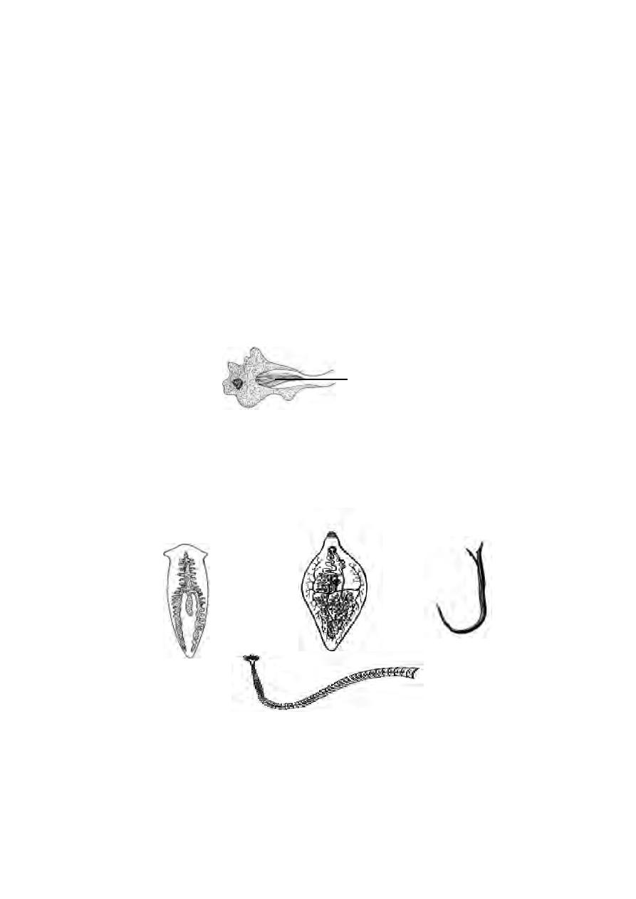
Phylum: Platyhelminthes :-
This phylum includes flatworms. These are acoelomates, without a
body cavity called coelom. The alimentary canal is either absent or very
simple. Excretion and osmoregulation occur through flame cells.These worms
are mostly hermophrodites, having both male and female reproductive organs
in a single individual. Most of the members are parasites. It is divided into
three classes, namely Turbellaria, Trematoda and Cestoda.
Class Turbellaria :- These are free living aquatic flatworms. The Planaria of
this class shows characteristic regeneration.
Class Trematoda :- These are flukes living as parasites inside a host (en-
doparasites). A protective cuticle covers the outer surface of the body. Flukes
have suckers for attachment to the host tissues. The examples are Fasciola
(liver fluke), Schistosoma (blood fluke).
Fig. 1.2.7 A flame cell
flagella
Fig. 1.2.8 Platyhelminth worms
Blood fluke
17
Class Cestoda :- It includes all tape worms. These are internal parasites
with a complex life history. The life cycle involves two hosts.
Their body characters are adaptations for parasitic life. Mouth and
alimentary canal are absent. Food is absorbed through general body surface.
The head is called the scolex. It has a ring of hooks and suckers for attach-
ment to the host tissue. The body consists of several segments called
Proglottids. (eg) sheep and cattle tape worms.
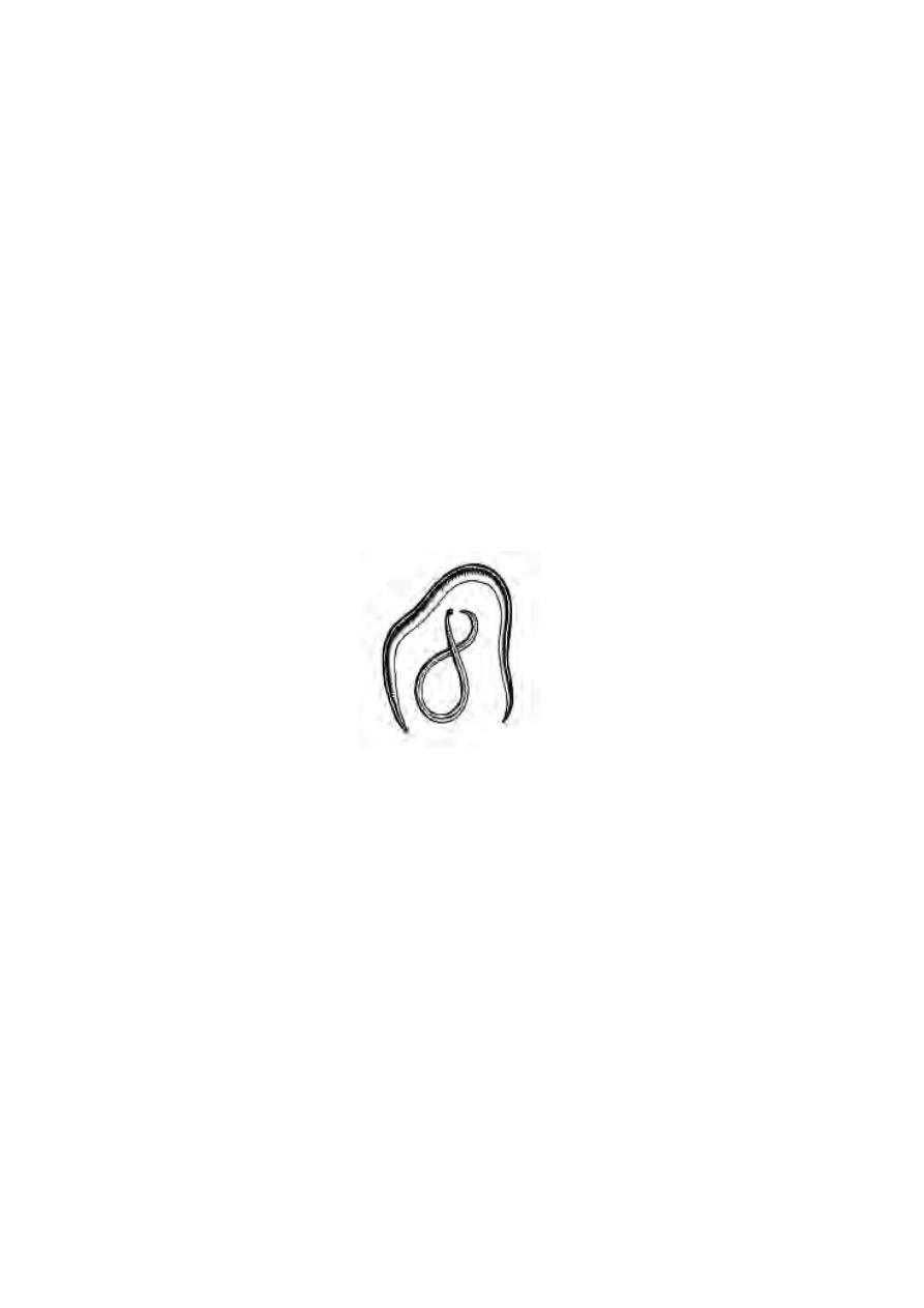
Phylum : Nematoda :-
These are the popular round worms. The body is narrow and pointed
at both the ends. There are no body segments. The body is covered by a thin
cuticle. The body cavity is considered as a pseudocoelom. The alimentary
canal is a straight tube. They reproduce sexually and the sexes are seperate.
There are several free living soil nematodes. Others are parasites.
(eg) Ascaris lumbricoides.
In subsequent Phyla the animals show following general
characters
1. There is a coelom within the mesoderm. Hence these are called as
coelomates.
2. The body consists of a series of compartments. This phenomenon is
called as metameric segmentation. They have a circulatory system pro-
viding internal transport.
Phylum: Annelida :-
These are worm like animals. The body segments are rings externally.
Internally the segments are seperated by septa. Externally the body is
protected by a cuticle. Excretion and osmoregulation are acheived by ciliated
tubules called nephridia. There is a central nervous system. The brain is
Fig. 1.2.9 Ascaris
18
Fig. 1.2.11 Annelids
Leech
Nereis
formed of ganglia in the head region. The nerve cord is ventral in position.
For the first time head formation or cephalization happens. These are bi-
sexual and hermophroditic. The larva is called the trochophore.
This phylum includes three Classes, namely Polychaeta, Oligochaeta
and Hirudinia. The polychaetes are marine worms. They have a distinct head.
There are pairs of lateral projections called parapodia. The examples are
Nereis (ragworms), Arenicola (lugworm).
Earthworms are included in the Class Oligochaeta. The Class:
Hirudinia includes leeches. These are blood suckers and ectoparasites. They
have well developed suckers for attachement at anterior and posterior ends.
Phylum : Arthropoda :-
These are the most successful group of animals. They outnumber all
other animals in population strength. The body is segmented. It is covered by
a hard exoskeleton made of chitin. During growth the exoskeleton is shed
(moulting of ecdysis). The legs or paired appendages are jointed. The head
Fig. 1.2.10 Annelida
Trochophore larva
Nephridium
19
Millipede
region has a pair of prominent compound eyes. Each compound eye is made
up of several photoreceptor sub units called Ommatidia.
They have an open circulatory system without vessels. The body
cavity is filled with a fluid called haemolymph. Such body cavity is known as
haemocoel. These are unisexual, exhibiting sexual dimorphism. The young
forms produced are invariably called the larvae. The larvae undergo
metamorphosis and develop into adults.
This Phylum comprises five Classes, Class Onychophora: It includes
small worm like Peripatus. Peripatus shows Annelidan and Arthropoda
characters. Hence this may be considered as a connecting link between the
two groups.
Class Crustacea :- The examples for this class are prawns, crabs and
lobsters. The dorsal body surface is covered by a sheild like carapace.
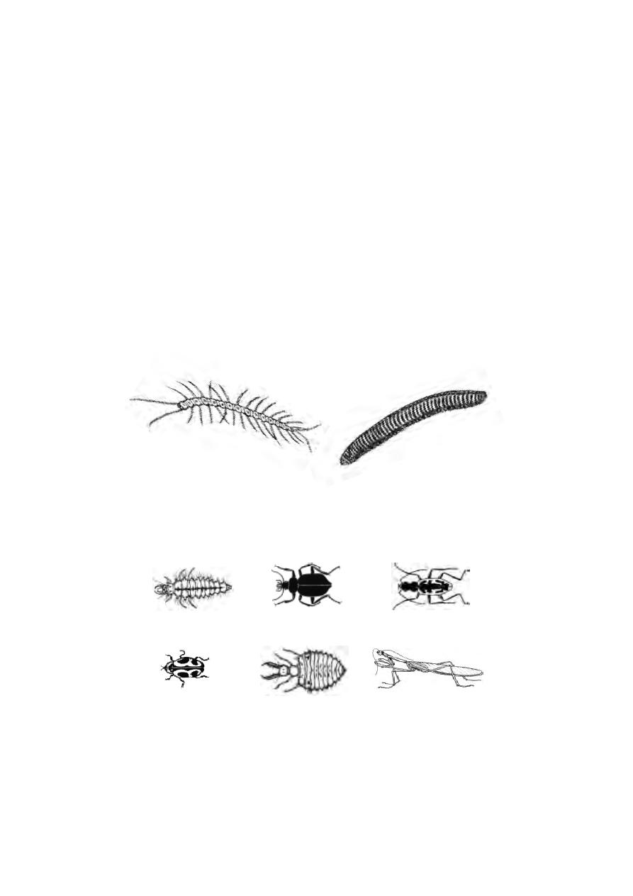
Class Myriapoda :- It includes centepedes and millipedes. These organ-
isms have a distinct head and simple eyes. The centepedes have a pair of
poison claws. The body consists of numerous segments, bearing pairs of legs.
Centipede
Fig.1.2.12 Myriapods
Fig. 1.2.13 Insects
1,2 & 3 Insect parasites
Ladybird beetle
Antlion
Praying mantis
Aphis lion
Ground beetle
Tiger beetle
20
Class Insecta :- It comprises the common insects. The body is divided into
head thorax and abdomen.In several insects, the adults have two pairs of wings
on the thorax. Respiration happens through the tracheal system.
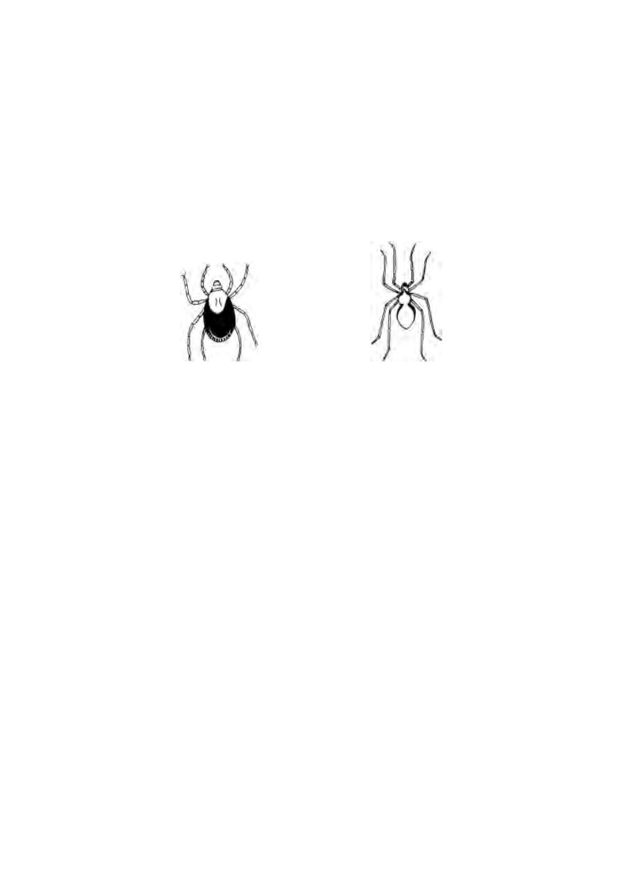
Class Arachnida :- It includes scorpions, spiders, ticks and mites. The
body is divided into cephalothorax and abdomen. There are four pairs of
legs attached to the cephalothorax.
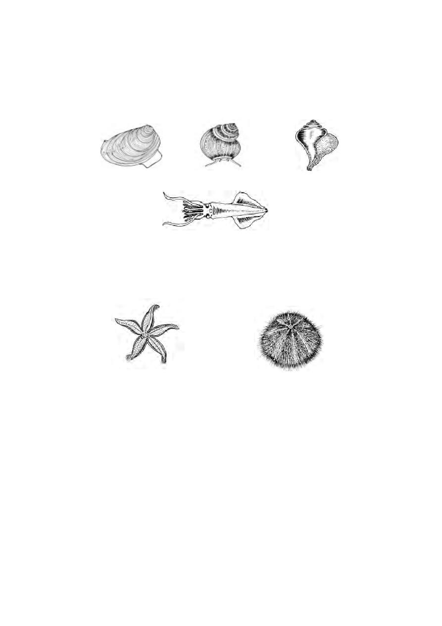
Phylum Mollusca :-
It is a very successful and diverse group of animals.
Considered to be the second largest group of animals with regard to species
number. These are soft bodied animals without segmentation. The body is
divided into head, muscular foot and visceral mass. The body is covered by
a mantle and a shell.
Respiration happens through gills (ctinidia) in the mantle cavity. The
most common larva is a trochophore larva.
There are seven classes of which three are more prominent.
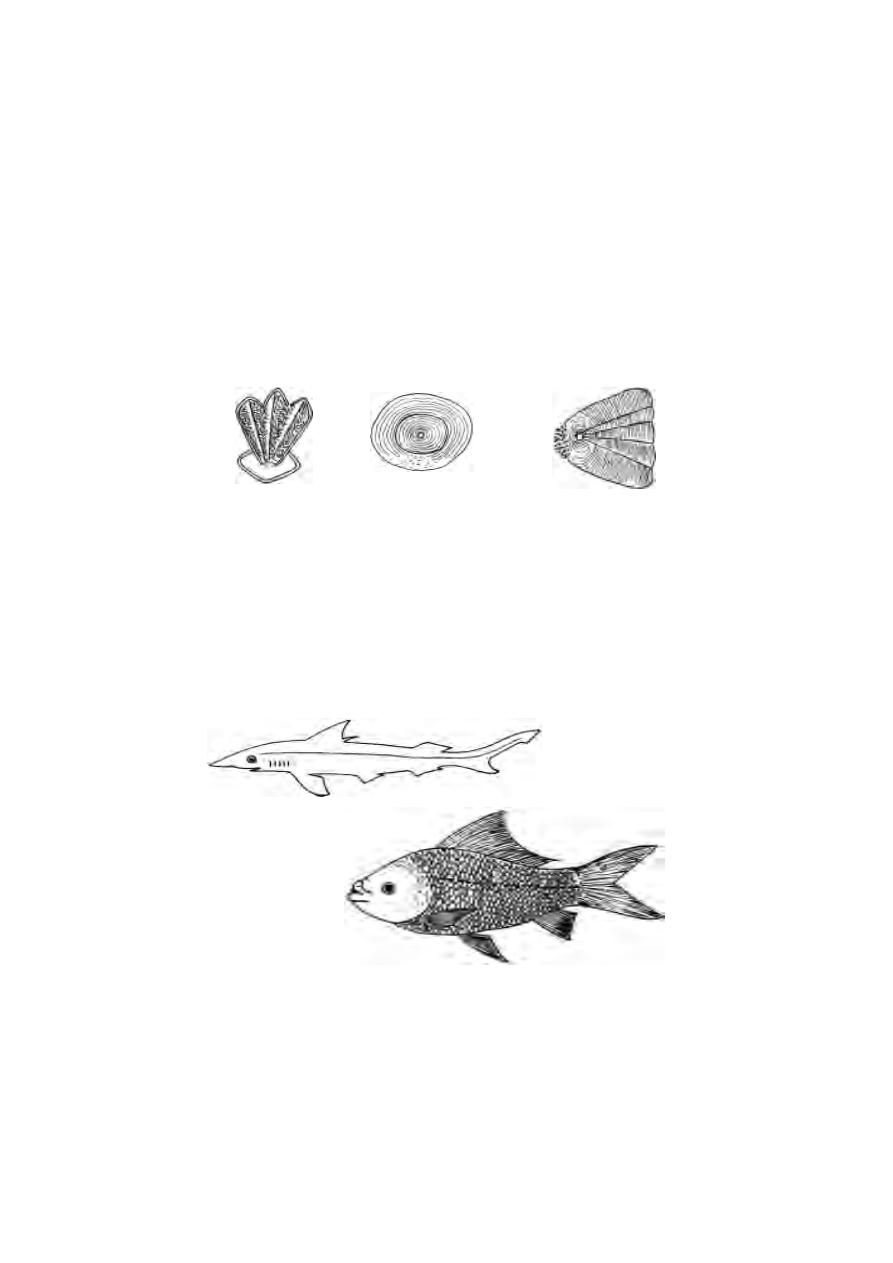
Class Pelecypoda or Bivalvia :- These are aquatic molluscs having bivalves.
They burrow in mud and sand. The body is laterally compressed.
(eg) mussels, clams, oysters.
Class Gastropoda :- These are either aquatic or terrestrial molluscs. They
posses a spiral shell.
The foot is large and flat. They have well developed head with
tentacles and eyes. (eg) snails, slugs, and limpets.
Class Cephalopoda :- These are mostly marine. They are adapted for
swimming. The foot is modified into eight to ten long tentacles in the head
region. The shell is either internal or absent. (eg) Octopus, Loligo, Sepia.
Fig. 1.2.14 Arachnids - Spiders
Tick
House spider
21
Phylum Echinodermata :- These are marine organisms. While the adults are
radially symmetrical the larvae remain bilaterally symmetrical. The mouth is
on the lower surface. They have a water vascular system with tube feet.
eg. star fishes, brittle stars, sea urchins and sea-cucumbers.
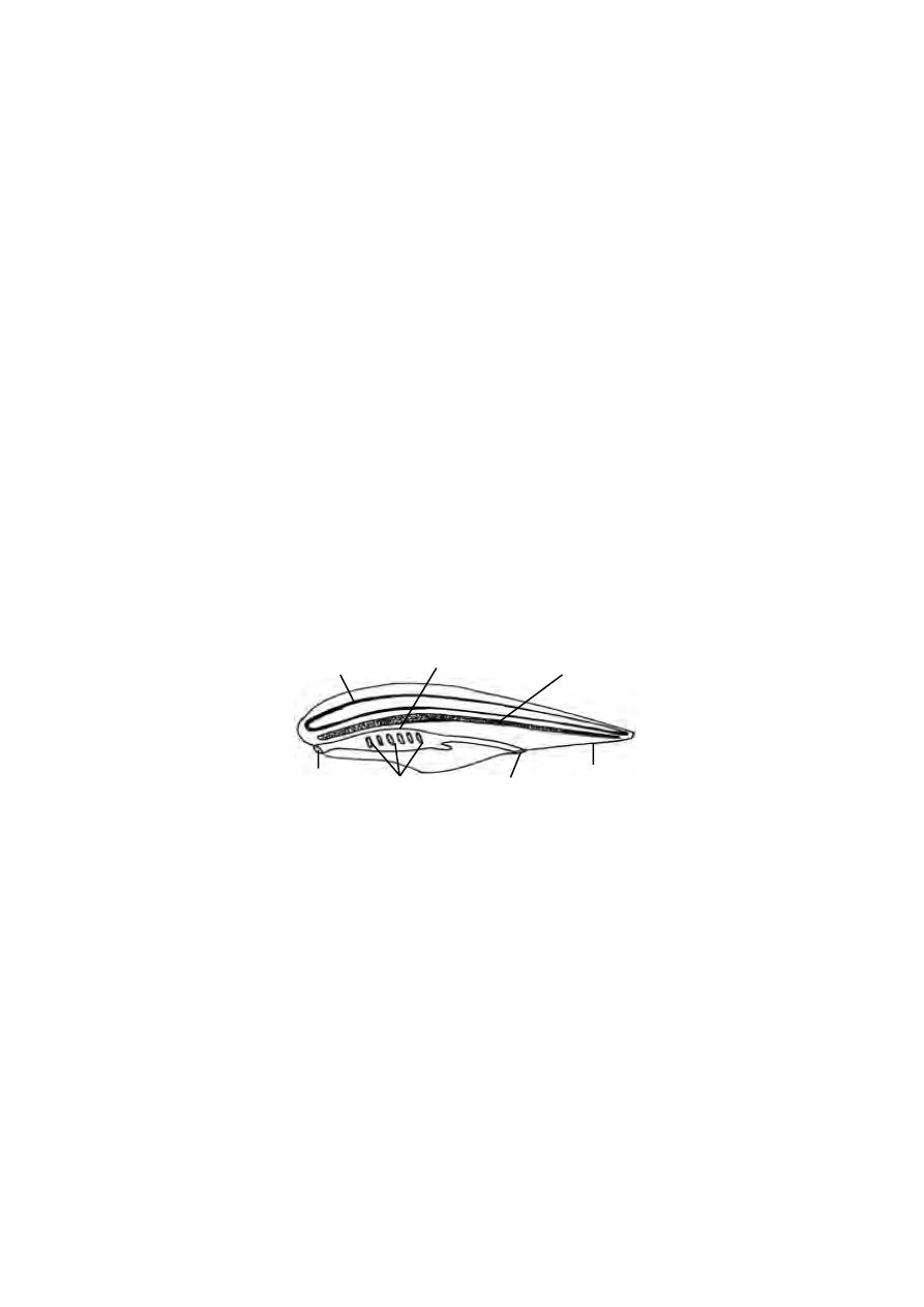
Phylum Chordata
This phylum derives its name from one of the common characteristics
of this group namely the notochord (Gr. noton, back + L. chorda, cord). The
animals belonging to all other phyla of the Animal Kingdom are often termed
‘the non -chordates’ or ‘the invertebrates’ since they have neither notochord
nor backbone in their body.
The backboned animals (vertebrates), together with a few closely re-
lated animals which do not possess a backbone, are included in this phylum.
Most of the living chordates are familiar vertebrate animals. The chordates
are of primary interest because human beings are members of this group.
Fig. 1.2.15 Molluscs
Freshwater mussel
Apple snail
Loligo
Chunk
Fig. 1.2.16 Echinoderms
Star fish
Sea-urchin
22
Diversity of Chordates
The chordates exhibit an astonishing diversity in form, physiology and
habits. The number of chordate species is limited. About 49,000 species are
on record which are only half of the living species of molluscs and less than
one tenth of arthropods. Despite their modest number of species, the chor-
dates make remarkable contribution to the bio-mass of the earth. Nearly all of
them are medium to large in size. The vertebrates in particular are consider-
ably larger and many of them are among the largest of living animals. The
gigantic blue whale which is 35 meters long and 120 tons in weight is the
biggest known animal. The smallest vertebrate , philippine goby is a fish,
measuring only 10 mm in length. The chordates are able to occupy various
kinds of habitats. They have adapted themselves to more modes of existence
than any other group. They are found in the sea, in freshwater, in the air and
on all parts of land from the poles to the equator.
General Characters :
The three distinctive characteristics of the chordates are the presence
of notochord, dorsal tubular nerve cord and pharyngeal gill slits.
1. Notochord :
During the embryonic development of a chordate there appears a sup-
porting rod called the notochord. It lies dorsal to the alimentary canal and
ventral to the nerve cord. In some chordates this structure persists throughout
life. In others it is partially or completely replaced by a ‘backbone’. It is
made up of separate bony elements or vertebrae. Structurally it is com-
posed of large number of specialized vacuolated cells. It is surrounded by
fibrous and elastic sheath. The stiffness of the notochord is due to the tur-
gidity of fluid-filled cells and surrounding connective tissue sheath.
Fig. 1.2.17 Chordata - a diagrammatic structure.
nerve cord
notochord
mouth
anus
tail
pharynx
gill-slits

23
2. Dorsal tubular nerve cord
The nerve cord lies just above the notochord and remains entirely out-
side the coelom. It is a tubular structure having a small hollow canal running
from one end to the other. The dorsal hollow nerve cord persits
throughout the adult life of almost all chordates.
3. Gill slits or Pharyngeal clefts
These are paired lateral clefts leading from the pharynx to the exte-
rior. They are present throughout life in fishes and a few tailed amphibians. In
amphibians, like frogs and toads it is found only in the larval stages. In higher
vertebrates (reptiles, birds and mammals) they are embryonic and non-func-
tional.
4. Ventral heart
The heart is chambered. It is located ventral to the alimentary canal.
5. Closed blood vascular system
In chordates, the blood passes through a continuous system of tubes
namely arteries, capillaries and veins.
6. Hepatic portal system
In chordates, the food laden blood from the digestive tract passes
through the capillary net work in the liver, before reaching the heart. Thus the
veins originating from the digestive tract as capillaries and ending in the liver
again as capillaries constitute the hepatic portal system.
Classification.
The Phylum Chordata is classified into four sub phyla:
Sub phylum 1. Hemichordata,
Sub phylum 2. Cephalochordata
Sub phylum 3. Urochordata
Sub phylum 4. Vertebrata.
First three sub phyla are collectively known as Protochordates. Since
the members of these sub phyla do not have a cranium or skull they are also
referred to as Acrania.
24
Protochordata (Acrania)
The protochoradates are considered as the fore runners of vertebrata
The classification of the protochordates is based on the nature of the noto-
chord.
Sub phylum : Hemichordata.
These are exclusively marine organisms. They are solitary or colonial
forms. They mostly remain as tubiculous forms. The body is soft, vermiform,
unsegmented,bilaterally symmetrical and triploblastic. The body is divisible into
three distinct regions namely proboscis, collar and trunk. The body wall is
composed of single layer of epidermal cells. The dermis is absent. They have
no endoskeleton. A projection from pharynx, projecting inside the
proboscis may be considered as notochord. They have a spacious coelom
lined by coelomic epithelium. The alimentary canal is a straight tube running
between mouth and anus. They are ciliary feeders. Sexes are separate.
Examples : Balanoglossus, Saccoglossus.
Sub phylum : Cephalochordata.
Cephalochordates are small fish like marine chordates. The persistent
notochord extends forward beyond the brain. Hence these are called
cephalochordates. The epidermis is single layered. Paired fins are absent.
Muscles, nephridia and gonads are segmentally arranged. The pharynx is large
with numerous gills. It is a filter feeder.
Example : Amphioxus.
Fig. 1.2.18 Balanoglossus
Fig. 1.2.19 Amphioxus
Fig. 1.2.20 An ascidian

25
Sub phylum : Urochordata
This taxon constitutes a unique group of animals exhibiting diversity in
form and habit. In Urochordata the notochord is confined to the tail re-
gion of the larva. The adults are mostly degenerate, sessile forms. The body
is enveloped by a tunic or test. The free end of the body bears two openings,
the mouth and the atriopore. The proximal part of the alimentary canal is
greatly enlarged to form a spacious pharynx. They are hermaphroditic ani-
mals. The development occurs through free swimming tadpole like larva.
Example : Ascidia, Doliolum, Salpa.
Sub phylum : Vertebrata (Craniata)
This group is characterized by the presence of brain case or cranium
and a vertebral column which forms the chief skeletal axis of the body.
The notochord is an embryonic structure. It is replaced in the adult
stage by a cartilaginous or bony vertebral column. The body is covered
with an integument having an outer epidermis and an inner dermis. The
skin has many modifications such as glands, scales, feathers, claws horns and
hairs.
The digestive system is ventral to the vertebral column. It is provided
with a large liver and pancreas. The circulatory system consists of the ven-
tral, chamberd heart. The circulatory system is of a closed type with arteries,
veins and capillaries. The blood plasma contains red and white blood cor-
puscles. Gill slits are limited in number (usually 5 pairs). There are two pairs
of appendages. The anterior part of the nerve cord becomes differentiated
into brain and spinal cord. The special organs of sense like the nose, eyes and
ears are closely connected with the brain. Urinary and genital systems are
closely connected to form an urinogenital system.
Fig. 1.2.21 A vertebrate- diagrammatic structure.
heart
brain
spinal cord
vertebral column
mouth
gill - slits
urinary bladder
kidney
cloaca
liver
pancreas
gonad
lungs
26
The sub phylum vertebrata may be classified into two groups
(i) Pisces and (ii). Tetrapoda.
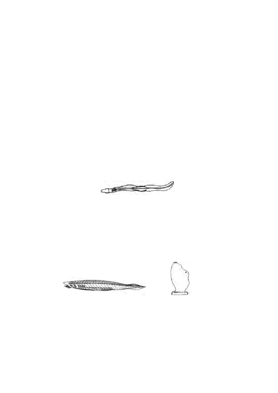
Class : Pisces
Fishes are poikilothermic, aquatic vertebrates with jaws. The body
is streamlined. It is differentiated into head, trunk and tail. Between head and
trunk, the neck is absent. Locomotion is effected by paired and median fins.
The body has a covering of scales. They are of various types like
placoid, cycloid, ctenoid and ganoid scales. The body muscles are arranged
into segments called myotomes.
The Alimentary canal consists of a definite stomach and pancreas
and terminates into cloaca or anus. Respiration is performed by gills. Gill
slits are 5-7 pairs. They may be naked or covered by an operculum. The
heart is two chambered (an auricle and a ventricle).
Fig. 1.2.22 Scales
placoid
cycloid
ctenoid
Fig. 1.2.23 Fishes
Shark
Catla
27
Sinus venosus and renal portal system are present. The red blood
corpuscles are nucleated. The functional kidney of the adult is of meso-
nephric type. The external nostrils do not communicate with the buccal
cavity. Lateral line sense organs are well developed. Sexes are separate.
Fertilization is either internal or external . Examples: Shark, Catla.
Tetrapoda
The vertebrates with two pairs of limbs adapted for locomotion on
land are known as tetrapods. The limbs are of pentadactyl type. The tetra-
pods are identified by a cornified outer layer of skin and nasal passages
communicating with mouth cavity and lungs. The super class Tetrapoda is
divided into four classes namely. Amphibia, Reptilia, Aves and Mammalia.
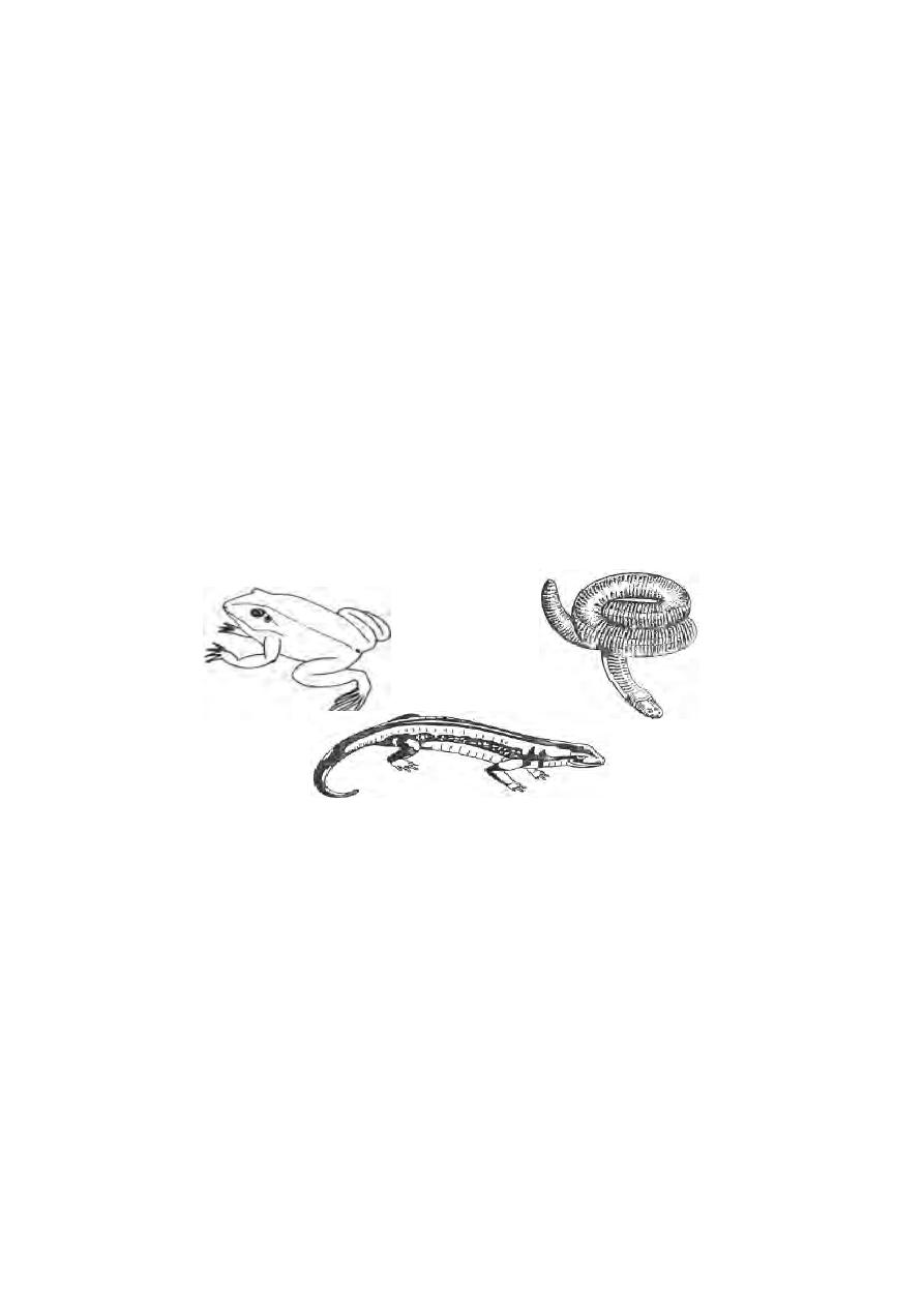
Class : Amphibia
The living representatives of this class include frogs, toads, newts,
salamanders and limbless caecilians.
The transition from aquatic to terrestrial living is clearly indicated in
the class Amphibia. These were the first vertebrates to live on land. Amphib-
ians are not completely land adapted. They hover between aquatic and land
environments. This double life is expressed in their name, amphibia. It is
because of, these reasons ‘the amphibians are considered, a defeated
group’.
The body forms vary greatly from an elongated trunk with distinct
head, neck and tail to a compact, depressed body with fused head and trunk
and no intervening neck. The forelimbs of frogs and toads are smaller than
hind limbs. In frogs, hindlimbs have webbed feet. The surface of the skin is
Fig. 1.2.24 Amphibians
Frog
Caecilian
Salamander
28
smooth and slimy. The slimy nature is due to the presence of mucous secret-
ing glands. Scales are practically absent.
The mouth is usually large with small teeth in upper or both jaws. The
external nostrils communicate into the anterior part of the mouth cavity.
Respiration is effected by gills, lungs, skin and pharyngeal region. The heart is
three chambered with two auricles and a single ventricle. The skeleton is mostly
bony, with varying number of vertebrae; exoskeleton is absent. Sexes are
separate. Fertilization is either external or internal. The tadpole Metamorphoses
into adult.
Examples : Frog, Toad, Salamander, Caecilian
Amniota
The tetrapods like reptiles, birds and mammals are referred to as
amniotes. The amniotes have certain membranes associated with embryos
inside the egg. It is an adaptation in terrestrial forms during development.
These membranes are the amnion, chorion and allantois.
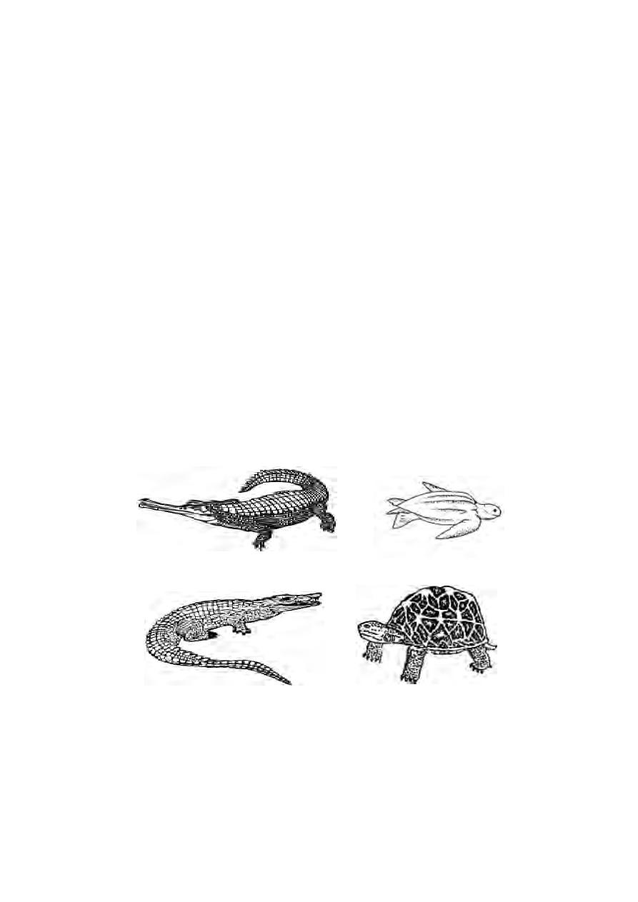
Class : Reptilia
Reptiles are represented by lizards, snakes, turtles, tortoises, alliga-
tors, crocodiles and the tuatara lizard, Sphenodon punctatum.
Fig. 1.2.25 Reptiles
Marsh crocodile
Gangetic crocodile
Star turtle
Leatherback turtle

29
The body is variable in shape. It is covered with an exoskeleton of
horny imbricate epidermal scales. Skin glands are practically absent. The limbs
are of pentadactyl type adapted for climbing, running and paddling. The en-
doskeleton is well ossified. Respiration is by lungs. The heart is three cham-
bered (In crocodiles it is four chambered). The functional kidney of the adult
are metanephros. The Sexes are separate. Fertilization is internal. The eggs
are covered with leathery shells. Reptiles have developed some form of copu-
latory organ to transfer the sperms into the cloaca of the female.
Example : Garden lizard, Cobra, Monitor lizard, Crocodile, Turtle.
Class : Aves
Birds are one of the most intersting and widely known group of ani-
mals. There are more than 8600 species of birds distributed all over the world.
Birds as a group exhibit a characteristic uniformity in structure.
Aves are warm blooded vertebrates with an exoskeleton of feathers
forming a non-conducting covering to keep the body warm. The feet are cov-
ered with scales. The forelimbs are modified as wings and provided with
feathers for flight. The hindlimbs are attached far forwards to balance the
weight of the body. The bones are spongy, containing air-cavities rendering
the body light. There is a fusion of bones and this is especially seen in the
vertebral column. Only three digits are present in the forelimbs. In the hindlimbs
there are four toes with the first directed backwards. A horny beak is present.
The alimentary canal ends in a cloaca. Inside the body air sacs are
present and some of them communicate with air cavities in the bones. The
heart is four chambered. The red blood corpuscles are oval and nucleated.
The kidneys are three lobed. The ureters open into the cloaca. Urine is semi-
solid and contains uric acid. The nervous system is well developed. Eyes are
usually powerful and a specialized structure called pecten is present inside
the eye ball to help in accomodation. Sexes are separate, Fertilization is inter-
nal. Eggs are provided with large amount of yolk. The egg is covered by a
hard calcareous shell. In spite of several advanced features the birds have
certain reptilian characters. Hence they are known as “glorified reptiles”.
Examples : Pigeon, parrot, crow, sparrow, peakcock, ostrich, penguin.
Class : Mammalia
The term “mammalia” was given by Linnaeus (1758) to that group of
animals which are nourished by milk from the breasts of the mother. They are
a successful group, for they adapt themselves readily to new situations and to
new food habits.
30
The body is generally covered with epidermal hairs. The integument
is provided with sweat, sebaceous and scent glands. The mammary glands
are modified integumentary glands. The external ear or the pinna is present in
most of the mammals. A muscular diaphragm is present in between thoracic
and abdominal cavities. It helps in respiration. The red blood corpuscles are
non-nucleated, biconcave and usually circular in form. The heart is four cham-
bered. Only the left aortic arch is present. In brain cerebral hemispheres
are very large and highly convoluted.
Corpus callosum, a transverse band of nerve fibres connecting the two ce-
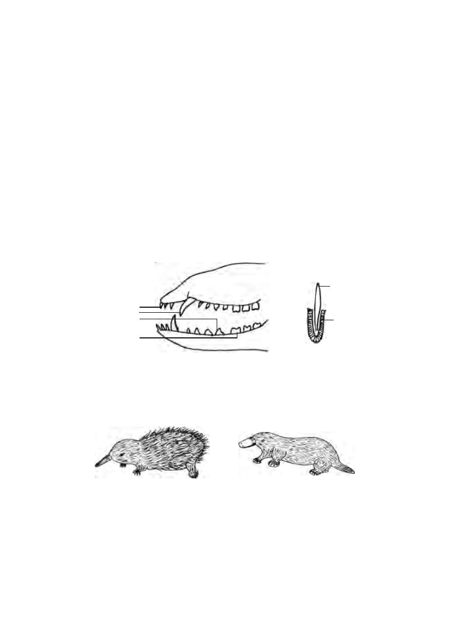
rebral hemispheres, is present. Dentition is thecodont, heterodont and di-
phyodont. Cloaca is absent. Testes lie outside the body cavity, enclosed in
scrotal sacs. Eggs are small with little or no yolk. Fertilization is always inter-
nal. Mammals are Viviparous ie., they give birth to alive young ones. Pla-
centa is usually present.
The class Mammalia is subdivided into three subclasses namely
Monotremata, Marsupialia and Placentalia.
1. Sub class : Monotremata or Prototheria
Fig. 1.2.27 Egg laying mammals
Platypus
Ant-eater
Fig. 1.2.26 Mammalian teeth
Heterodont
Thecodont
incisor
canine
pre molar
molar
tooth
socket
31
These are primitive egg laying mammals Example : Spiny ant-eater,
duck billed platypus.
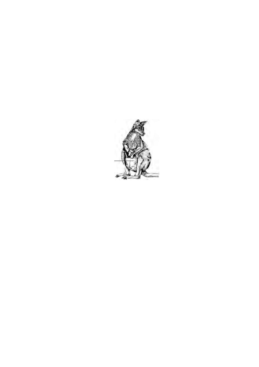
2. Sub class : Marsupialia or Metatheria
These are popularly called as marsupials or pouched mammals. The
young ones are born in an immature stage and migrate into the pouch on the
mother’s body. Further development is completed in the pouch or marsu-
pium.
Example : Kangaroo
3. Sub class : Placentalia or Eutheria
In this group eggs develop within the uterus. The developing embryo
receives nutrition through maternal blood circulation via the placenta.
Example : Elephant, tiger, lion, man, monkey, dog, cat , rat, bat.
Order Primates :
It is an order coming under the subclass Eutheria. This order is of
interest because it includes man, besides lemurs, tarsiers, monkeys and
apes. They inhabit chiefly the warmer parts of the world. This group stands
first in the animal kingdom in brain development. However, most of them are
unspecialized and tree dwelling (arboreal). Primates are omnivorous in
habit. The body is covered with hairs except palm, sole and parts of face.
The neck is mobile. The forelimbs are shorter than the hindlimb. The limbs
have five digits and all the digits end in flat nail. The pollex or thumb or first
toe are smaller than other digits and are opposable (except the hallux of
man). The brain is highly developed. The cerebral hemispheres are much
marsupium
Fig. 1.2.28 Pouched mammal

32
convoluted and cover the cerebellum. The eyes are directed forward and
the vision is binocular and stereoscopic. Mammae are two and thoracic
in position.
To know
Invertebrates
Scientific Names
Earthworm (k©òG)
Lampito mauritii
Cockroach (fu¥gh‹ ó¢Á)
Periplaneta americana
House fly (<¡fŸ)
Musca nebula
Locust (bt£L¡»ë)
Schistocera gregaria
Bed bug (_£il¥ ó¢Á)
Cimex hemipterus
Leaf insect (Ïiy¥ó¢Á)
Phyllium sps
Stick insect (F¢Á¥ó¢Á)
Carausius sps
Water-scorpion (Ú®¤njŸ)
Nepa sps
Butterfly (t©z¤J¥ ó¢Á)
Pieris sps
Rat flea (vè bjŸS¥ó¢Á)
Xenopsylla cheopis
Scorpion (njŸ)
Palamnaeus swammerdami
King crab (uh# e©L)
Limulus sps
Spider (ÁyªÂ¥ó¢Á)
Aranea sps
Apple snail (M¥ÃŸ e¤ij)
Pila globosa
Freshwater mussel (e‹Ü® e¤ij)
Lamellidens marginalis
Star fish (e£r¤Âu Û‹)
Asterias rubens
Vertebrates
Angel fish (VŠrš Û‹)
Pterophyllum scalare
Guppy (f¥Ã Û‹)
Poecilia reticulata

33
Frog (jtis)
Rana hexadactyla
Garden lizard (Xzh‹)
Calotoes versicolor
Cobra (ešy gh«ò)
Naja naja
Peacock (kæš)
Pavo cristatus
Crow (fhf«)
Corvus splendens
Sparrow (FUé)
Passer domesticus
Parrot (ȑ)
Psittacula Krameri
Rat (vè)
Rattus rattus
Dog (ehŒ)
Canis familiaris
Cat (óid)
Felis domesticus
Tiger (òè)
Panthera tigris
Lion (Á§f«)
Panthera leo
Elephant (MÁa ahid)
Elephas maximus
Man (kåj‹)
Homo sapiens
Monkey (Fu§F)
Macaca radiata
Mongoose (Ñç¥ÃŸis)
Herpestes edwardsii
Bear (fuo)
Ursus arctos
Fruit bat (gHªÂ‹å btsthš)
Cynopterus sphinx
Donkey (fGij)
Equus hemionus
Rhinoceros (fh©lhäUf«)
Rhinoceros unicornis
Spotted deer (òŸë kh‹)
Axis axis
Man (kåj‹)
Homo sapiens

34
1.3. Type study - 1. Plasmodium
Phylum
-
Protozoa
Class
-
Sporozoa
Order
-
Haemosporidia
Members of the genus Plasmodium are collectively known as ma-
larial parasites. They cause a febrile disease called malaria. Malaria as a
chill and fever disease is known to mankind for a long time. Eradication of
malaria is an important problem in public health. For a long time it was
believed that malaria was caused by harmful vapours produced in marshy
land (Gr. Malo-bad+air). Charles Laveran, a french military surgeon, for
the first time, noticed Plasmodium in the blood of a malarial patient, in 1880.
Its connection with the intermediate host and the modes of transmission
were experimentally worked out in Calcutta by Sir Ronald Ross in 1889. For
this discovery he was awarded the nobel prize for medicine in 1902. Grassi
(1890) provided absolute scientific proof for the specific relationship between
Anopheles mosquito and the human malarial parasite.
Plasmodium
: The plasmodium is an intracellular sporozoan blood parasite.
For the completion of life cycle it requires two hosts, a vertebrate and a blood
sucking invertebrate. Transference of the parasite is effected by the inverte-
brate host. In man, the infection takes place by the inoculation of the slender,
sickle shaped nucleated sporozoite in the blood by the bite of an infected
female mosquito belonging to the genus Anopheles. At least four species of
Plasmodium, P. vivax, P. falciparum, P. malariae and P. ovale, are known
to attack man causing different kinds of malaria.
The life cycle of the malarial parasite involves two hosts, the man and
the mosquito. The modes of development in these two hosts are different. In
man the mode of reproduction is asexual and in mosquito it is sexual. Man is
the intermediate host and the mosquito is the definitive host.
Life cycle in Man - Schizogony
There are two phases in the life cycle of malarial parasite in man.
They are (1) Pre erythrocytic cycle or Exoerythrocytic cycle (in liver cells)
and (2). Erythrocytic cycle or Endo-erythrocytic cycle (inside the red blood
corpuscles)
35
Pre-erythrocytic cycle:
The pre-erythrocytic cycle comprises the asexual reproduction of the
parasite in the liver. When an infected female Anopheles mosquito bites a
person, thousands of slender, sickle shaped nucleated sporozoites are in-
jected in the blood. The sporozoites first enter the capillary vessels of the skin
and then enter the general circulation. These parasites circulate in the blood
for about 30 minutes and enter into the pre-erythrocytic cycle in the reticu-
loendothelial cells of the liver.
The sporozoites penetrate the liver cells and develop into forms known
as cryptozoites. A cryptozoite has a compact nucleus and no pigment or
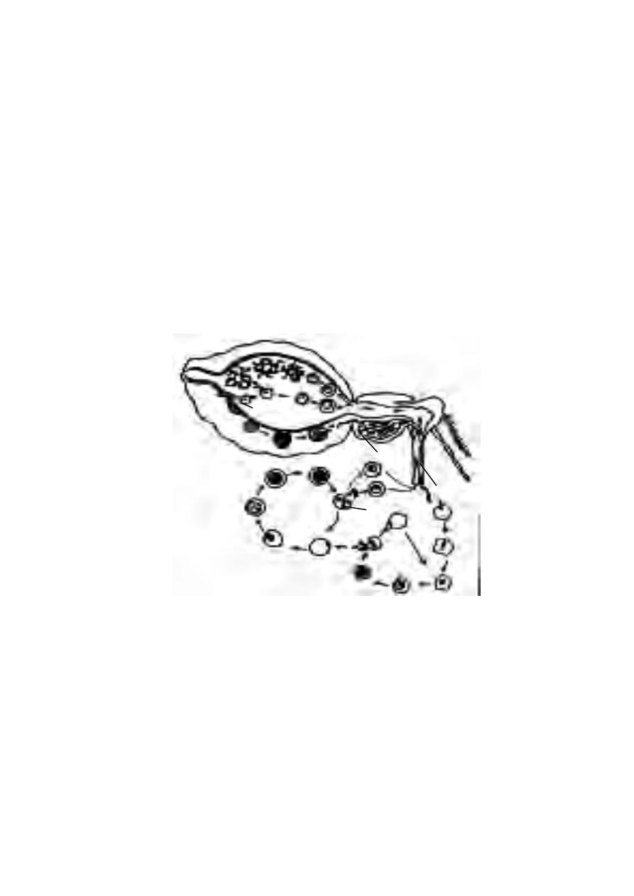
Fig. 1.3.1 Life cycle of malarial parasite
Sporogony
Gametogony
Schizogony
merozoite
Endo-erythro-
cytic cycle
RBC
microgamete
marcrogamete
sporozoi-
livercell
ookinete
oocyst
early
schizont
rupture of
RBC
salivary gland
salivary duct
food channel
exflagellation
zygote
spore
formation
signet ring
stomach
crypto-schizont
crypto-merozoites
36
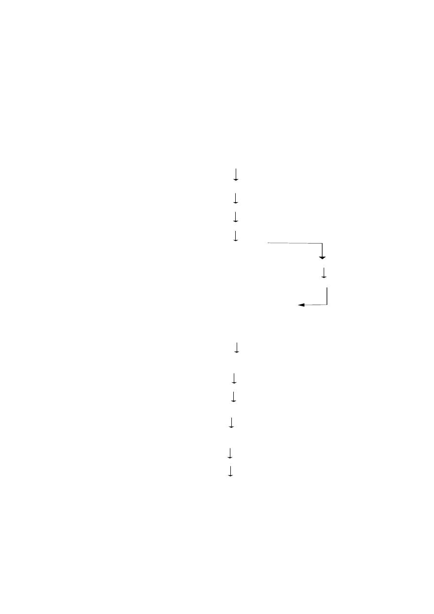
Life history of Plasmodium (the malarial parasite)
Life cycle in Man (Schizogony)
Mosquito
In liver :- Pre or
Exoerythrocytic Cycle
Sporozoites
Cryptozoites
Cryptomerozoites
In RBC :- Erythrocytic or
Trophozoites
Endo-erythrocytic cycle or
In blood plasma
Schizonts
Cycle of golgi
(febrile attacks)
Merozoites
Gametocytes(in RBC)
Life cycle in Female anopheles mosquito (Sporogony) or Cycle of Ross
Inside the gut
Gametocytes (in RBC)
Gametes [male(micro)
and female (macro)]
Zygote
Ookinete
In the wall of the gut
Oocyst
Sporozoites
Man

37
vacuoles. Cryptozoites rapidly grow feeding on the liver cells. When a crypto-
zoite has reached its full growth it fills the entire cell. In this stage it is known
as the crypto-schizont. It undergoes schizogony and the resulting cells known
as crypto-merozoites are set free in the blood by the rupture of the liver
cells. The released crypto-merozoites invade fresh liver cells or red blood
corpuscles. This cycle is considered as a period of incubation before the para-
sites could start the erythrocytic cycle. During this period of 7 - 17 days, the
parasites are not seen in the blood stream.
Erythrocytic or Endo-erythrocytic cycle.
Each cryptomerozoite makes its way into a red blood corpuscle and
feeds on its contents. After some time, the parasite gets an amoeboid shape.
This growing stage is known as the trophozoite stage. Soon it develops a
vacuole which gradually increases in size. Thus the nucleus is pushed to one
side. This stage is called the signet ring stage. With further growth the vacu-
ole disappears and the amoebula occupies the entire interior of the corpuscle.
This stage is known as the schizont stage.
In the schizont, the nucleus breaks up into bits (6-24) and each be-
comes surrounded by a small amount of cytoplasm. These cells are known as
merozoites. By the rupture of the wall of the red blood corpuscles the mero-
zoites along with wastes(haemozoin) are released into the blood. This causes
the malarial fever. The liberated merozoites attack another set of corpuscles
and start the life cycle anew. This method of infection is known as autoinfec-
tion. The life cycle in the blood of man is called the cycle of Golgi or
schizogony or endoerythrocytic cycle.
Schizogony keeps up the multiplication of the parasites and their main-
tenance in the blood.
After schizogony has taken place for several generations some of the
meroziotes which invade the red corpuscles, instead of developing into tro-
phozoites and schizogonts, develop into gametocytes. The gametocytes are
of two types - marco-gametocytes and micro-gametocytes. The macro-
gametocyte has a small nucleus and a dense food laden cytoplasm. The mi-
cro-gametocyte has a relatively large nucleus and clear cytoplasm. Their fur-
ther development depends on their entry into the stomach of a female anoph-
eles. If it does not take place they disintegrate.

38
Life cycle in the mosquito - sporogony
When a female anopheles mosquito bites an infected person, it sucks
blood along with all the stages of parasite. But in the gut of the mosquito, only
the mature gametocytes survive and the rest of the stages are destroyed.
From the gametocytes develop gametes. The process of development of ga-
metes from gametocytes is known as gametogony.
Gametogony :
The nucleus of the micro-gametocyte divides into many fragments and
the cytoplasm is thrown into flagellated structures. There may be as many
cytoplasmic structures as there are nuclei. This process is known as exflag-
ellation. The resultant cells are called the microgametes. The nucleus of
the macro-gametocycte divides equally into two. The cytoplasm divides un-
equally. So among the resulting cells one is bigger and the other is smaller.
The small cell is thrown out. This process is known as maturation. The re-
sulting bigger cell is known as female gamete or macrogamete.
Syngamy and sporogony :
Inside the stomach of the mosquito the microgamete and the macro-
gametes come into union and nuclear fusion takes place. This kind of union is
called syngamy and the resultant form is known as zygote.
The zygote assumes an elongated form and is capable of movement.
It is known as ookinete. It pierces the wall of the stomach and comes to lie
under the outer layer of stomach wall. There, it ceases to move, becomes
round and forms a membranous cyst-wall. This stationary zygote enclosed in
a cyst-wall is known as oocyst. It grows in size absorbing the nourishment
from the host.
The nucleus of the oocyst divides repeatedy, each being surrounded
by a fragment of cytoplasm. Thus inside the oocyst, a large number of cells
develop into minute, slender, sickle shaped bodies called sporozoites. The
cyst wall breaks, liberating the sporozoites into the body cavity (haemocoel)
of the host. They wriggle forward and enter the salivary gland. When such
an infected female anopheles mosquito bites a healthy person, it injects into
his blood a stream of sporozoites. This kind of transmission is called
inoculation.
Types of Malaria :
The disease caused by Plasmodium is known as malarial fever. It is
charcterised by recurring bouts of fever, each lasting several hours. The

39
Different species of Plasmodium
S.No
Species of Plasmodium
Type of fever
1.
Plasmodium vivax
benign tertian fever
2.
P. falciparum
malignant tertian fever
3.
P. malariae
qurtan fever
4.
P.ovale
Ovale or mild tertian fever
Stages in the Life History of Plasmodium
S.No
Stage
Occurrence
1.
Sporozoites
In the blood stream of man
2.
Cryptozoite
Liver (man)
3.
Trophozoite
Blood (man)
4.
Amoebula stage
RBCs (man)
5.
Signet ring stage
RBCs (man)
6.
Merozoite
RBCs (man)
7.
Macrogamete
Stomach of female
anopheles mosquito
8.
Microgamete
Stomach of female
anopheles mosquito
9.
Ookinete
Stomach of female
anopheles mosquito

40
febrile condition in man is due to toxins liberated into the blood along with the
merozoites when the corpuscle is ruptured at the end of schizogony.
There are four species of Plasmodium known to cause malaria in
man. The commonest and most widely distributed species is
P. vivax. It causes benign tertian malaria in which the fever recurs
every third day (every 48 hours). P.falciparum is largely limited to the
tropics and subtropics and causes the malignant tertian or subtertian
malaria. This type of malaria has a high death rate. Blood corpuscle parasitised
by this species tend to clump together and block up small blood vessels and
damage the essential organs. It is a dangerous species and the disease often
appears in an epidemic scale. P. malariae causes quartan malaria with
feverish fits every fourth day (every 72 hours). The fourth species is P. ovale.
It is principally found in west Africa but occassionally in S. America, Russia
and Palestine. It causes benign tertian malaria in which the fever recurs
every third day (every 48 hours).
These four species differ from each other in the details of structure,
time needed to complete the schiogzony, the incubation period, number of
merozoites released and duration of sexual cycle.
Control of Malaria
The control measures fall under the following three categories.
Treatment of infected patient
(1) Plasmodium does not produce antitoxins or antibodies in human blood.
Therefore malaria cannot be treated by inoculation or vaccination with
immune sera. It can only be treated with drugs that may kill all stages of the
parasite without poisoning the patient.Quinine, which is extracted from the
bark of cinchona trees, had been used effectively for the past 300 years to
cure malaria. The various synthetic drugs, such as Paludrine, Atabrin,
Camoquin, Chloroquine, Resochin, Pamaquin etc are used as suppressants of
various stages of the parasites.
(2) Prevention of infection :
It can be effected in two ways.
(i) using protective measures such as mosquito nets, anti-mosquito creams
(repellants) and coils.
(ii) use of the prophylactic drugs; small daily dose of anti-malarial drugs will
kill the parasite either in the sporozoite or merozoite stage.

41
(3) Control of vector
It is perfectly clear that if the vector is completely exterminated the
infection cannot be transmitted from one person to another. It is the most
effective and surest way of controlling malaria. It is achieved by using effec-
tive insecticides and by draining swamps. It destroys the breeding places of
mosquitoes.
Adult mosquito can be most effectively controlled by spraying DDT,
malathion or any other insecticide in the houses; fumigating pyrethrum cresol
and other compounds of naptha; sterilization of male mosquitoes. The young
stages of mosquito can be controlled by introducing larvivorous fishes like
Gambusia and Lebistes in ponds, lakes, canals and tanks.
Type study - 2. Earthworm
Phylum
-
Annelida
Class
-
Chaetopoda
Order
-
Oligochaeta
Type
-
Lampito mauritii
Earthworms are nocturnal animals. They lie in the burrows during the
day and come out at night for food. Earthworms leave the burrow only during
the rainy season when their burrows are flooded with water.
External features
Lampito (Megascolex) mauritii is a common earthworm found in South
India. The body is long, slender, cylindical and bliaterally symmetrical. It is
about 8 to 21 cm long and 3 to 4 mm in thickness. The dorsal surface is dark
purplish brown, and the ventral surface is paler in colour. It is marked by a
series of segments. The segments are separated from one another by inter-
segmental grooves. The division is both external and internal. Inside the body,
each cavity of the segment is separated from the next, by a thin partition
called the septum. All the segments look alike. This kind of repetitive ar-
rangement of the segments is called metamerism.
The mouth is found in the centre of the first segment of the body,
called the peristomium. Overhanging the mouth is a small flap called the
upperlip or prostomium. The last segment has the anus. It is called the
pygidium. In mature worms, segments 14 to 17 may be found swollen with a
glandular thickening of the skin called clitellum.
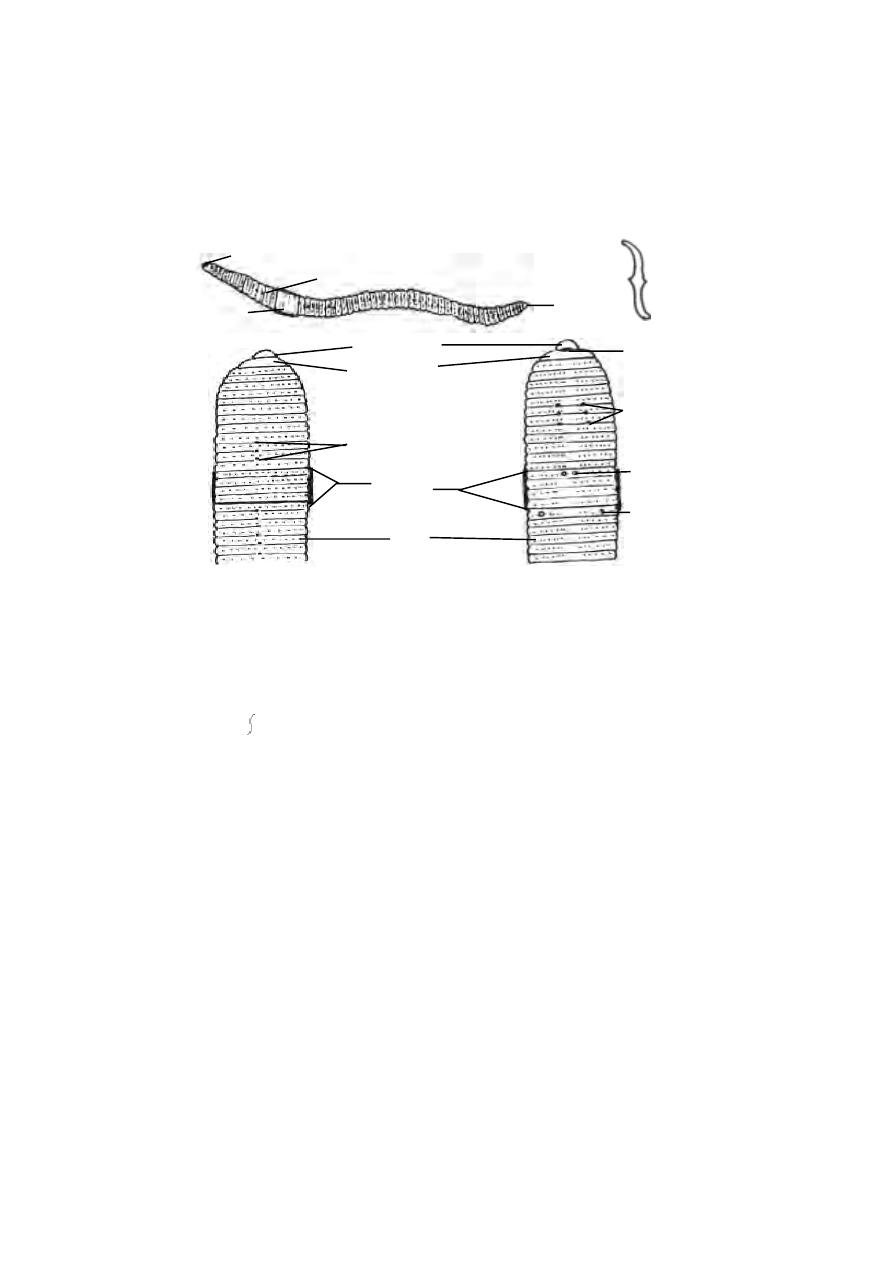
42
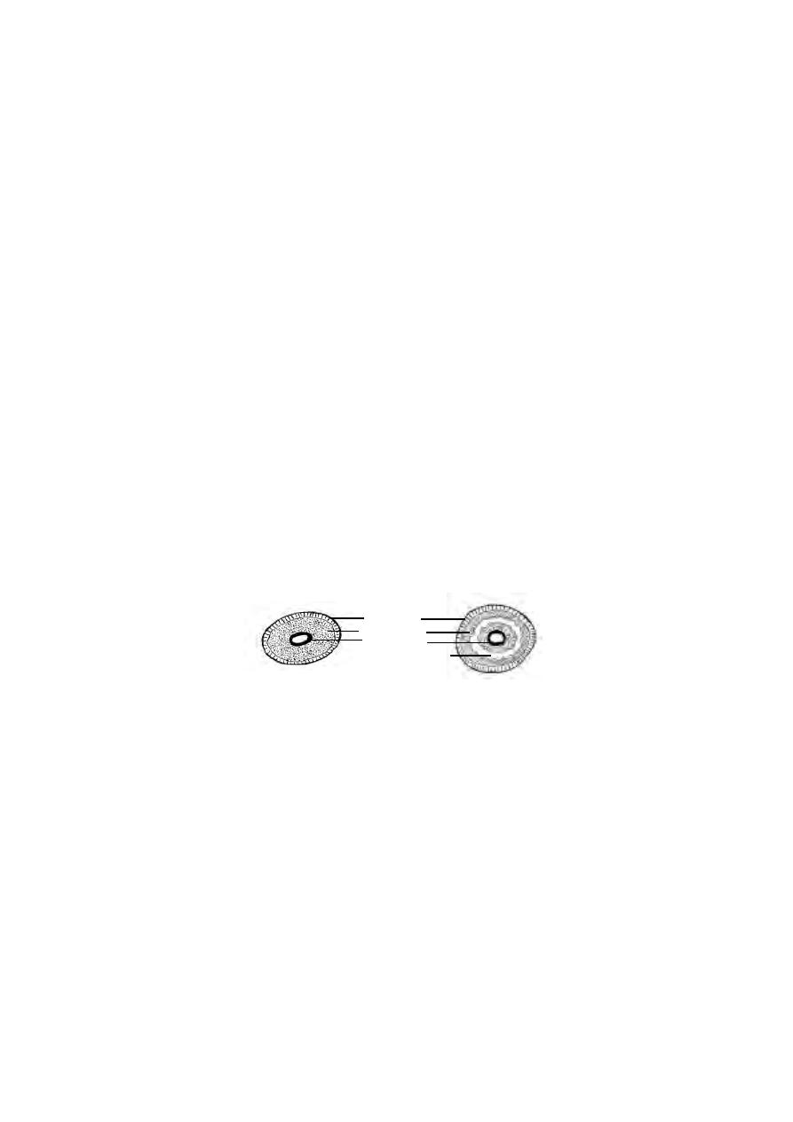
Body setae
Tiny curved bristles called setae are found embedded in small pits of
the body wall. These pits are called the setigerous pits. The setae are
arranged around the body. They are made of chitin and have a swollen middle
part and pointed curved ends. The setae resemble the mathematical
symbol ‘ ’. They can be moved in any direction and extended or withdrawn
by the action of muscles. They are used for locomotion.
External apertures :
(i). Dorsal pores : These are minute openings situated in the mid dorsal line
in the intersegmental grooves commencing from the 10th segment. The co-
elom communicates to the exterior through these pores and keep the body
surface moist and free from harmful micro organisms.
(ii). Spermathecal openings : Three pairs of openings are situated ventrolat-
erally in the intersegmental grooves between segments six and seven, seven
and eight and eight and nine. These opening can be easily seen in mature
worms.
(iii). Openings of oviduct : These are a pair of apertures lying close together
on the ventral surface of the 14th segment.
7
peristomium
openings of
spermathecae
Fig. 1.3.2. Earthworm-External
Entire
Dorsal view
Body setae
Ventral view
prostomium
clitellum
setae
dorsal pores
anus
prostomium
dorsal pore
opening of
oviduct
opening of
vas
deferens
clitellum
mouth
14
17
8
10
6
14
18

43
(iv). Openings of Spermiduct : A pair of apertures are situated on the
lowerside of the 18th segment.
(v). Nephridiopores : Numerous minute openings scattered on the body wall
from 14
th
segment onwards.
Body wall :
The body wall of earthworm is thin soft and moist. It consists of the
following layers arranged from outside.
Cuticle : It is a thin, transparent, iridescent layer secreted by the underlying
epidermis.
Epidermis : It is in the form of a single layer of columnar cells. This layer
contains gland cells and receptor cells.
Dermis : It is a very thin sheet of connective tissue forming a basement for
the epithelial cells on the outside and muscles on the inside.
Muscles : The muscles are arranged in two layers, namely the outer circular
and inner longitudinal.
Coelomic epithelium : It is the inner most layer of the body wall forming the
lining of the body cavity.
Body Cavity :
A spacious body cavity called the coelom is seen between the alimen-
tary canal and the body wall. It is divided into a series of compartments by the
transverse partitions of connective tissue called the septa. The coelom is lined
with the coelomic epithelium and filled with coelomic fluid. It is a colourless
fluid with amoeboid coelomic corpuscles floating in it. The fluid oozes out
through the dorsal pores. It keeps body surface moist as a condition quite
Fig. 1.3.3. Earthworm-T.S.
setae
cuticle
epidermis
circular muscle
longitudinal muscle
typhlosole
ventral nerve cord
ventral vessel
dorsal vessel
chloragogen cells
coelom
nephridium
intestine
endodermal lining
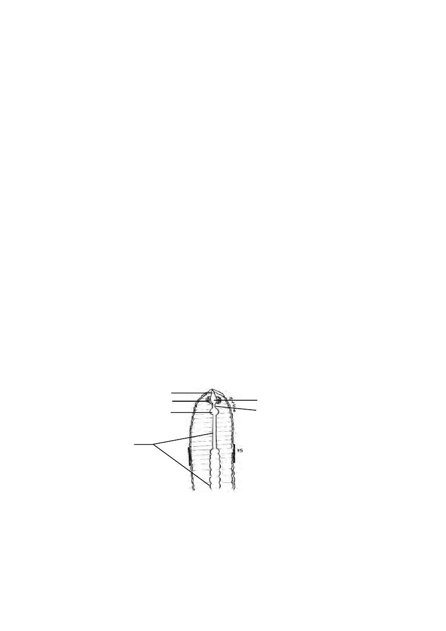
44
essential for respiration. The coelomic cavity communicates to the exterior
through reproductive and excretory apertures. The germ cells are budded off
from the wall of this cavity.
Locomotion :
Earthworms move about by contraction and expansion of its body wall.
When the circular muscles of the body wall contract, the body becomes thin
and elongated. This process results in the forward extension of the body. Then
it fixes itself firmly to the ground with help of the body setae and mouth.
Subsequently when the longitudinal muscles contract, the body becomes thick
and shortened. As a result, the body is drawn forward towards the anterior
end which is already fixed to the ground. Thus by a repeated process of alter-
nate contraction and expansion of muscular body wall locomotion is effected.
Digestive System :
The digestive system runs as a straight tube from mouth to anus. The
mouth is situated in the first segment. The mouth opens into the buccal cav-
ity which occupies segments 1 and 2. The buccal cavity in turn leads into a
thick muscular Pharynx. The pharynx occupies segments 3 and 4 and is sur-
rounded by the pharyngeal glands. The oesophagus is a short narrow tube
lying in 5
th
segment. It leads into the gizzard lying in the 6
th
segment. Its inner
surface has a chitinous lining. The intestine is a large tube extending from the
gizzard to the anus. The intestine upto the 14
th
segment is narrow and the
remaining part is sacculated. The dorsal wall of the intestine is folded into the
cavity as the typhlosole. This fold contains blood vessels. It increases the
absorptive area of the intestine. The inner epithelium consits of columnar cells
and glandular cells.
Fig. 1.3.4. Earthworm-Digestive system
pharyngeal gland
buccal cavity
gizzard
intestine
pharynx
oesophagus
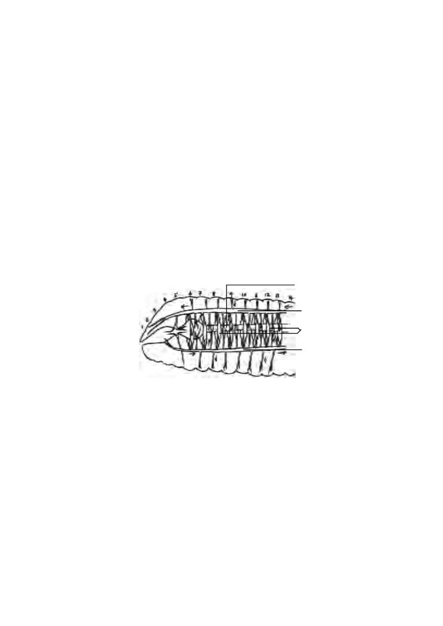
45
Feeding :
The earthworm feeds on decaying organic materials contained in the
soil. It takes the soil into its alimentary canal where the organic matter is
digested and absorbed. The unwanted soil particles are sent out as worm
casts.
Circulatory System :
In the body of earthworm there are two median longitudinal vessels.
The dorsal longitudinal vessel runs above the alimentary canal. The ven-
tral longitudinal vessel runs below the alimentary canal. The dorsal vessel
is contractile and blood flows forwards in it. There are paired valves inside
this vessel which prevent the backward flow of the blood. The ventral vessel
is non contractile and blood flows backwards in it. The ventral vessel has no
valves. In the anterior part of the body the dorsal vessel is connected with the
ventral vessel by eight pairs of commissural vessels or the lateral hearts
lying in the segments 6 to 13. These vessels run on either side of the alimen-
tary canal and pump blood from the dorsal vessel to the ventral vessel.
The dorsal vessel receives blood from various organs in the body.
The ventral vessel supplies blood to the various organs.
Excretory System :
Excretion is the process of elimination of metabolic waste products
from the body. In earthworm, excretion is effected by minute paired tubes
called nephridia. These are found, one pair, in each segment.
A typical nephridium has an internal funnel like opening called the
nephrostome. It is fully ciliated. The nephrostome is in one segment and the
rest of the tube will be in the succeeding segment. This tube has three distinct
Fig. 1.3.5. Earthworm - Circulatory system-Lateral view
alimentary canal
dorsal vessel
ventral vessel
lateral hearts
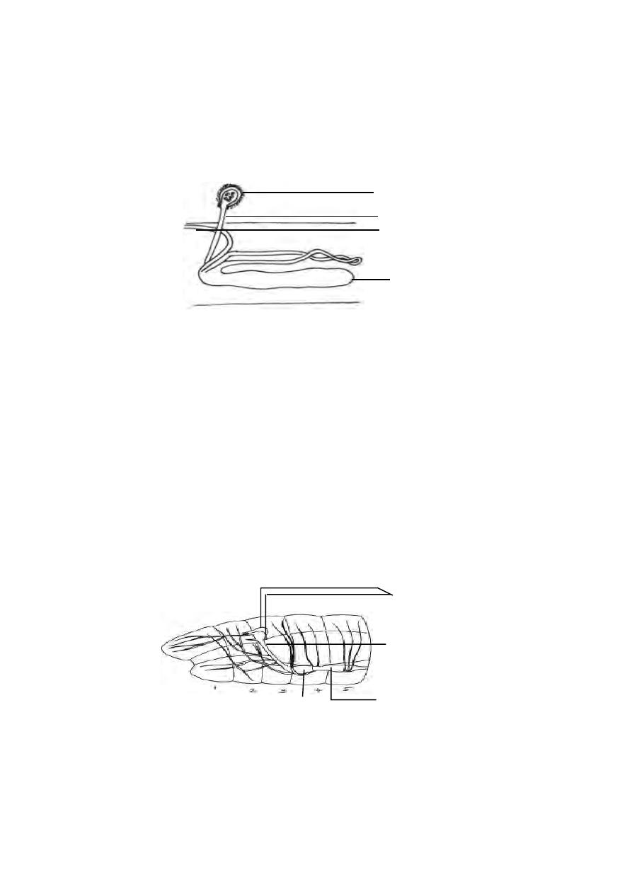
46
divisions. The first part following the nephrostome is ciliated inside. This is
called the ciliated region. The next part is wider and is thrown into coils.
This part has glands on its wall.It is called the glandular region. The last part
has neither cilia nor glands. It is called the muscular region. This region
opens outside by an aperture called the nephridiopore. The waste material
is collected from the body cavity by the ciliated funnel. The ciliated region
pushes the waste into the nephridium. The glandular part extracts waste from
the blood and add it on to the waste inside. Finally the waste goes out through
the nephridiopore.
In the South Indian earthworm, Megascolex, there are certain modifi-
cations. There are three types of nephridia in the megascolex. They are : (i).
Meganephridia, (ii). micronephridia, (iii). Pharyngeal nephridia.
Besides nephridia there are some special cells on the wall of the intes-
tine called Chlorogogen cells. They collect the waste and then drop down
into the body cavity. These are then sent out through nephridia.
Nervous System :
Fig. 1.3.6. Earthworm - nephridium
nephrostome
segment
segment
septal excretory canal
body of nephridium
ciliated region
ventral nerve cord
Fig. 1.3.7. Earthworm - Nervous system
sub pharyngeal ganglion
brain
circum pharyngeal
connective

47
The brain is formed of the supra pharyngeal ganglia. It is a bilobed
mass of nervous tissue situated on the dorsal wall of the pharynx in 3
rd
seg-
ment. The ganglia found below the pharynx in the 4
th
segments is called the
subpharyngeal ganglia. The brain and the subpharyngeal ganglia are con-
nected by a pair of circum pharyngeal connectives.They run one on each
side of the pharynx. Thus a nerve ring is formed around the anterior region of
the alimentary canal. The double, solid ventral nerve cord runs backwards
from the subpharyngeal ganglia, in the mid ventral line to the hind end of the
body. The ventral nerve cord has segmental ganglion one in each segment.
From the brain nerves are given off to the peristomium. From each ganglion of
the ventral nerve cord, three pairs of nerves are given off to the body wall and
other organs.
Receptors which are stimulated by the sense of touch(tactile re-
ceptors), Chemical changes(chemoreceptors) and changes in temperature
(thermoreceptors) are present in the body wall. These receptors are in the
form of groups of slender columnar cells with short hairs projecting at the free
end and connected with sensory fibres at the inner end. Receptors stimulated
by changes in the intensity of light (Photoreceptors) are found on the dorsal
surface of the body. Gustatory receptors (sense of taste) and
olfactory receptors (sense of smell) are found in the buccal cavity.
Reproductive System :
Both male and female reproductive organs are present in the same
worm. Hence the earthworms are known as hermaphrodites. Since the
sperms develop earlier than production of ova, self-fertilization is avoided. It
is known as protandry.
Male reproductive organs :
The male sex organs are formed of two pairs of testes and a pair of
vasa deferentia. Testes are found in segments 10 and 11. They are tufts of
finger shaped processes attached to the anterior septa of segments 10 and 11.
There are two pairs of seminal versicles formed as outgrowths of the testicu-
lar segments. Further two pairs of seminal funnels called ciliary rosettes
are situated in the same segment as the testes. The ciliated funnels of the
same side are connected to a long tube called vas deferens. The two vasa
deferentia of both sides run backwards along the ventral body wall upto the
18
th
segment where they open to the exterior through the male gential aper-
ture. Male genital apertures contain penial setae for copulation. A pair of
prostate glands, each in the form of a much coiled tube are situated in seg-
ments 18 and 19. The prostate glands open to the exterior along with the vas
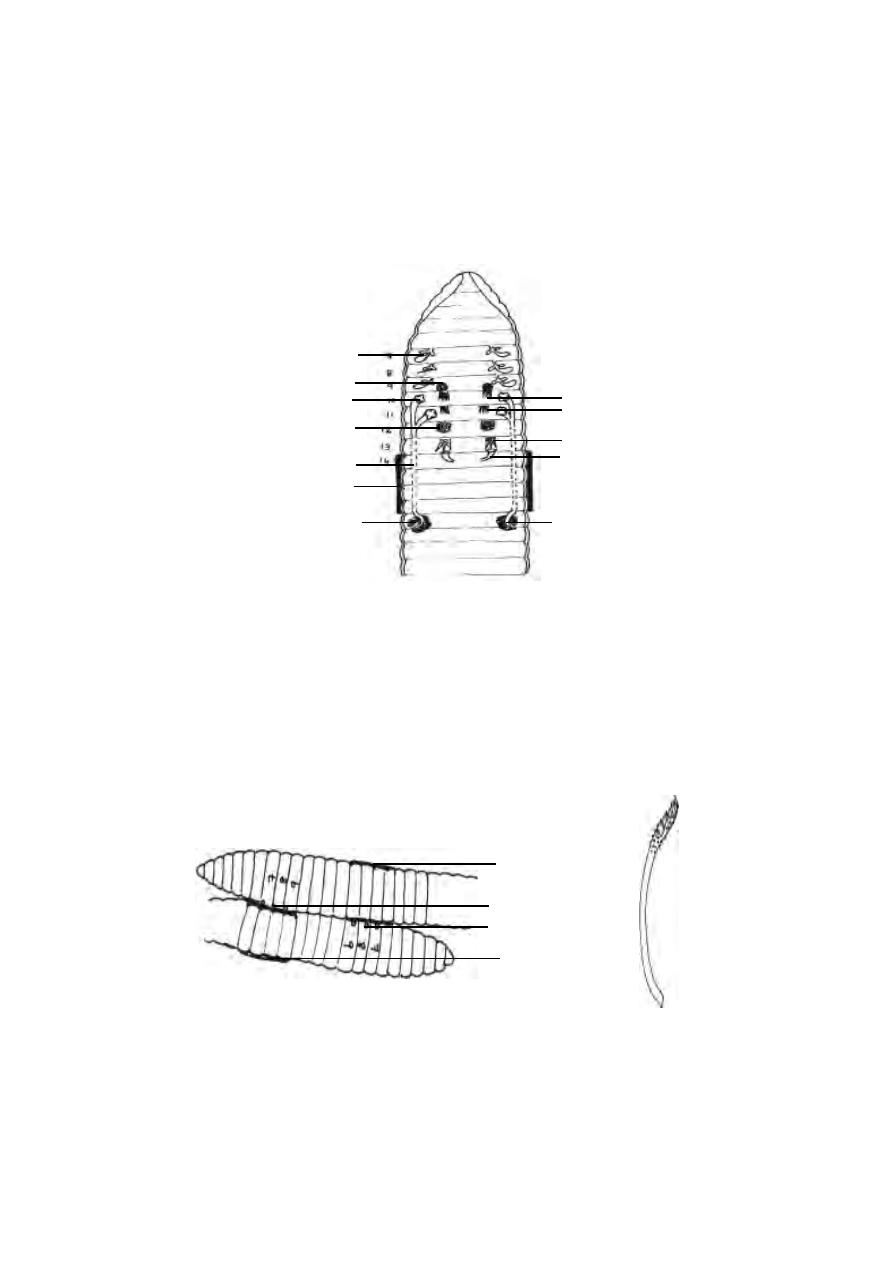
48
Fig. 1.3.8. Earthworm - Reproductive system
seminal vesicle
seminal funnel
seminal vesicle
vas deferens
clitellum
spermatheca
testis
testis
ovary
oviduct
prostate gland
prostate duct
deferens. The secretion of the prostate glands help to arrange the sperms into
bundles called spermatophores.
Female reproductive organs.
A pair of ovaries are found lying in segment 13. They are attached to
the anterior septum of the 13
th
segment. Each ovary is a flat structure with a
number of finger like processes. The ova are arranged in a linear order in the
ovaries. There are a pair of oviducts. They open internally into the 13
th
seg-
ment and externally on the ventral surface of the 14
th
segment. Three pairs of
spermathecae are present in segments 7, 8 and 9. These external openings
are situated in the intersegmental grooves of segments 6 and 7, 7 and 8, and 8
and 9. The spermatozoa received from another individual during copulation
are stored in spermathecae .
Copulation :
Fig. 1.3.9. Earthworm - copulation
spermathecal opening
spermathecal opening
clitellum
clitellum
Penial setae
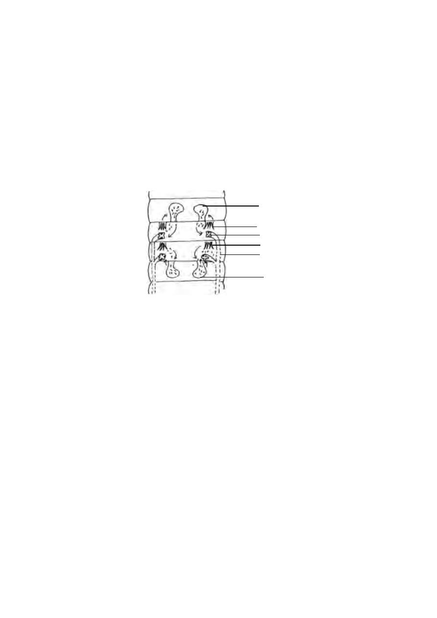
49
During copulation the head ends of the two worms are directed in the opposite
directions and the clitellum of one worm is opposite to the spermathecal seg-
ments of the other. The spermatozoa of one worm pass into the spermathecae
of the other worms. The worms separate after the mutual exchange of sper-
matozoa.
Later the glandular cells of the clitellum secrete a thick fluid which
hardens into a girdle surrounding the clitellum.
The girdle is moved forward by the wriggling movements of the body. As the
girdle is moved forwards it receives the ova and spermatozoa. The girdle con-
taining the germ cells (ova and sperms) and the nutrient albuminous fluid is
slipped off at the anterior end and it becomes a closed sac called the
cocoon. Fertilization and the development of the eggs into worms takes place
within the cocoon. Young worms come out of the cocoon after complete
development.
Type study 3. Amphioxus
Phylum
: Chordata
Sub Phylum : Cephalochordata
Type
: Amphioxus
This group comprises an assemblage of small fish like
marine organisms belonging to two genera, Branchiostoma and
Asymmetron. Amphioxus, the lancelet (genus Branchiostoma) has been
the object of much study because it illustrates the three fundamental
chordate characters in simple form. Thus, it may be considered as a
Fig. 1.3.10. Earthworm - Sperm transfer, storage and fertilization
seminal vesicle
seminal vesicle
testis
testis
seminal funnel
vas deferens
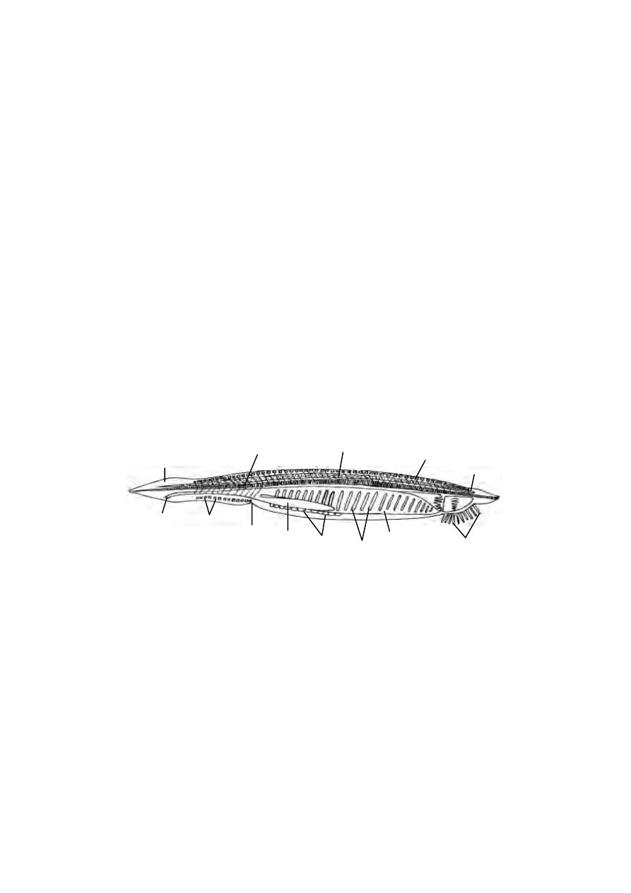
50
blue print of the phylum. Amphioxus has a long notochord which persists
throughout life, a dorsal hollow nerve cord and many gill slits in the pharynx.
Distribution :-
Amphioxus is a marine organism having an almost world wide
distribution. It is found on the sandy shores of tropical and warm temperate
seas.
Habit and Habitat :-
Amphioxus is usually found buried in the sand with the fore or
anterior end projecting outside. In this portion, it draws small organisms into
the mouth along with the current setup by the ciliary apparatus at the fore
end of the body. Occasionally, it emerges out of the burrow and swims about
by the sinuous movements of the body. As soon as it senses some distur-
bance it buries itself in sand again either by the head end or by the tail end.
External features :-
Amphioxus is a small translucent animal and 0.5 to 3 cm long. The
body is long laterally compressed and pointed at both ends (hence called
lancelet). It lacks a head. The body bears fins. The dorsal fin runs along the
whole length of the mid dorsal line. It expands around the tail as caudal fin
or tail fin and then continues ventrally as a ventral fin, extending for about
one-third of the length of the body. The dorsal and ventral fins are supported
by fin rays. The anterior two-thirds of the body is triangular in section. It is
because the two lateral sides project down as flaps called metapleural folds.
gill slits
tail fin
myotome
nerve cord
anus
ventral finray
liver
atriopore
gonad
pharynx
Fig. 1.3.10 (a) Amphioxus - Entire
notochord
dorsal fin rays
buccal cirri

51
The posterior one-third is oval in out line. The metapleural folds are con-
tinuous anteriorly with the oral hood.
Below the anterior end of the body, there is a median funnel shaped
cavity called the vestibule or preoral chamber. The vestibule is surrounded
by a membrane called oral hood. The oral hood is provided with 22
delicate, but stiff tentacle-like processes called oral cirri, guarding the
opening.
The mouth is situated at the bottom of the vestibule in the middle of a
membrane called velum. The velum is fringed with 12 velar tentacles
directed backwards into the pharynx. On the inner side of the oral hood, the
epithelial lining is thrown into ciliated finger-shaped processes. As the
movements of these processes cause a flow of water towards the mouth and
give the appearance of wheel-like movement, the tract is called wheel
organ. On the roof of the oral hood there is a ciliated pit called the Hatschek’s
pit. The anus lies in front of the tail fin, being placed slightly towards the left
side.
Atrium :-
In Amphioxus, the gill slits instead of opening directly to the
outside, open into a special cavity called the atrium. It is a space between
the pharyngeal wall and the body wall. It is lined by ectoderm and is
enclosed by the lateral extensions of the body wall. It surrounds the pharynx
laterally and ventrally but not dorsally. The cavity encroaches into the body-
cavity almost completely replacing the coelom. The atrium opens outside by
a small aperture called the atriopore, lying in front of the anus. The atrium
gives protection to the gills, preventing them from being choked up with sand.
Body wall :- The body wall is soft, thin and translucent. It is composed of
outer epidermis and inner dermis. The epidermis consists of a single layer
of columnar epidermal cells, bearing at places sensory hairs and the
unicellular glands. The dermis consists of an outer layer of connective
tissue (cutis) containing fibres and an inner thick layer of matrix (sub cutis)
invaded by few fibres, blood channels and nerve endings. Beneath the
dermis is the muscular layer arranged segmentally as muscle blocks, called
the myotomes or myomeres. The number of myotomes may vary from 60
or more. The muscles are thickened dorsally than ventrally.
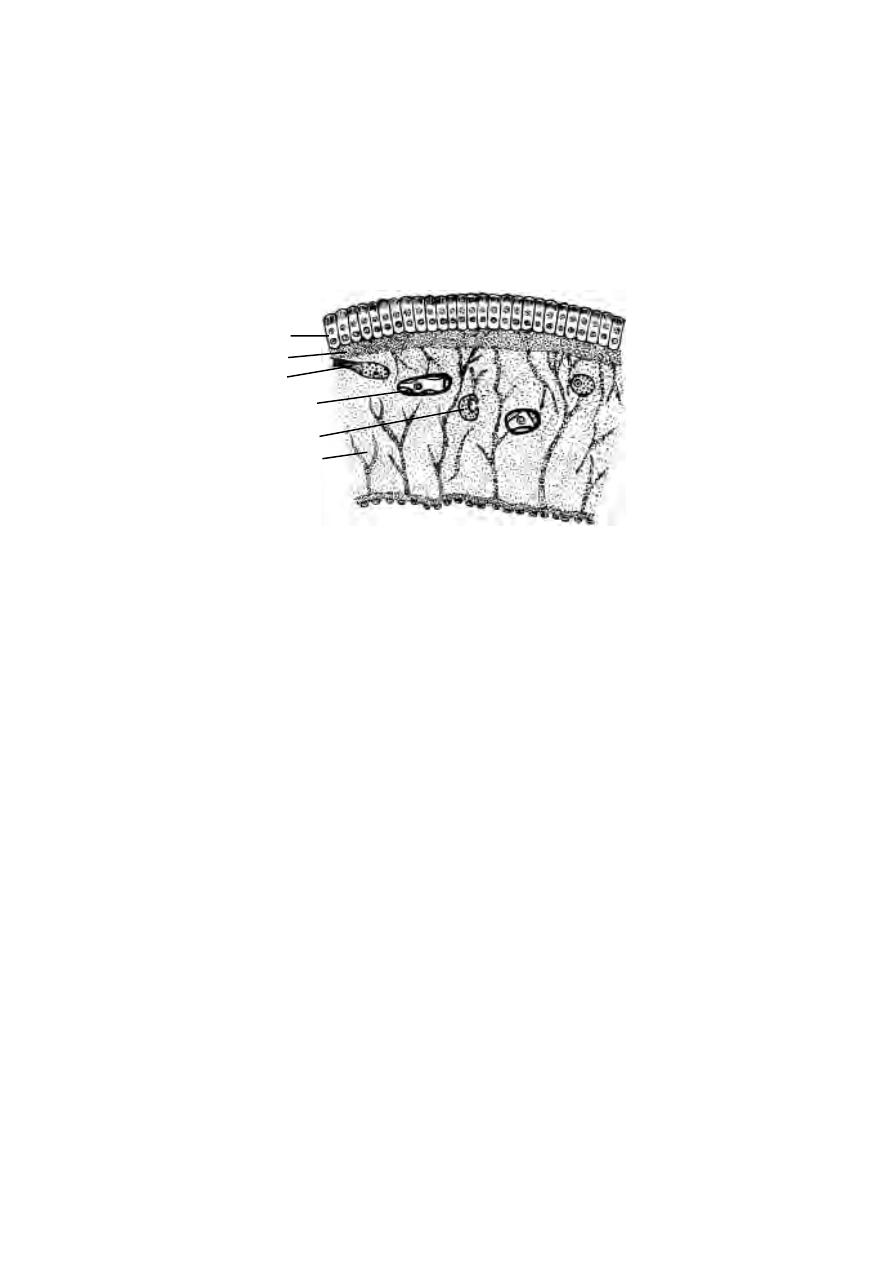
52
Coelom :- Coelom of amphioxus is distorted and restricted by the immense
size of the atrium.
Skeleton :- The skeleton consists of a peculiar tissue called notochordal
tissue and of gelatinous material. The chief supporting structure is the
notochord. Other skeletal structures are connected with the oral hood, the
fins and Pharynx.
Digestive system :- The mouth lies at the bottom of the vestibule. It leads
into a large laterally compressed chamber, the Pharynx which occupies the
most anterior part of the body cavity. The wall of the pharynx is perforated
by numerous gill slits (as many as 180 or more) on each side. From the
pharynx the narrow tubular midgut or intestine extends backwards to the
anus. On the ventral side of the intestine is a large diverticulum, the liver
diverticulum or hepatic caecum.
Running along the inner wall of the pharynx both on the dorsal and
ventral middle lines are two grooves lined with cilia ; the dorsal groove is the
hyperbranchial groove and ventral groove is the endo style. The dorsal
groove leads into the intestine. The ventral groove is composed of four tracts
of mucous glands separated by tracts of ciliated cells. The glands secrete
a sticky mucous which entangle the food materials. Anteriorly the endo style
and the hyperbranchial grooves are connected by two ciliated tracts, which
Fig. 1.3.10 (b) Amphioxus V.S. of body wall
sub-cutis
blood vessel
nerve
epidermis
cutis
nerve
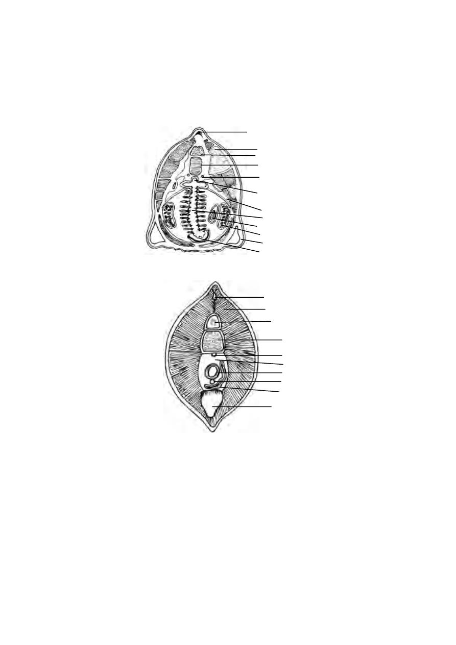
53
encircle the pharynx just behind the mouth. These ciliated tracts are called
peripharyngeal bands. The inner wall of the pharynx is lined with cilia.
Feeding mechanism :- Amphioxus feeds on minute organisms found
suspended in the water. While feeding, a continuous current of water enters
the mouth and expelled out through atriopore via pharynx and atrium. The
Fig. 1.3.10 (d) Amphioxus - T.S. through the intestinal region
fin ray
myotome
nerve cord
notocord
dorsal aorta
intestine
ventral aorta
atrium
finray
coelom
Fig. 1.3.10 (c) Amphioxus - T.S. through pharynx
dorsal finray
myotome
nerve cord
notocord
dorsal aorta
epipharyngeal groove
pharynx
gonad
liver
metapleural fold
endostyle
dorsal coelom
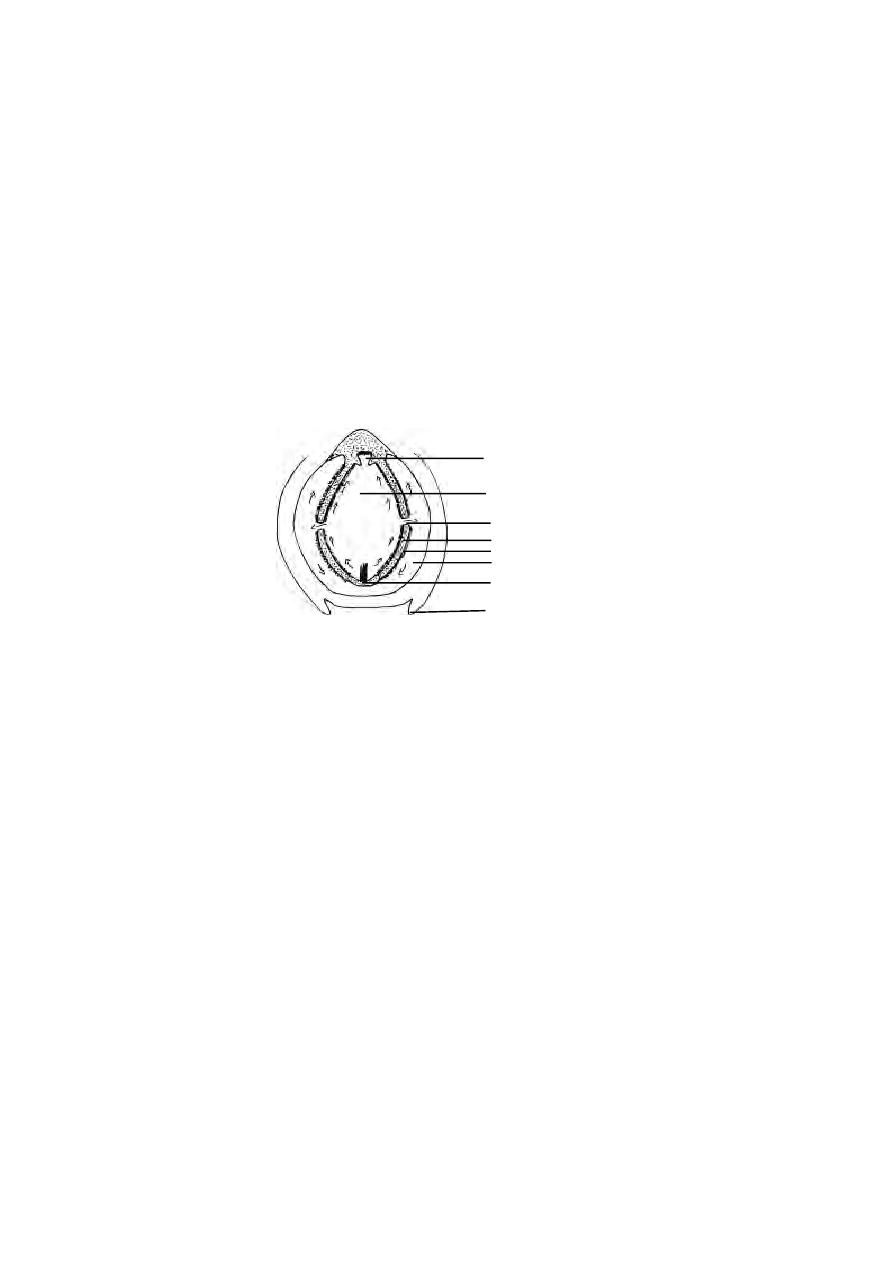
54
water current is maintained by the actions of cilia in the wheel organ and the
gill bars of the pharynx. The velar tentacles behind the mouth fold across
and strain off the sand particles. Small organisms are brought into the
endostyle and get entangled in the mucous. The cilia drive them along the gill
bars and peripharyngeal bands into hyper branchial groove and thence to the
intestine. Digestion takes place mainly in the midgut. Digestive enzymes are
secreted in the midgut diverticulum and poured into the midgut. The
digested food is also absorbed in the midgut.
Respiration :- Gaseous exchanges take place in the gill bars when the
water passes through the gill slits. The oxygen diffuses into the blood flowing
in the channels of gill bars and the carbon di oxide from the blood diffuses
into the running water.
Blood vascular system :- There is no specialized heart. The blood is
colourless. Respiratory pigments are also absent. There is a median
contractile vessel (ventral aorta) in the ventral wall of the pharynx below
the endostyle. It ends anteriorly by branching in the snout. The ventral aorta
sends paired afferent branchial vessels into the primary gill bars. From
the gill bars (Primary and secondary bars) the blood is collected by
efferent-branchial vessels which open into the right and left lateral dorsal
aortae one on each side of the pharynx above the hyperbranchial groove.
The lateral dorsal aortae unite behind the pharynx forming a median
dorsal aorta which runs behind and gives branches and breaks up into
capillaries in the intestine. The blood from the intestine and the posterior
Fig. 1.3.10 (e) Amphioxus - Course of feeding current
epipharyngeal groove
pharyngeal cavity
gill slit
gill bar
atrium
endostyle
atrial cilia
metapleural folds
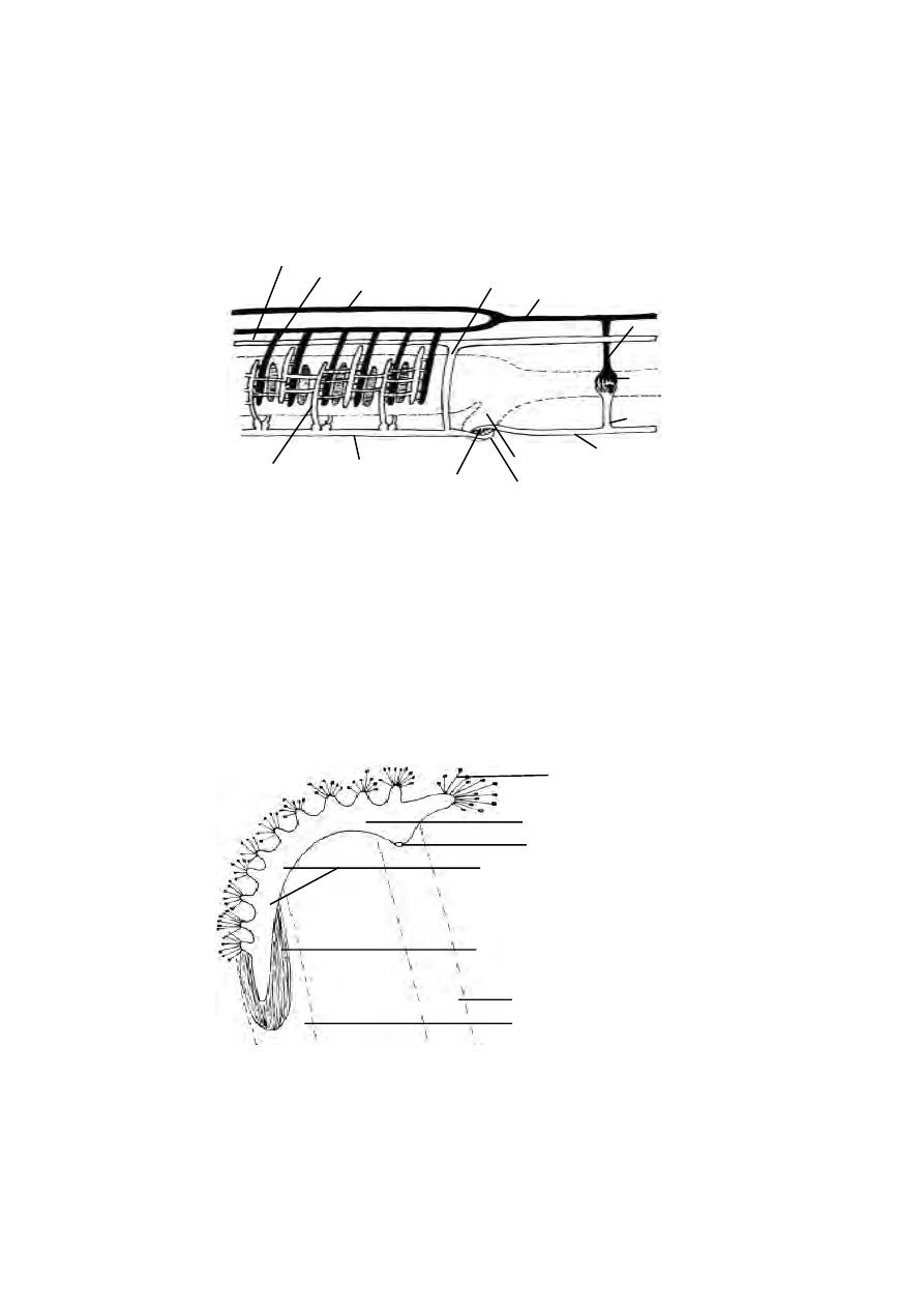
55
region of the body is collected by a median subintestinal vessel, which lies
below the intestine. This vessel extends forward to break up into capillaries
in the liver diverticulum forming hepatic portal system. From the liver the
blood is collected by the hepatic vein which joins the ventral aorta. There
are two cordinal veins, one on each side of the dorsal body wall, which
collects blood from the muscles and body wall. These are connected with
the ventral aorta by the cuvierian ducts.
Excretory system :- The excretory organs are the paired nephridia (about
90 pairs) of ectodermal origin.
cardinal vein
efferent branchial vessel
lateral dorsal aorta
median dorsal aorta
cuvierian duct
vessel to the
alimentary canal capillary
veins from the
sub intestinal vessel
liver
hepatic portal vein hepatic vein
ventral aorta
afferent branchial vessel
alimentary canal
alimentary
canal
system in the alimentary
canal
Fig. 1.3.10 (f) Amphioxus - Diagram of the vascular system
Fig. 1.3.10 (g) Amphioxus - A nephridium
external opening of nephridium
horizontal limb of nephridium
vertical limb of nephridium
coelomic space in primary bar
secondary gill bar
primary gill bar
solenocytes

56
These are segmentally arranged on each side of the pharynx in its
dorsolateral walls. Each nephridium is a closed, bent tube. Each tube has
an anterior vertical and posterior horizontal limb. The vertical limb lies in the
coelomic canal of the primary gill bar. It ends blindly. The horizontal limb
projects into the dorsal coelom. It opens to the exterior through
nephridiopore. Both limbs of the nephridia are provided with groups of
flame cells or solenocytes. A nephridium bears about 500 solenocytes.
Each solenocyte is a hollow cell with a flagellum hanging down from the wall.
There are blood vessels passing to the nephridia from afferent branchial
vessels. The nitrogenous waste matter is collected by the solenocytes from
the blood as well as coelomic fluid and is passed on to the body of the
nephridium by the flagella. From this it is discharged into the atrium. It finally
goes through atriopore.
Nervous system :- The nervous system of amphioxus consists of a median,
hollow nerve cord lying above the notochord and below the fin rays. It
extends from the anterior to the posterior end of the body. It is contained in
a canal called neural canal. A small longitudinal cleft, the dorsal fissure
extends throughout the length of the nerve cord. The nerve cord is not
differentiated into the brain and spinal cord.
Reproductive system :- In amphioxus the two sexes are separate, but there
is no sexual dimorphism. There are about 26 pairs of gonads arranged
segmentally. These are simple hollow sacs and are mesodermal in origin.
There are no gonoducts. The gonads bulge conspicuously into the atrial
cavity. When the germ cells ripe, the sacs burst and the genital elements are
set free into the atrial cavity from where they pass outside by atriopore.
Fertilization and development take place in sea water.
Type study - 4. Pigeon
Sub phylum
-
Vertebrata
Class
-
Av e s
Order
-
Columbiformes
Type
-
Columba livia
Birds are easily recongnised group of vertebrates. In birds every part
of the body is modified to suit their aerial mode of life. Birds possess feathers,
beak and feet modified in relation to their aerial life.
The Pigeons are flying birds(carinate). They are known both as wild
and domesticated forms. The Pigeons are seen both in tropical and temperate
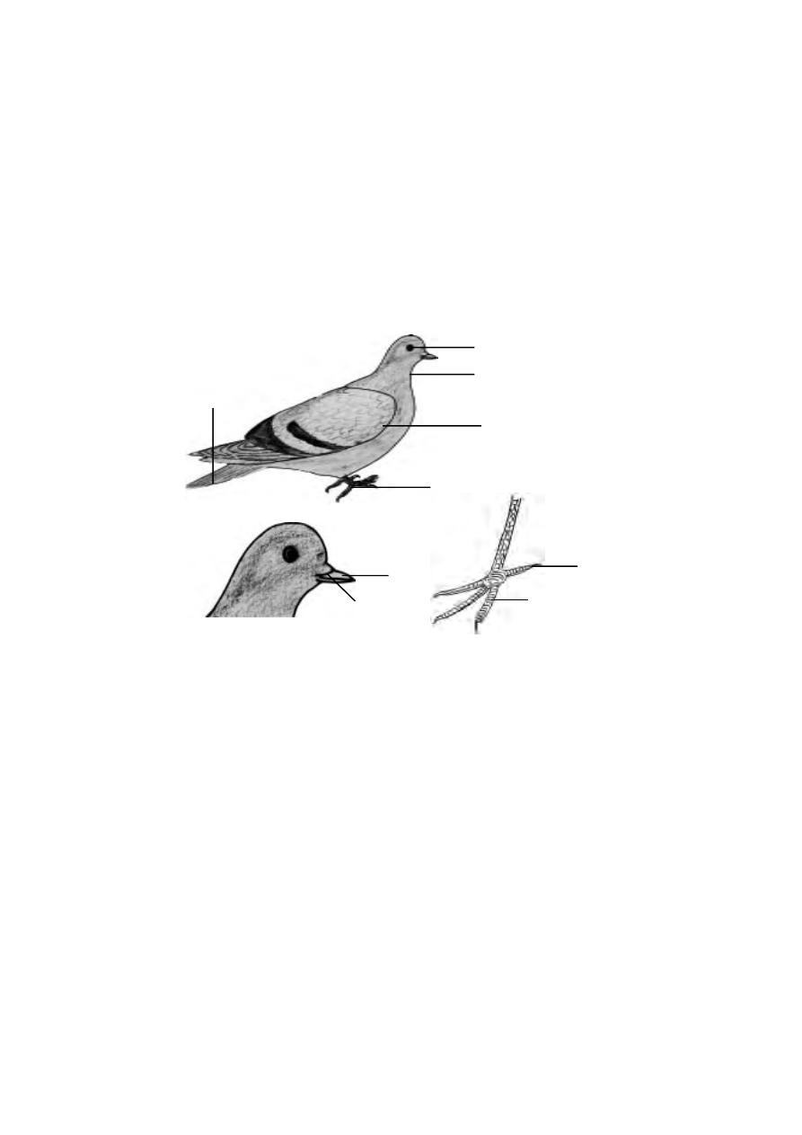
57
zones. About 10 species of Pigeons are found in India. The pigeons fly in
flocks and roost together. The domestic pigeons have many varieties, namely
panter, fantail and tumblers. They differ in size, colouration and feather ar-
rangement. All of them are, however, descendants of the rock pigeon-columba
livia.
External features.
The Body is spindle shaped. Their size varies from 20-25 cm. They
are covered by coloured feathers leaving beak and a small portion of the
hindlimbs.
The body is divisible into head, neck, trunk and a small, conical tail.
The head is round and drawn out anteriorly into a strong, hard, pointed beak.
The mouth is a terminal wide gape, guarded by elongated upper and lower
beaks. The beaks are covered with a horny sheath or rhampotheca. A swol-
len area of soft skin, the cere, surrounds the nostril. It is present on each side
of the upper beak. The eyes are large and guarded by upper and lower eye-
lids and a transparent nictitating membrane. A pair of ear openings are
situated at a short distance behind the eyes. Each opening leads into a short
external auditory meatus, ending in the tympanic membrane forming the
ear drum.
Fig. 1.3.11. Pigeon - External
Entire
Head
Leg
beak
cere
eye
neck
leg
tail further
wing
claw
toe
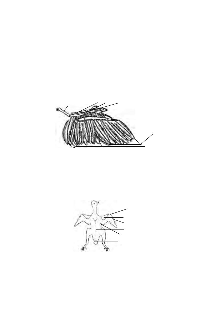
58
Fig. 1.3.13. Pigeon(feathers removed)
prepatagium
thumb
post patagium
cloaca
wing
trunk
tail
The neck is long and mobile. It helps in the movement of the head in
various directions. The trunk is compact, heavy and bears a pair of wings and
a pair of legs. The cloacal aperture is at its hind end on the lower surface.
Projecting behind the cloacal aperture is the tail. Above the tail is a knob on
which opens an oil gland or preen gland or uropygeal gland. It secretes a
fluid used for preening the feathers.
The wings :-
The forelimbs as modified wings are located in the anterior region of
the trunk. The limbs are of the pentadactyl type. The wing has three typical
divisions as - the upper arm, forearm and hand. The hand has three imper-
fectly marked digits. While the pigeon is at rest the three divisions of the
wing are bent upon one another in the form of the letter ‘Z’. During flight the
wings are straightened and extended. A fold of skin the alar membrane or
prepatagium, stretches between the upper and forearm along the
anterior border of the limb. A smaller fold known as postpatagium is present
between the trunk and upperarm.
Fig. 1.3.12. Pigeon - a wing showing feather arrangement
humerus
radius
ulna
alaspuria
primaries
secondaries
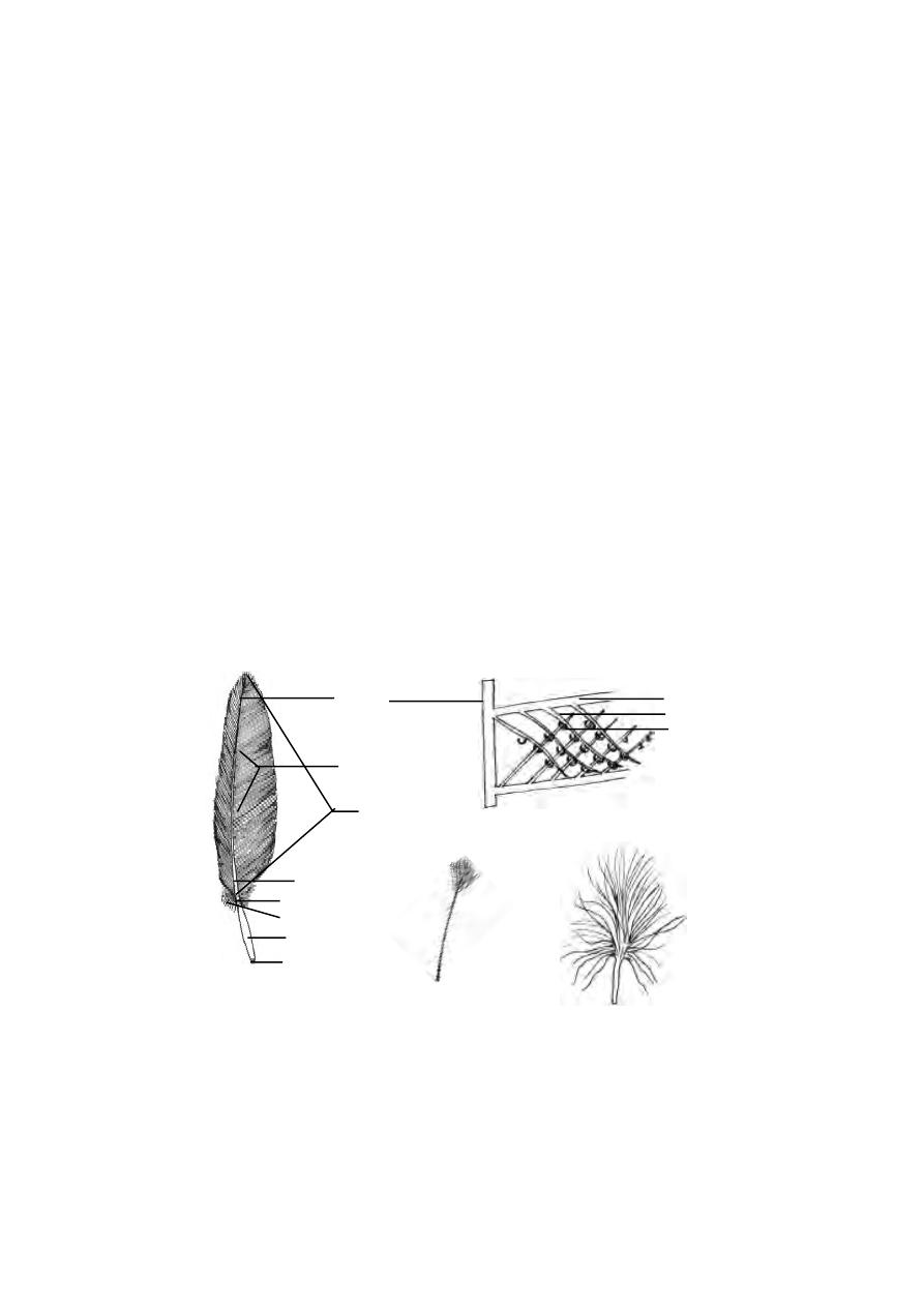
59
While the pigeon is not flying the whole weight of the body has to be
supported by the hind limbs, In order to balance the heavy trunk the hindlimbs
are attached for forwards. Each hindlimb or leg has three typical divisions, the
thigh, shank and foot. The thigh without being free is enclosed within the
boundaries of the trunk. Each hindlimb has four digits. The first toe is directed
backward. The feet are naked and covered with horny epidermal scales.
Each digit is provided with a horny claw. The tail is small and concealed by the
feathers of the trunk. It bears the tail feathers or rectrices.
Exoskeleton :-
The feathers are integumentary structures. They are characteristic of
birds. The feathers are derived from the epidermis. They are seasonally or
periodically cast off and the new one being developed from the old papillae.
They are arranged on the skin in definite tracts, called feather tracts or ptery-
lae. The interspaces without feathers are known as apteria or featherless
tracts.
There are three types of feathers in pigeon. They are the large quill
feathers found on wings and tail for flight, the contour feathers, forming a
covering for the body and the filoplumes, lying between the contour feath-
ers.
Fig. 1.3.14. Pigeon - Feather types
A quill feather
A filoplume
A down feather
Barbs and Barbules
rachis
vane or vexillum
superior umbilicus
inferior umbilicus
quill or calamus
aftershaft
barbs
stem
barb
barbule
hooklet

60
Quill feather :
Each quill feather has a central stem or scapus. It is divided into
lower hollow part called the quill or calamus and a solid upper part termed
rachis. The quill has at its lower end an opening called inferior umbilicus,
through which vascular processes or papilla of the dermis project into the
growing feather. Another opening the Superior umbilicus occurs at the junc-
tion of quill and the rachis on the inner face of feather. Close to this opening,
there is a small tuft of soft feathers called aftershaft. Attached to the rachis
are small filaments or barbs. The rachis with the barbs constitute the vane
or vexillum. Each barb is provided with barbules and hooklets. The barbs
remain attached with one another to form a continuous blade for striking the
air in flight.
There are twenty three quill feathers or remiges in each wing. Eleven
of these known as primaries. are attached to the hand. The remaining twelve
fixed on the forearm are called secondaries. Attached to the thumb is a
small tuft of feathers known as ala spuria or bastard wing. The tail bears
twelve tail feathers or rectrices which are arranged in the form of a fan.
The contour feathers are soft and the barbs are plume like with no
interlocking mechanisms. These help to keep the body warm and lock air pock-
ets. The filoplumes have delictae hair like long axis and a few barbs devoid
of barbules. Down feathers have small axis and a few barbs devoid of lock-
ing structures at the distal end. Nestlings are covered with down feathers.
Endoskeleton :-
The endoskeleton of pigeon is strong but lightly built. The texture of
the bone is often spongy. Bone marrow is absent. The air spaces from the
lungs may continue into the bones, making them light. The bones are more or
less devoid of bone marrow. These are called Pneumatic bones. Most of the
bones except those of the tail, forearm, hand and hind limb contain air spaces.
In general there is a tendency for the reduction and fusion of bones. It gives
rigidity to the skeleton.
Flight muscles :-
The wings are the modified forelimbs. They are organs of flight. The
musculature of the forelimbs are greatly modified in response to the function
they perform. Flight is the coordinated effort of a number of paired muscles of
which the following are most important.

61
Pectoralis major
(Depressor muscles)
These are the largest breast muscles. They are about one fifth of the
body weight. By the contraction of this muscle the wings are lowered during
flight.
Pectoralis minor or subclavius :-
These are smaller but longer than pectoralis major. By their contrac-
tion the wings are raised in flight.
Coracobrachialis :-
These small flight muscles pull the wing downwards in flight.
Digestive system :-
The two jaws of the mouth are modified into beak. Both the jaws are
devoid of teeth. The mouth leads into the buccal cavity. The floor of the
buccal cavity is provided with a narrow, triangular tongue. It has a horny
covering and is provided with sensory papillae. The buccal cavity narrows
behind into the Pharynx. The salivary glands are absent in the buccal cavity.
Three pairs of buccal glands are present in the mouth. Their secretion is
mainly mucous.
The alimentary canal proper starts from the Pharynx. The Pharynx
leads into a long oesophagus that runs back through the neck. At the base of
the neck region, it enlarges into a thin walled, distensible sac known as crop
containing mucous glands. It serves as a store house for the food. The crop is
followed by the stomach. The stomach is divisible into two parts, the
anterior tubular proventriculus containing gastric glands and a posterior
laterally compressed gizzard. The gizzard has a thick muscular wall and a
horny inner lining. Its cavity is small and contains small stones which are
helpful to grind the food. Thus the gizzard acts as a grinding mill. This type
of arrangement is necessary because of the absence of teeth in the buccal
cavity. The intestine arises from the right side of the gizzard. It is divisible into
an anterior U- shaped duodenum, and a posterior long coiled ileum. The
ileum enlarges posteriorly into a short rectum or large intestine. Anteriorly,
the rectum bears a pair of small rectal caeca. The rectum opens to the exte-
rior by the cloaca.
Internally the cloaca is divided into three chambers, the anterior
coprodaeum, the middle urodaeum and the posterior proctodaeum. The
rectum opens into the coprodaem. The urinogenital ducts open into the
urodaem. The proctodaem opens to the exterior by a transverse slit like
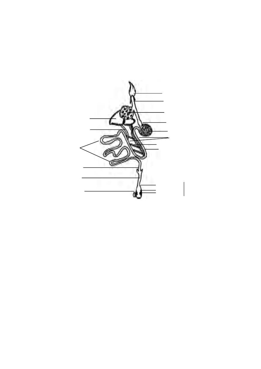
62
aperture called cloaca. At the proctodacum, there is a dorsal glandular sac
known as Bursa of Fabricii. Its function is unknown.
The digestive glands associated with the alimentary canal are the liver
and the pancreas. The liver is bilobed with a large right and a small left lobe.
It is devoid of gall bladder. There are two bile ducts. They are forms one
from each lobe. They open into the duodenum independently. The pancreas
lies between the two limbs of the duodenum. It has three ducts, all opening
into the distal limb of the duodenum.
Respiratory System :-
The flight activity requires a continuous and abundant supply of oxy-
gen. Hence, the respiratory system of pigeon is highly
developed and well differentiated. The respiratory system consists of external
nostrils, glottis, larynx, trachea, bronchus and lungs.
The external nostrils are a pair of slit like apertures occurring at the
base of upper beak. They communicate to the pharynx by internal nostrils. A
glottis lies behind the tongue. It opens into the larynx. The larynx opens into
a trachea. The trachea is a long, cylindrical and flexible tube running back-
Fig. 1.3.15. Pigeon Digestive system
oesophagus
crop
proventriculus
gizzard
duodenum
pancreas
pancreatic duct
liver
bile duct
ileum
caecum
rectum
coprodaeum
urodaeum
proctodaeum
cloaca
bursa of fabricii
head
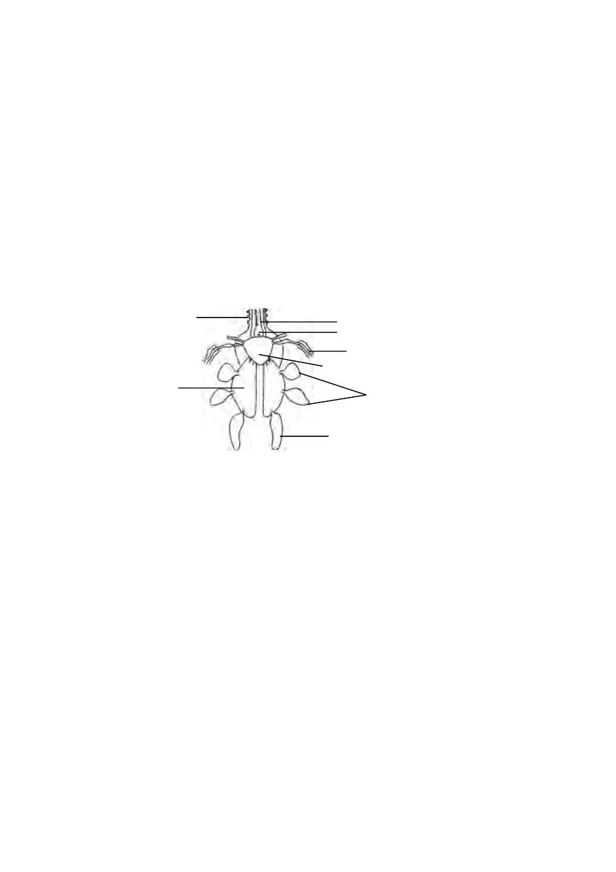
63
ward through the neck. On entering the thoracic cavity, the trachea
expands into a syrinx or voice box. Later it divides into two bronchi, one for
each lung. The walls of tracheal and bronchial tubes are supported by a series
of closely set cartilagenous rings. Each bronchus enters a bright red lung. The
bronchus divides and subdivides into smaller branches, ultimately ending in
fine air capillaries. Lungs are solid spongy organs. They do not hang freely in
the thoracic cavity but are lodged firmly in the ribs. Some of the branchial
tubes pass through the lungs and communicate with the air cavities in the
bone. There are nine air sacs. They are a median interclavicular, a pair of
cervical, two pairs of thoracic and a pair of abdominal air sacs.
The air sacs help to maintain high body temperatures. They make the
body lighter and help in flight.
Mechanism of Respiration :-
In birds the expiration is an active process. The process of inspiration
is passive. In a resting bird, the sternum is moved up and down with the help
of intercostal and the abdominal muscles.
During flight, the sternum is rendered immovable due to the support
of wings, but the body cavity is raised and lowered by the action of wings and
by the lowering of the vertebral column.
Circulatory system :-
The heart is four chambered, with two auricles and two ventricles.
There is complete separation of the oxygenated and non-oxygenated blood.
Birds have two distinct circulations as arterial and venous systems.
Fig. 1.3.16. Pigeon - Lungs and air sacs
trachea
syrinx
cervical air sac
inter clavicular air sac
air sac in cavity of humerus
thoracic air sacs
lung
abdominal air sac
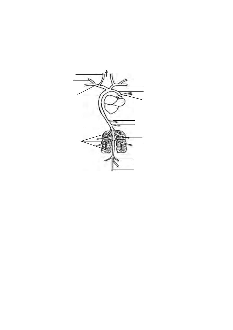
64
Arterial system :-
The arteries are blood vessels that carry blood away from the heart.
Since the heart pumps blood with force into the arteries, the vessels have
slightly muscular and strong walls.
The two major arteries that leave the heart of pigeon are the
pulmonary artery and systemic artery. The pulmonary artery originates
from the right ventricle. It carries deoxygenated blood towards the lungs for
oxygenation. As this artery leaves the heart, it divides into right and left
pulmonary arteries carrying blood to right and left lungs.
The systemic artery is a part of the right aortic arch. In birds the left
aortic arch is absent. The right aortic arch begins from the left ventricle.
The left ventricle pumps oxygenated blood. The right systemic artery is formed
as a constituent of the right aortic arch.
Fig. 1.3.17. Pigeon - arterial system
carotid artery
brachial artery
pectoral artery
subclavian artery
innominate artery
systemic artery
pulmonary artery
coeliacomesenteric artery
anterior mesenteric artery
dorsal aorta
femoral artery
sciatic artery
renal arteries
iliac artery
posterior mesenteric artery
caudal artery

65
The systemic artery forms right and left innominate arteries. Each
innominate artery forms a carotid artery and a subclavian artery. The
subclavian arteries inturn divide into brachial and pectoral arteries. They
carry blood to the muscles around the thoracic region and the flight muscles.
The carotid arteries carry blood to the head region.
The right systemic artery, further runs posteriorly and forms the
dorsal aorta. The dorsal aorta is a major artery supplying blood to the
abdominal organs and the posterior regions of the body. The most prominent
blood vessels are
1. The coeliaco-mesenteric artery and anterior mesenteric artery -
these arteries carry blood to various regions of the alimentary canal and
certain visceral organs.
2. Renal arteries - carry blood to the kidneys.
3. Femoral arteries (2) - supply pure blood to pelvic muscles and external
thigh region.
4. Sciatic arteries (2) - supply pure blood to internal thigh region.
5. Iliac arteries (2) - supply blood to hip region.
6. Posterior mesenteric artery - supplies blood to the most
posterior visceral organs.
7. Caudal artery - carries blood to the tail region.
Venous system :-
The deoxygenated blood from various regions of the body are
collected by several veins. Finally these veins take the blood to the right
auricle through the two precaval and a single postcaval veins.
The precaval and postcaval veins receive blood from several veins
originating from various organs of the body.
66
From the tail region the blood is drained by the caudal vein. It runs
upto the renal portal veins. Each portal vein runs through the kidney and
joins the femoral vein. The renal portal vein provides renal veins. It also
receives a pair of sciatic veins draining blood from legs. The right and left
femoral veins are united to form the posterior part of the post caval vein.
The precaval veins (2) are formed by the jugular and brachial veins
from the neck shoulder and head regions.
Fig. 1.3.18. Pigeon - venous system
jugular vein
brachial vein
pectoral vein
precaval vein
post caval vein
hepatic veins
hepatic vein
epigastric vein
iliac vein
femoral vein
sciatic vein
renal portal vein
internal iliac vein
caudal vein
67
Nervous system :-
The brain is divisible into the fore-, mid- and hind brains. The
cerebral hemispheres are distinct. They are round and large in size. The
olfactory lobes are very small and they do not contain cavities. The
diencephalon is hidden from the view by the forward prolongation of the
cerebellum. The diencephalon has the pineal body dorsally and
infundibulum and pituitary body ventrally. The optic lobes are lateral in
position owing to the large size of the cerebral hemispheres and cerebellum.
The medulla oblongata instead of being continued backwards as in other
tetrapods, descends almost vertically from the cerebellum
Sense organ :-
In pigeon the olfactory sense is poor. There is no external ear. The
tympanum is slightly sunken from the surface of the skin.
The eyes are large. During flight the eyes and their shape are pro-
tected by unique sclerotic plates of the outer eye layer. The nictitating
membrane slides over the eyeball and presumably protects the cornea by
Fig. 1.3.19. Pigeon - Brain
Dorsal view
Lateral view
olfactory lobe
cerebral hemisphere
optic lobe
flocculus
cerebellum
medulla oblongata
optic chiasma
cerebellum
optic lobe
medulla
oblongata
Fig. 1.3.20. Eye of Pigeon
sclerotic plate
cornea
aqueous chamber
iris
lens
ciliary process
pecten
optic nerve
retina
choroid coat
sclerotic coat
68
closing it, during flight. Inside the eye, a vascular pigmented process projects
into the vitreous body. It is know as the pecten. It arises from the point of
entry of the optic nerve into the eye ball. Its function is not definitely known,
but possibly it may help in long distance vision.
Urinogenital system :-
The excretory organs are a pair of kidneys. They are dark red, three
lobed structures. They open separately into the urodaeum of the cloaca through
two different ureters. There is no urinary bladder.The urine is excreted in
the form of uric acid, a semi solid white mass discharged along with faeces
through the cloacal aperture.
Male Reproductive system :-
The male has a pair of oval testes. From each testis, a duct, the
vasdeferens, passes back and opens into the cloaca. The vas deferens is
dilated at its posterior end into a seminal vesicle. There is no copulatory
organ.
Female reproductive organs :-
Only the left ovary persists in the adult. The right ovary disappears
during development. The ovary and the oviduct of only oneside are
functional during breeding season.
Fig. 1.3.21. Pigeon - Urinogenital system
kidney
ureter
testis
vasdeferens
cloaca
seminal vesicle
rectum
right oviduct
oviduct
oviducal funnel
Male
Female
ovary

69
Unit - 1
Part I
Choose the right answer :-
1. The theme of the world summit on environment held in Johennesberg in 2002
was
a) only one earth
b) our common future
c) sustainable earth
d) energy crisis
2. The introduction of phylogeny in taxonomy was made by
a) Cuvier
b) Lamarck
c) Charles Darwin
d) Linnaeus
3. The Concept of Numerical Taxonomy was developed by.
(a) Cuvier
(b) Michael Adamson
(c) Lamarck
(d) Charles Darwin
4. If two or more workers describe the same organism using different names it
results in
a) homonyms
b) synonyms
c) a valid name
d) nomenclature
5) The class Rhizopoda includes
(a) Protozoans with pseudopodia
(b) Animals with jointed legs
(c) Animals with setae
(d) Animals with fore limbs.
6. Which among the following is a pseudocoelomate ?
a) Platyhelminthes
b) Annelids
c) Mollusca
d) Nematoda
7. Five Kingdom concept does not include
a) fungi
b) viruses
c) flowering plants
d) bacteria
8. Excretion in Platyhelminthes is effected by
(A) flame cells
(B) nephridia
(C) kidneys
(D) gills
9. Which among the following is considered ‘a blood fluke’
a) Schistostoma
b) Taenia solium
c) Fasciola
d) Ascaris

70
10. The head formation or cephalization happened for the first time in Phylum
a) Arthropoda
b) Annelida
c) Mollusca
d) Echinodermata
11. Metameric Segmentation is seen in
(a) Protozoans
(b) Arthropods
(c) Annelids
(d) Mollusca
12. Which among the following is considered as a connecting link between
Annelida and Arthropoda
a) Centipede
b) Cockroach
c) Peripatus
d) Spiders
13. The characteristic feature of echinoderms is the possession of
a) compound eye
b) absence of coelom
c) diploblastic condition
d) water vascular system
14. The water vascular system is seen in
(a) Echinodermata
(b) Annelida
(c) Mollusca
(d) Nematoda
15. Acrania refers to
a) fishes
b) birds
c) chordates
d) prochordates
16. Kidney in amniotes is
(a) Metanephric
(b) Mesonephric
(c) Anephric
(d) Pronephric
17. Viviparous animals are
(a) Fishes
(b) Mammals
(c) Aves
(d) Reptiles
18. The segmented body muscles in fishes are called as
a) myotomes
b) smooth muscles
c) skeletal muscles
d) visceral muscles
19. Which among the following is considered as a defeated group
a) amphibians
b) reptiles
c) mammals
d) fishes

71
20. The amniotes are characterised by the presence of
a) four legs
b) amphibious life
c) egg membranes
d) metamorphosis
21. Which among the following is not a character of mammals
a) diaphragm
b) right aortic arch
c) mammary glands
d) corpus collosum
22. The infective stage of plasmodium is
a) cryptozoite
b) amoebula
c) sporozoite
d) merozoite
23. In earthworm the clitellum is present in segments
a) 5 to 10
b) 14 to 17
c) 13 to 18
d) 20 to 25
24. The muscle fold found in the dorsal wall in the intestine of the earthworm is
a) diaphragm
b) Typhlosole
c) myotome
d) ommatidium
25. The chlorogogen cells on the wall of the intestine in earthworm is meant for
a) digestion
b) circulation
c) excretion
d) reproduction
26. Amphioxus has a
a) long notochord
b) short notochord
c) tubular notochord
d) all the above
27. Amphioxus is usually found
a) floating in water
b) buried in sand
c) in shallow pits
d) in deep water
28. In Amphioxus the metapleural folds are continuous anteriorly with the
a) Velum
b) Oral hood
c) Vestibule
d) Oral cirri
29. In Amphioxus the velum is fringed with
a) 8 velar tentacles
b) 12 velar tentacles
c) 20 velar tentacles
d) 10 velar tentacles

72
30. The roof of the oral hood in Amphioxus has a ciliated pit called
a) gastric pit
b) intestinal pit
c) Hatschek’s pit
d) pancreatic pit
31. The number of myotomes in amphioxus may vary from
a) 80 or more
b) 60 or more
c) 100 or more
d) 50 or more
32. In Amphioxus the skeleton consists of a peculiar tissue called
a) skeletal tissue
b) nervous tissue
c) notochordal tissue
d) perivisceral tissue
33. In Amphioxus the wall of the pharynx is perforated by
a) 180 or more gill slits
b) 150 or more gill slits
c) 200 or more gill slits
d) 80 or more gill slits
34. The ventral side of the intestine in Amphioxus has
a) intestinal diverticulum
b) caecal diverticulum
c) liver diverticulum
d) all the above
35. In Amphioxus the mucous glands secrete a sticky mucous for
a) entangling the food materials
b) capturing the food materials
c) digesting the food materials
d) engulfing the food materials
36. In Amphioxus the inner wall of the pharynx is lined with
a) cilia
b) tentacles
c) cartilage
d) cirri
37. The blood in amphioxus is
a) coloured
b) watery
c) colourless
d) all the above
38. The excretory organs in amphioxus are
a) kidneys
b) flame cells
c) nephridia
d) green glands
39. The nephridium of amphioxus bears about
a) 500 solenocytes
b) 200 solenocytes
c) 600 solenocytes
d) 800 solenocytes

73
40. In Amphioxus there are about
a) 24 pairs of gonads
b) 26 pairs of gonads
c) 10 pairs of gonads
d) 30 pairs of gonads
41. The uropygeal gland of pigeon are found
a) above the tail
b) in the alimentary canal
c) in the brain
d) in the reproductive system
42. The hollow bones of birds are called as
a) pneumatic bones
b) fused bones
c) skull bones
d) limb bones
43. Limbless amphibians are
(a) Frogs
(b) Toads
(c) Salamanders
(d) Caecilians
Part II
Give very short answers.
1. Define biosphere?
2. Why do we consider Aristotle as the father of biological classification?
3. What is the characteristic feature of biological taxonomy?
4. Define Taxonomy
5. What is a taxon?
6. What are triploblastic animals?
7. Why do we call nematocysts as stinging cells?
8. What is a trochophore?
9. What are amniotes?
10. What is a diploblastic animal?.give an example
11. Write short notes on scolex
12. Name the two types of setae in an earthworm
13. Name the three types of nephridia in earthworm
14. What are chlorogogen cells
15. Mention the habitat of amphioxus.
16. What are metapleural folds?
17. Write short notes on a) oral hood b) oral cirri
18. Write short notes on myotomes.

74
19. What is hepatic caecum?
20. What is endostyle? what is its function?
21.Write short notes on quill feather.
22.What is syrinx?
23.What are flight muscles ?
24. Why do we call birds as ‘glorified reptiles’?
25. Name the subclasses under mammalia.
26. Mention any one characteristic feature of the primates.
27. What are cryptozoites?
28. What is a Ookinete?
29. What is tertian malaria?
30. What are ‘lateral hearts’?
31. What is ‘pecten’?
Part III
Answer breifly
1. Provide a breif account on the biodiversity in India.
2. What are the various concepts of ‘species’?
3. Give an account of the basic principles of nomenclature.
4. Differentiate bracketed and indented keys for identifieation with a suitable
example.
5. Give an account of urinary or excretory system of Pigeon.
6. Explain the structure of the nephridium
7. Draw a neat labelled diagram of TS of the earthworm.
8. Write notes on Phylum Annelida.
9. Give a brief account on Protochordates.
10. Write notes on Primates.
11. What are the types of malaria?
12. Explain the male reproductive organs of earth worm.
13. Draw a neat labelled sketch to show the external morphology of amphioxus.
14. Explain the excretory system of amphioxus.
15.Draw and label the parts in a cross-section of amphioxus through
pharyngeal region.
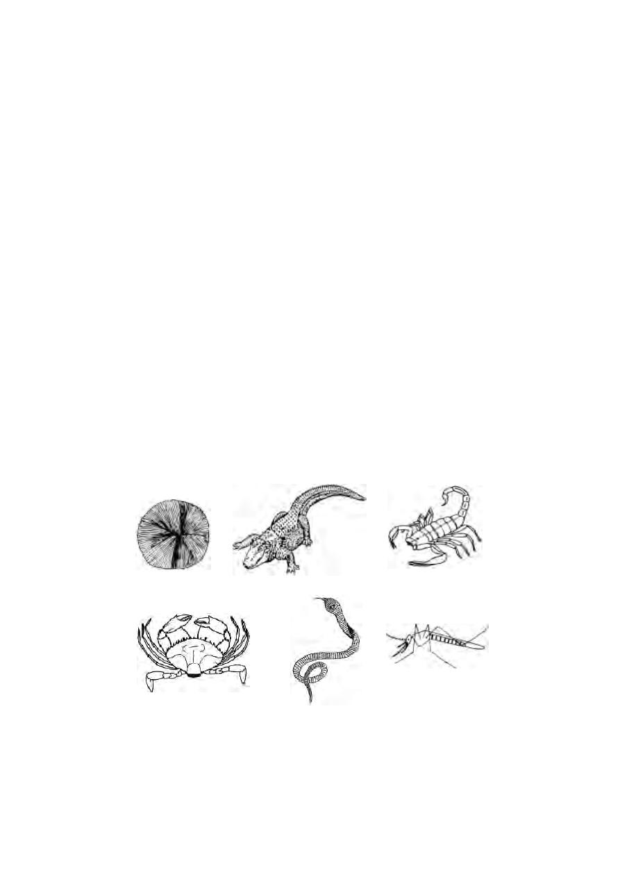
75
16. Describe the structure of the brain of the pigeon.
17. Provide an account on external openings on the body of earthworm .
18. Write notes on flight muscles in pigeon.
Part IV
Answer in detail
1. Define species and provide an account on various animal groups.
2. Write an essay on the various methods of taxonomy.
3. Explain the digestive system of earthworm.
4. Give a detailed account on the general characters of chordates.
5. Explain the life cycle of Plasmodium in man.
6. Describe the external features of Columba livia.
7. Give a detailed account on the reproductive system and the process of
reproduction in earthworms.
8. Explain the digestive system of amphioxus with diagram.
9. Describe the blood vascular system of amphioxus with the aid of a neat
diagram.
10. Describe the arterial system of Pigeon
11. Explain the female reproductive system of pigeon
Classify giving reasons.

2. Cell Biology
‘All living things are made up of cells’. ‘The cell is the basic unit of
structure and function’. These generalised statements are known as Cell
theory. This theory was forwarded by Mathias Schleiden and Theodor
Schwann in 1838 - 39.
Today the cell theory includes four more ideas :
the cells are the building block of structures in living things
the cell is derived from other cells by division
the cell contains information that is used as instructions for growth,
development and functioning
the cell is the functioning unit of life; the chemical reactions of life
take place within cells.
The idea and concept of cell biology evolved during the 19th century
as a result of gradual advancement in the field of microscopy and biochemis-
try. Today the study of the structure of cells (cytology) is part of a major
branch of biology known as cell biology. Due to its wide application many
new branches have sprung up in biology. Some of the new branches related to
cytology are, Cytotaxonomy, Cytogenetics, Cell physiology,
Cytochemistry, Molecular Biology, Cytopathology and Cytoecology.
2.1 Microscopy
The cells are very minute and complex organisations. The small
dimensions and transparent nature of cell and its organelles pose problems to
cell biologists trying to understand its organisation and functioning. Various
instruments and techniques have been developed to study cell structure,
molecular organization and function.
The diameters of majority of cells range from 5-500 µm, but most are
between 10-150 µm. The systeme International (SI) units of length are
1 metre (m)
= 1000 millimetres (mm)
1 mm (10
-3
m)
= 1000 micrometres (µm)
1 µm (10
-6
m)
= 1000 nanometres (nm)
1 nm (10
-9
m)
= 1000 picometres (pm)
76

The Angstrom (Å) is 10
-10
m. It is sometimes used to record the
thickness of cell membranes and the sizes of macromolecules.
While viewing objects, human eyes have limited distinguishing or re-
solving power. The ability to reveal minute details is expressed in terms of
limit of resolution. It is “the smalllest distance that may separate two points
on an object and still permit their observation as distinct separate points”. The
resolving power of naked human eye is 0.1 mm or 100 µm. It means that we
cannot differentiate any points that are closer than this. Hence we need in-
struments capable of high resolution to see smaller objects.
Power of magnification is different from resolving power. Magnifi-
cation is ‘the increase in size of optical image over the size of the object being
viewed’. Increased magnification without improved resolution results only in
a large blurred image. The human eye has no power of magnification.
The first useful compound microscope was invented by Francis
Janssen and Zacharias Janssen in 1590. It had two lenses with magnifica-
tion powers between 10x and 30x. Galileo Galilei (1564-1642) invented a
simple microscope to study the compound eye of insects. His microscope had
only one magnifying lens. Marcello Malpighi (1628-1694) an Italian
microanatomist used a microscope to study organ tissues of animals. Robert
Hooke an English microscopist in 1665 examined a slice of cork tissue under
a compound microscope built by him. He coined the term “cells” to honey
comb of cells in cork tissue.
Fig.2.1.1. Van Leeuwenhoek’s simple microscope
lens
specimen holder
adjustment screw
Front view
Side view
viewer’s
eye
77

Anton van Leeuwenhoek (1632-1723) improved the quality of lenses
used in microscopes. His microscopes achieved magnification upto 300x. He
was the first to observe free living cells.
Further advancements in cell biology were made by improving the
quality of compound microscopes.
Compound light microscope
This microscope uses visible light for illuminating the object. It
contains glass lenses that magnify the image of the object and focus the light
on the retina of the observer’s eye. It has two lenses one at each end of a
hollow tube. The lens closer to the object being viewed is called objective
lens. The lens closer to the eye is called ocular lens or eyepiece. The object
is illuminated by light beneath it. A third lens called condenser lens is
located between the object and the light source and it serve to focus the light
on the object.
Dark field microscope
This type of microscope is useful for viewing suspensions of bacteria.
It has a special condenser that allows only rays of light scattered by struc-
tures within specimen. The result is an image that appears bright against a
dark background, with a high degree of contrast. The process is similar to
seeing dust particles floating in a sunbeam.
Phase contrast microscope
The phase contrast microscope has special fitments to the objective
lens and sub stage condenser, the effect of which is to exaggerate the
structural differences between the cell components. As a consequence, the
structures within living, unstained cells become visible in high contrast and
with good resolution. Phase contrast microscopy avoids the need to kill cells
or to add dye to a specimen before it is observed microscopically.
Oil - immersion microscopy
In oil-immersion microscopy the light gathering properties of the ob-
jective lens are enhanced by placing oil in the space between the slide and
objective lens. Normally the technique is used to view permanently mounted
specimens.
The oil immersion lens gives higher magnification than the normal high-
power objective lens.
78
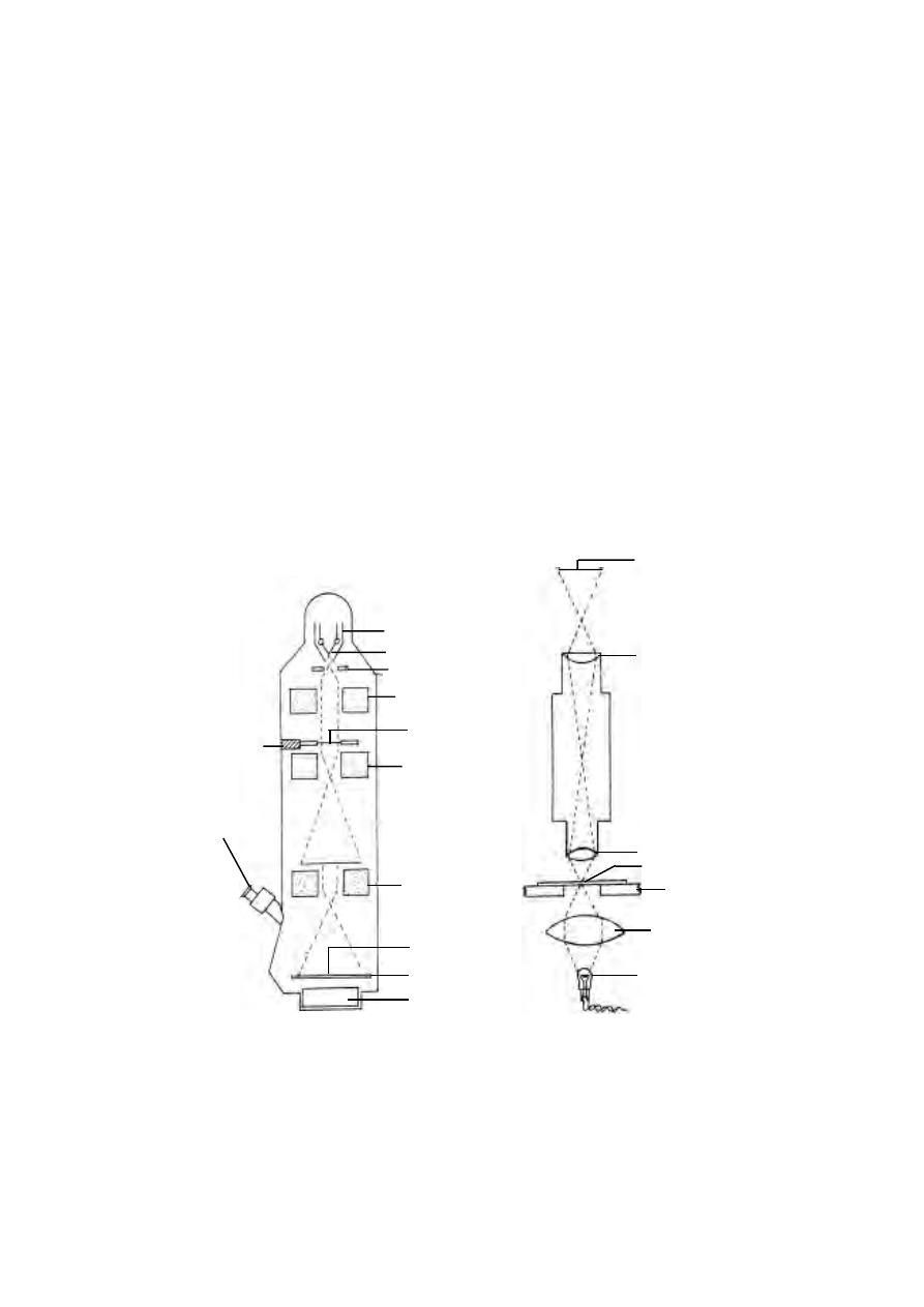
Electron microscopy.
The electron microscopy uses the much shorter wavelengths of elec-
trons to achieve resolutions as low as 3Å. Electromagnetic coils (ie., mag-
netic lenses) are used to control and focus a beam of electrons accelerated
from a heated metal wire by high voltages, in the range of 20,000 to 100,000
volts. The electrons are made to pass through the specimen. Electrons that do
passes out of the object are focused by an objective coil (‘lens’). Finally a
magnified image is produced by a projector coil or ‘lens’. The final image is
viewed on a screen or is recorded on photographic film to produce electron
micrograph. This type of electron microscope is called transmission
electron microscope (TEM)
In a compound light microscope, the image is formed due to
differences in light absorption. The electron microscope forms images as a
result of differences in the way electrons are scattered by various regions of
the object.
eyepiece lens
objective lens
stage
condenser
lens
tungsten
f i l a m e n t
lamp
projector lens
fluorescent screen
camera
binocular
viewer
specimen
holder
cathode shield
tungsten filament
anode
condenser lens
objective lens
specimen
image
Electron microscope
Fig.2.1.2. Comparison of the components and pathways of radiation
Light microscope
image
specimen
79

The degree to which electrons are scattered is determined by the thick-
ness and atomic density of the object. Hence the specimens used in electron
microscopy must be extremely thin. Living cells which are wet cannot be
viewed in electron microscope.
Scanning electron microscopy (SEM)
This microscope has less resolution power than the TEM (ie., about
200Å). However it is a very effective tool to study the surface topography of
a specimen. The whole specimen is scanned by a beam of electrons. An im-
age is created by the electrons reflected from the surface of the specimen.
Scanning electron micrographs show depth of focus and a three dimensional
image of the object.
2.1.2 Cytological Techniques
Cells are transparent and optically homogeneous organisations. They
can be observed either directly or after preservation. For direct observation,
the specimen needs sufficient contrast. Direct observation is possible by
using vital stains.
Vital stains :- Vital dyes or stains are taken up by living cells without killing
them. They selectively stain intracellular structures without affecting cellular
metabolism and function. For example, Janus green B selectively stains
mitochondria, Golgi apparatus, nuclear chromatin in a dividing cell can be stained
by methylene blue; Neutral Red dye or Congo Red dye can be used to
stain yeast cells.
Preserved and stained tissues :- For detailed microscopic study, tissues
containing cells are passed through various stages. The stages of cell prepa-
ration on a glass slide involves killing, fixation, dehydration, embedding,
sectioning, staining and mounting.
1). Killing and fixation :- This process causes sudden death of cells or
tissues and preserves freshly killed tissues in as lifelike a condition as pos-
sible. A good fixative prevents bacterial decay and autolysis. It will also make
80
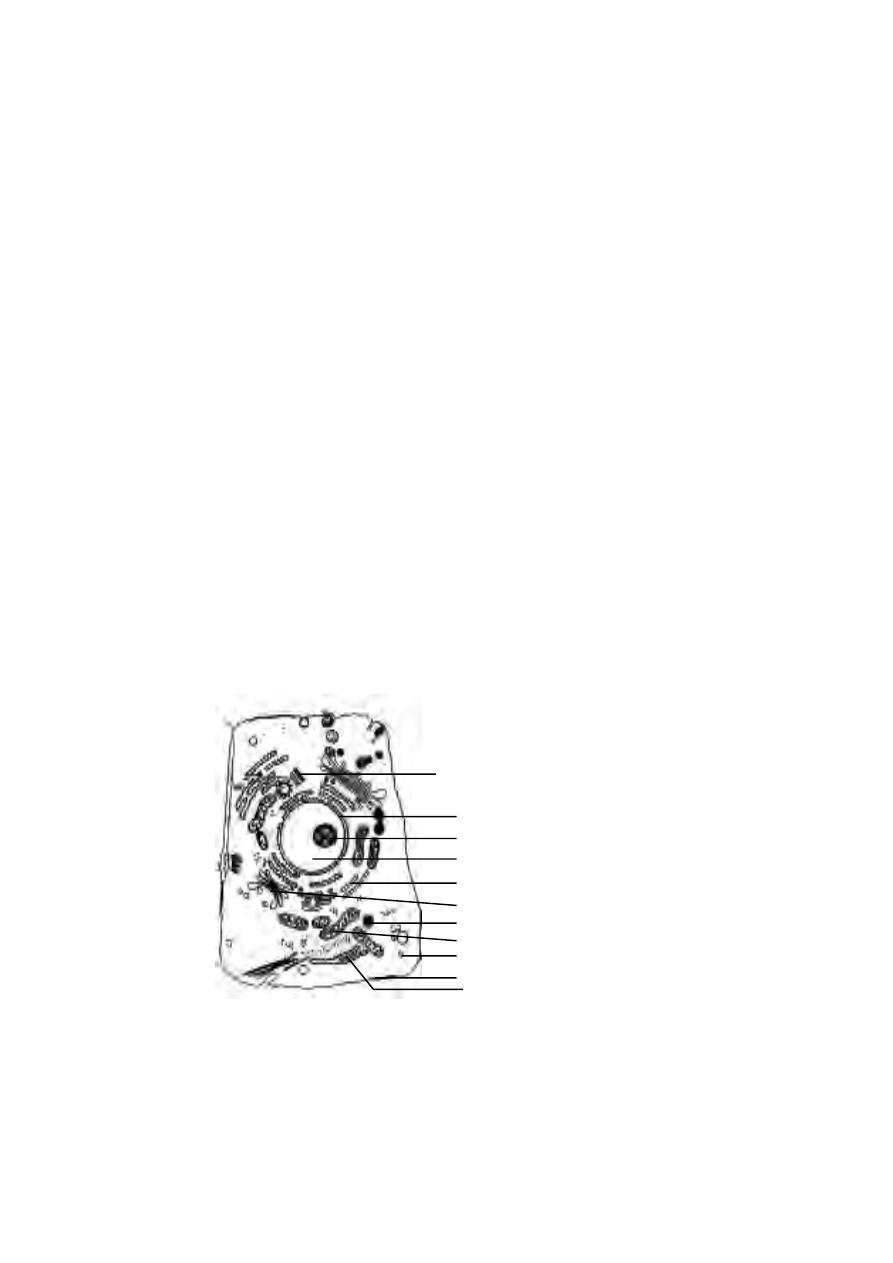
plasma membrane
golgi apparatus
nuclear envelope
rough endoplasmic reticulum
nucleus
mitochondrion
peroxisome
nucleolus
lysosome
centrioles
smooth endoplasmic reticulum
different cell components more visible and prepare the cell for staining. The
commonly used fixatives are Acetic acid. Formaldehyde, Bouin’s solution and
Carnoy’s fluid.
2). Dehydration :- In this process water vapour are removed from cells or
tissues using chemical agents. It is done by using ethanol and benzene.
3). Embedding :- The tissues are infiltrated with molten paraffin wax. It hard-
ens up on cooling and provides enough support to allow thin sections. Very
thin sections need to be taken for electron microscopy. Hence plastics are
used for embedding.
4). Sectioning :- The embedding material is cut into thin sections of needed
thickness. It is done by using an instrument called microtome.
5). Staining :- The sections are immersed in dyes that stain some structures
better than others. For example, cytoplasm stains pink with eosin. Nucleus
stains blue with haematoxylin or red with safranin.
6). Dehydration :- Stained sections are immersed in ethanol to remove wa-
ter. The tissue becomes more transparent. Dehydration is done gradually by
using a series of increasing concentrations of ethanol in water. Finally the
section is placed in ‘absolute’ alcohol.
7). Mounting :- Cleaned sections are mounted on a slide using a suitable
medium like canada balsam. A cover slip is added and the medium is allowed
to dry.
Fig.2.1.3. Ultra structure of a typical animal cell
81
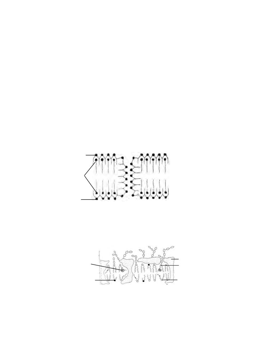
2.2.1 Plasma Membrane
It is the outer limiting membrane of both prokaryotic and eukaryotic
cells. It is an ultra thin, elastic, living membrane. Plasma membrane is a dy-
namic and selective transport barrier.
Since the plasma membrane is ultra thin, it could be observed only
under electron microscope. Structure of the membrane is studied by isolating
the same from the cell and conducting biochemical investigations.
In 1895 Overton suggested that the membrane is made of fatty sub-
stances. Other workers later concluded that two layers of lipid were present
in the cell membrane. According to a model proposed by Danielli and Davson
in 1935, the lipid bilayer of the membrane was coated on either side with
protein.
In 1960, Robertson using electronmicrographs proposed a unit
membrane hypothesis. According to this hypothesis the two outer layers of
protein are about 2 nm thick and appear densely granular. They enclose a
clear central area of about 3.5 nm wide consisting of lipids. The lipids are
mainly phospholipid molecules.
Fig.2.2.1. The Danielli-Davson model of the cell membrane
lipid bilayer
protein coat
protein coat
Fig.2.2.2. The fluid-mosaic model of the cell membrane
cell coat
lipoprotein bilayer
cholesterol
sphingolipid
phospholipid
glycolipid
glycoprotein
82

Singer and Nicholson (1972) have proposed a fluid mosaic model
for the plasma membrane. The fluid mosaic membrane is a dynamic
structure. In this structure much of the protein molecules float about. Some of
them are anchored to the organelles within the cell. Lipid molecules also move
about. ‘Fluid mosaic model’ is applied to all biological membranes in
general.
The cell membrane controls the passage of materials both into and out
of the cell. It regulates the passage of water and dissolved substances. Water
passes through the membrane by Osmosis. Water soluble substances cross
the membrane by diffusion or by active transport. Many water soluble sol-
utes are transported by carrier proteins. Lipid soluble compounds pass more
quickly by dissolving in the phospholipid layer.
2.2.2 Mitochondria
The mitochondria are filamentous or granular cytoplasmic organelles
of all aerobic cells of higher animals and plants. They are also found in micro
organisms including Algae, Protozoa and Fungi.
They were first observed by Kolliker in 1850 as granular structures
in the striated muscles. The name ‘mitochondria’ was given to them by Benda
(1897-98). Various steps of glycolysis in mitochondria was discovered by two
German biochemists Embden and Meyerhof. Embden got the Nobel Prize in
1922. Sir Hans Adolph Krebs, in 1937 found out various reactions of citric
acid cycle. Kennedy and Lehninger (1948-50) showed that Citric acid
cycle, oxidative phosphorylation and fatty acid oxidation took place in
the mitochondria.
The number of mitochondria in a cell depends on the type and func-
tional state of the cell. Certain cells contain large number of mitochondria
e.g., eggs of sea urchin contain 140,000-150,000 mitochondria. Oocytes of
amphibians contain 300,000 mitochondria. Liver cells of rat contain only 500-
1600 mitochondria. Some algal cells may contain only one mitochondrion.
The mitochondria may be filamentous or granular in shape. They vary
in size from 0.5 µm to 2.0 µm. Due to their minute nature they can not be seen
under light microscope.
Each mitochondrion is bound by two highly specialized membranes.
The outer membrane is smooth. It is separated from the inner membrane by a
6-8 nm wide space. The inner membrane is highly convoluted, forming a
series of inflodings known as cristae.
83
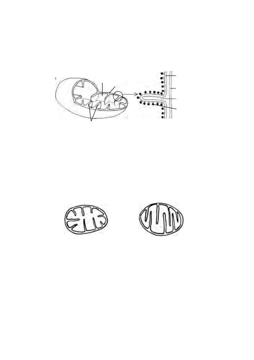
Thus mitochondria are double membrane envelopes. The inner mem-
brane divides the mitochondrial space into two distinct chambers. The outer
compartment is the peri-mitochondrial space. It is found between outer and
inner membranes. The inner compartment is the matrix space. It is filled
with a dense gel like substance called mitochondrial matrix. The matrix
contains lipids, proteins and circular DNA molecules.
The outer and inner membranes, intermembrane space and mitochon-
drial matrix contain several enzymes. Hence the mitochondria perform sev-
eral important functions such as oxidation, dehydrogenation, oxidative
phosphorylation and respiratory chain of the cell.
Since mitochondaria play a key role in the oxidation of carbohydrates
and fats, they are considered as the actual respiratory organs of the cells.
During such biological oxidations large amount of energy is released. The
energy is utilized by the mitochondria for synthesis of the energy rich com-
pound known as adenosine tri phosphate or ATP. Due to this function, the
mitochondria are also known as “power houses” of the cell. In animal cells
mitochondria produce 95 % of ATP molecules.
Fig.2.2.3. Three dimensional structure of a mitochondrion
matrix
intercristal space
oxysomes
membrane
membrane
cristae
A Crista
outer
inner
intercristal
space
Fig.2.2.4. Mitochondria
Plant cell
Animal cell
84
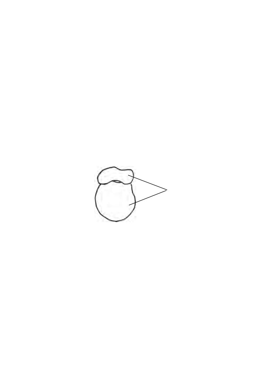
2.2.3 Ribosomes
The ribosomes are small dense, rounded and granular particles. They
contain ribonucleoprotein. They occur either freely in the matrix of the mi-
tochondria, chloroplast and cytoplasm or remain attached with the membrane
of the endoplasmic reticulum and nucleus.
The ribosomes were described by G.E.Palade in 1952. The name
‘ribosome’ was coined by R. B. Roberts in 1958.
The ribosomes occur in both prokaryotic and eukaryotic cells. In the
cells in which active protein synthesis takes place, the ribosomes remain at-
tached with the membranes of the endoplasmic reticulum. The cells where
such active synthesis happens are pancreatic cells, hepatic cells, osteoblasts,
serous cells of submaxillary gland, chief cells of the glandular stomach, thy-
roid cells and mammary gland cells.
The ribosomes are spheroid structures with a diameter of 150 to 250
Å. Each ribosome is composed of two subunits. One subunit is large in size
and has a dome like shape. The other ribosomal subunit is smaller in size and
it occurs above the larger subunit forming a cap-like structure.
The ribosomes are chemically composed of RNA and proteins. The
ribosomal RNA (rRNA) play a central role in the process of protein syn-
thesis. The ribosomal proteins enhance the catalytic function of the rRNA.
The functioning of rRNA is under genetic control.
Fig.2.2.5. Ribosome
sub-units
85
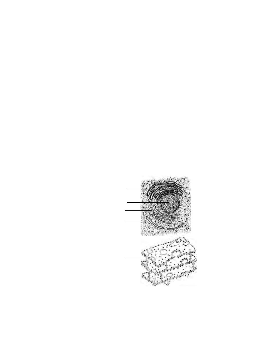
2.2.4 Endoplasmic Reticulum. (ER)
Electron microscopic study of sectioned cells has revealed the pres-
ence of a three dimensional network of sac-like and tubular cavities called
cisternae bounded by a unit membrane inside the cell. Since these structures
are concentrated in the endoplasmic portion of the cytoplasm, the entire
organisation is called the endoplasmic reticulum. This name was coined by
Porter in 1953.
The occurrence of ER varies from cell to cell. They are absent in
erythrocytes, egg cells and embryonic cells.
The ER is the site of specific enzyme controlled biochemical
reactions. Its outer surface carries numerous ribosomes. The presence of
ribosomes gives a granular appearance. In this condition ER is
described as rough endoplasmic reticulum (RER). RER is the site of syn-
thesis of proteins. Ribosomes are absent on smooth endoplasmic reticulum
(SER). SER is concerned with lipid metabolism.
Morphologically ER may occur in three forms namely 1. Lamellar form
2. Vesicular form and 3. Tubular form.
Fig.2.2.6. Endoplasmic recticulum
rough endoplasmic reticulum
smooth endoplasmic reticulum
nucleus
ribosome
sheet form of ER
86

Lamellar form or Cisternae :- These are long, flat, sac like tubules. Their
diameter is about 40-50 µm. The RER has a synthetic role. It is mostly seen
in cells of pancreas, notochord and brain.
Vesicles :- These are oval, vacuolar structures. Their diameter is about
25-500 µm. They occur in most of the cells.
Tubules :- These are branched structures forming the reticular system along
with the cisternae and vesicles. They have a diameter of 50-190 µm. They
occur in almost all cells.
Functions :-
1. It provides skeletal framework to the cell.
2. It facilitates exchange of molecules by the process of osmosis, diffusion
and active transport.
3. Enzymes of ER control several metabolic activities.
4. They serve as intracellular transporting system.
5. It conducts intra-cellular impulses.
6. It helps to form nuclear membrane after cell division.
7. SER synthesises lipids.
2.2.5 Golgi apparatus
The Golgi apparatus was discoverd by an Italian neurologist, Camillo
Golgi in 1873.
The Golgi apparatus occurs in almost all animal cells except red blood
cells. Animal cells usually have a single Golgi apparatus. Some cells have
more of Golgi apparatus.
In most of the ectodermal and endodermal cells it occurs in between
the nucleus and the periphery. In nerve cells it occupies a circum-nuclear
position.
The simplest unit of the Golgi apparatus is the cisterna. A cisterna is
about 1 µm in diameter. It has a membrane bound space. This space accumu-
lates secretions. Numerous such cisternae are associated with each other and
appear in a lamellar arrangement. In the lamellar arrangement the space
between each cisterna is 20-30 nm. A group of these cisternae is called the
dictyosome. A group of dictyosomes constitute the Golgi apparatus.
87
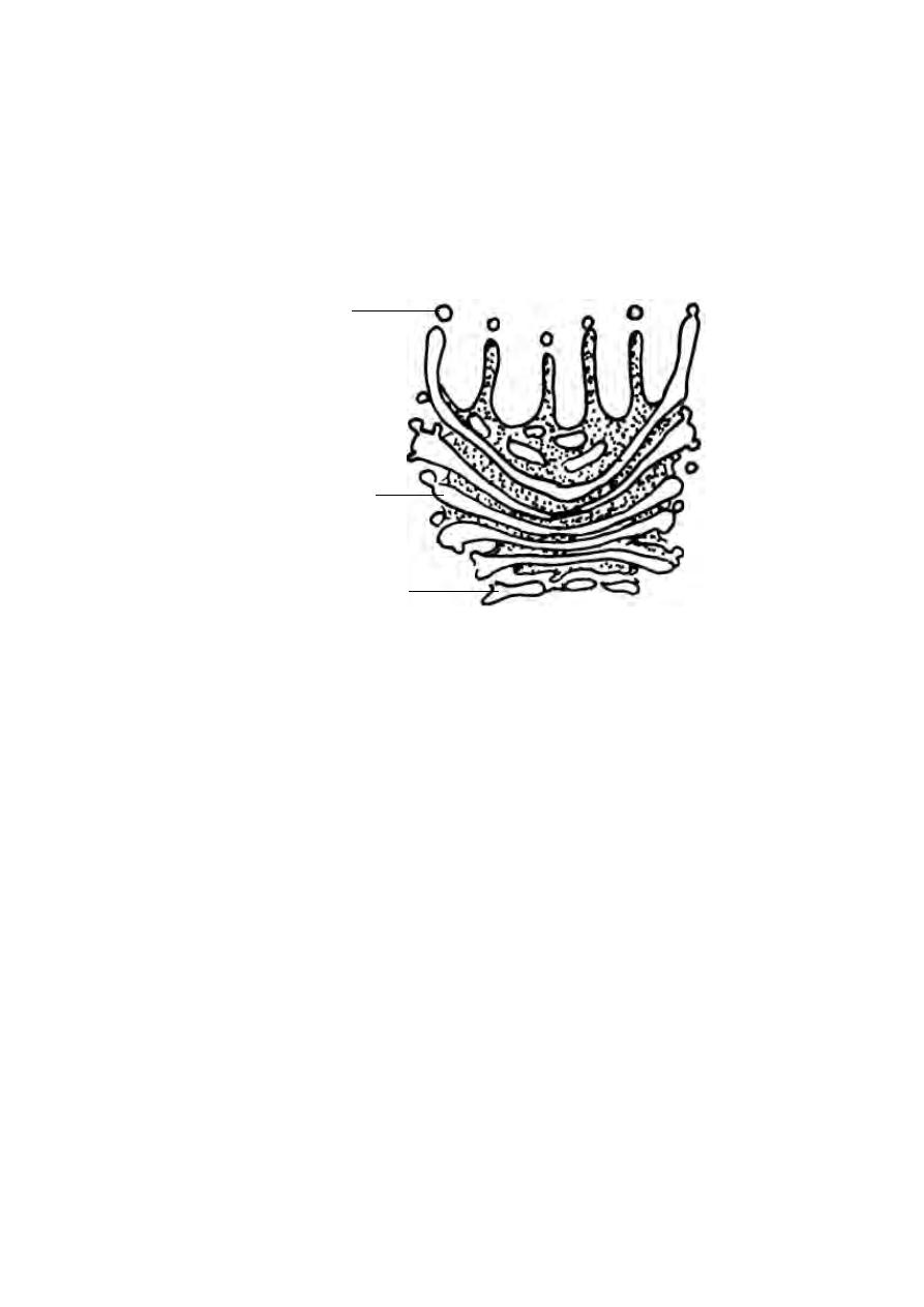
Typically a Golgi apparatus appears as a complex arrangement of in-
terconnecting tubules, vesicles and cisternae.
The Golgi apparatus is the site of synthesis of biochemicals. They also
collect proteins and lipids made in the ER and add additional substances.
2.2.6 Lysosomes
These are tiny vesicles surrounded by a membrane. Lysosomes are
involved in intracellular digestion and are primarily meant for destroying un-
wanted and aged organelles inside the cells.
Lysosomes were initially named as ‘perinuclear dense bodies’. The
name ‘lysosome’ was coined by C.de Duve in 1955.
Lysosomes occur in all animal cells. However they are not found in
mature mammalian erythrocytes. Muscle cells contain very few lysosomes.
They are numerous in epithelial cells of secretory and excretory organs.
Each lysosome is a round structure. It is filled with a dense material.
Their shapes and densities vary. Their size ranges from 0.2 to 5 µm.
Fig.2.2.7. Golgi apparatus
secretory vesicles
rough endoplasmic reticulum
layered cisternae
88
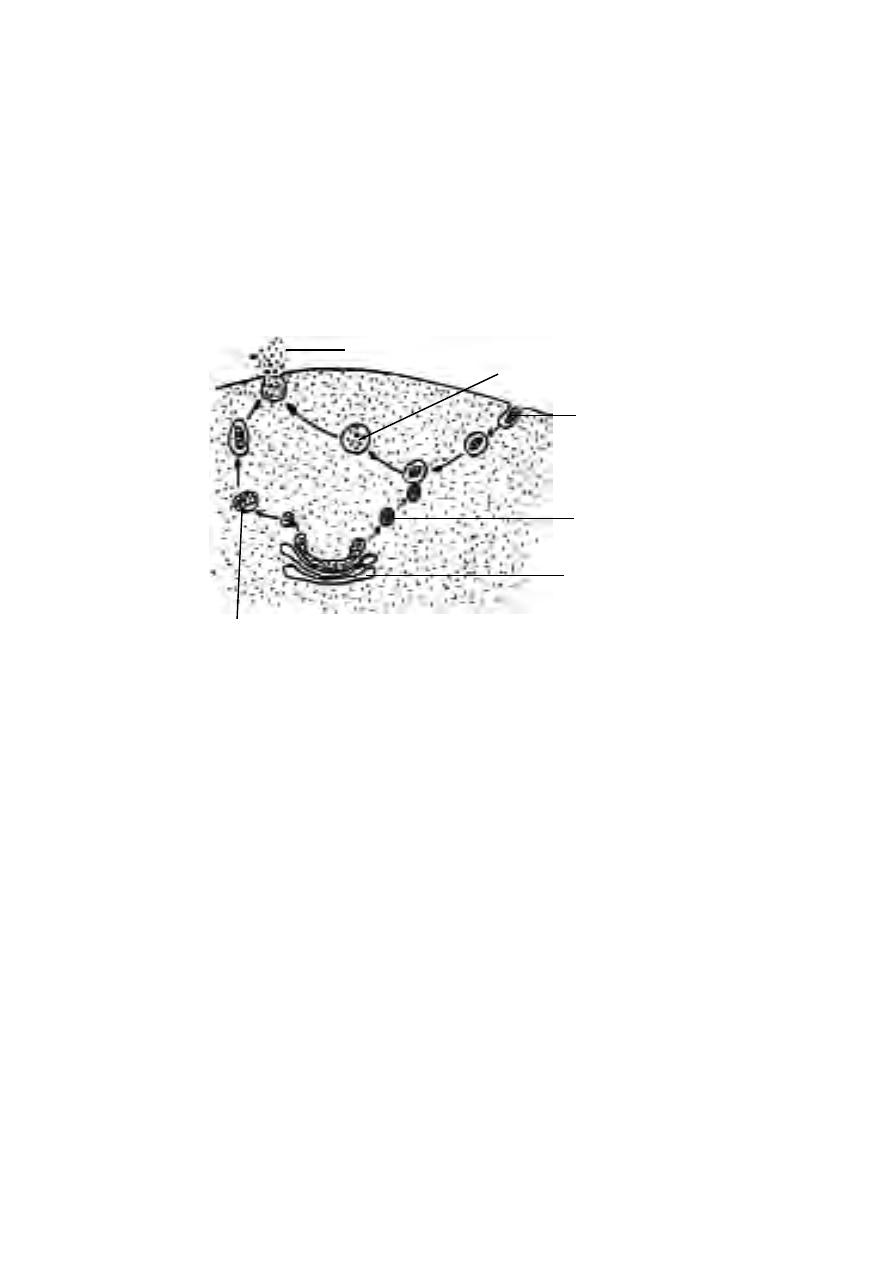
Recent studies reveal that lysosomes may contain upto 40 types of
hydrolytic enzymes.
The enzymes are mostly proteases, nucleases, glycosidases, li-
pases, phospholipases, phosphatases and sulphatases.
Lysosomes originate either from the Golgi apparatus or directly from
the endoplasmic reticulum. The enzymes they contain are used in the
dissolution and digestion of redundant structures or damaged macromol-
ecules from within or outside the cell. For example, when an animal cell in-
gests food into a food vacuole, lysomes fuse with the vacuole and break down
the contents. Their enzymes digest carbohydrates, fat and proteins. The glands
in some digestive organs package their digestive enzymes in lysosomes before
releasing them outside the membrane. When a cell dies its own lysosomes
release the enzymes that digest the remains of the cell in a process known as
autolysis.
Peroxisomes
These are spherical organelles bounded by a single membrane. They
are found in the cells of Protozoa, fungi, plants, liver and kidney of verte-
brates. The name peroxisomes was coined by C.de Duve and P. Baudhuin
(1966)
Peroxisomes contain catalase, an enzyme that catalyses the decom-
position of hydrogen peroxide to the harmless products, water and oxygen.
Fig.2.2.8. Lysosomes - in action
Lysosome
entry of solid matter
digestion
discharge
functionless organelle
Lysis
golgi apparatus
89

Hydrogen peroxide is a by- product of certain reactions of metabolism. It is
potentially a very harmful oxidising agent.
2.2.7 Centrioles
The centrioles are two cylindrical, microtubular structures found near
the nucleus. When a centriole supports a flagellum or cilium, it is called the
basal body.
The centrioles occur in most of the animal cells, algal cells and some
fern cells. They are absent in prokaryotes, red algae, yeast cells and flowering
plants and some non -flagellated or non-ciliated protozoans.
The centrioles range in size from 0.15-0.25 µm in diameter. They are
usually 0.3-0.7 µm in length.
Each centriole and basal body is formed of nine triplet microtubules
equally spaced around a perimeter. Each microtubule has a diameter of 200-
260 Å in diameter.
The microtubules are made up of a structural protein, tubulin, along
with lipid molecules.
It was initially considered that new centrioles arise by the division of
existing centrioles. This idea is no longer accepted. It appears that new cen-
trioles are produced de novo or are synthesized using an existing centriole as
a template.
In most of the animal cells the centrioles are the focal point for the
centrosome. The centrosome organizes cytoplasmic microtubules during
interphase in mitosis. It provides the two poles of the mitotic spindle.
The centrioles form the basal body and the cilia. In spermatozoon one
centriole gives rise to the tail fibre or flagellum. The centrioles are also in-
volved in ciliary and flagellar activity.
2.2.8 Nucleus
The nucleus is the most important organelle of cell. It controls all meta-
bolic processes and hereditary activities of the cell.
The nucleus was first discovered and named by Robert Brown in
1833. The occurrence of a nuclear membrane was first revealed by O. Hertwig
in 1893.
The nucleus is found in all the eukaryotic cells of plants and animals.
90
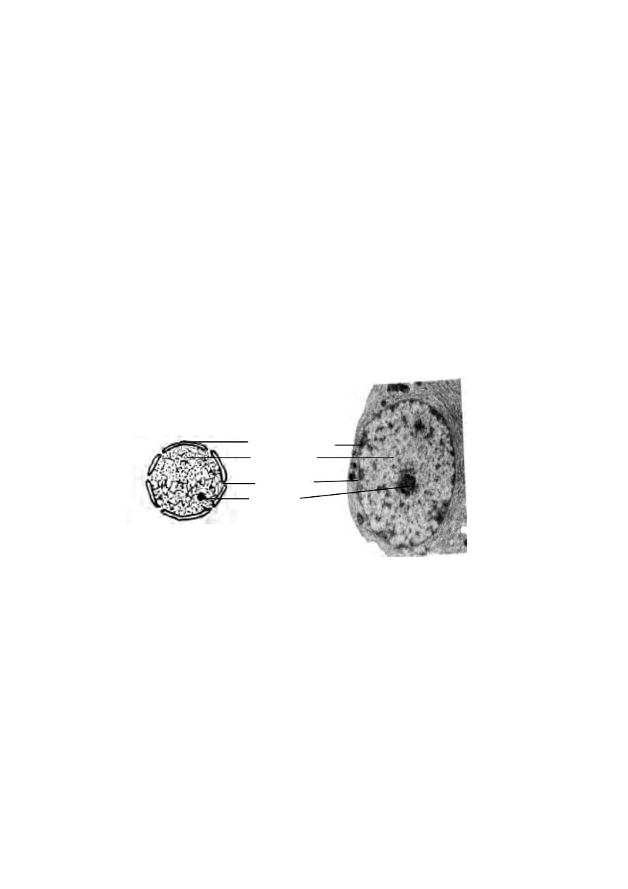
However some eukaryotic cells such as the sieve tubes of higher plants and
mammalian erythrocytes have no nucleus.
Usually the cells contain single nucleus (mononucleate). However
certain cells may have more than one nuclei. Accordingly they may be called
binucleate or polynucleate cells. The polynucleate cells of the animals are
called syncytial cells (Osteoblast cells)
The shape of the nucleus may be spherical, elliptical or discoidal.
In certain cells the nucleus is irregular in shape.
The size of the nucleus may vary from 3 µm to 25 µm in diameter. The
size is directly propotional to that of the cytoplasm. Nuclear size may also be
determined by the number of chromosomes or ploidy. The nucleus of the hap-
loid cells are smaller than that of the diploid cells.
The nucleus is surrounded by a nuclear envelope. This envelope is
comprised of two membranes of 5-10 nm thickness. The inner nuclear
membrane supports a fibrous sheath called the nuclear lamina. The inner
nuclear membrane is surrounded by the outer nuclear membrane. The space
between the inner and outer membranes is known as perinuclear space. It is
a 10 to 50 nm wide fluid filled compartment.
The nuclear lamina is a protein meshwork. It is a very dynamic
structure.
Fig.2.2.10. TEM of the Nucleus
Fig.2.2.9. Nucleus
Nucleopore
Nucleoplasm
Nucleolus
Nuclearmembrane
91

The nuclear envelope is perforated by nuclear pores. Each pore has
a diameter between 10 nm to 100 nm. It has been calculated that the nuclear
pores account for 5 to 15 percent of the surface area of the nuclear mem-
brane. There is continous movement of molecules across the nuclear enve-
lope through the pores.
The nucleus is filled with a transparent semisolid matrix known as
nucleoplasm or nuclear sap. The chromatin threads and the nucleolus re-
main suspendended in the nucleoplasm. The nucleoplasm is composed of nucle-
oproteins, proteins, enzymes and minerals.
The nucleoplasm contains several thread like coiled structures. These
are the chromatin fibres. During the cell division they become thick ribbon
like structures known as chromosomes. The chromatin is made up of
Deoxy - ribose nucleic acid (DNA) and proteins.
The nucleus contains one or more spherical colloidal structures called
nucleoli. The size of nucleolus is related to the synthetic activity of the cell.
The number of nucleoli in the cells may be one, two or four. Chemically, nucleo-
lus contains DNA of nucleolar origin, four types rRNA, 70 types of ribosomal
proteins, RNA binding proteins and RNA splicting nucleoproteins.
Ribosomal subunits are synthesized in the nucleolus. Initiation, pro-
duction and maturation stages of ribosomal formation happen in three dis-
tinct regions of the nucleolus.
Chromosomes
The chromatin fibres get condensed into chromosomes during cell di-
visions. They are capable of self-reproduction and they play an important rloe
in heredity.
The nucleus was first observed and described by karl Nagli (1842) in
the nuclei of plant cells. Chromosomes and their role in cell division was first
explained by A. Schneider (1873). In 1887 Benden and Bovery reported
that the number of chromosomes for each species is constant. T. H Morgan
and H. Muller in 1922 revealed the occurrence of nearly 2000 genetic fac-
tors on four chromosomes of Drosophila. In 1924, Robert Feulgen showed
that chromosomes contain DNA.
The number of chromosomes is constant for a particular species.
The reproductive cells such as sperm or ovum has one set of chromosomes
and it is known as the haploid set(n). It is also known as the genome. The
somatic or body cells contain two haploid set or genomes and are known as
92
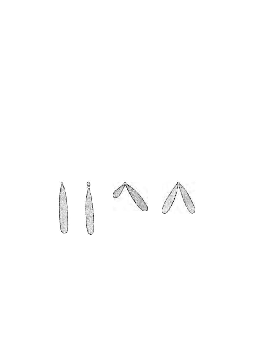
the diploid cells (2n). The diploid condition is arrived at by the union of the
haploid male and female gametes in the sexual reproduction.
Number of Chromosomes
Common name
Scientific name
Chromosome Num-
ber
Paramoecium
P. aurelia
30-40
Hydra
H. vulgaris
32
Housefly
Musca domestica
12
Fruit fly
Drosophila sps
8
Pigeon
Columba livia
80
Gorilla
Gorilla gorilla
48
Man
Homo sapiens
46
The size of a chromosome can be measured during mitotic metaphase.
It may range from 0.25 µm to 30 µm.
The shape of the chromosome changes from phase to phase. Each
chromosome has a clear zone, known as centromere or kinetocore along
their length. The centromere divides the chromosome into two parts. Each
part is called the chromosome arm. Thus according to the position of the cen-
tromere and nature of the chromosome arm, the chromosomes may be Telo-
centric, Acrocentric, Submetacentric and Metacentric.
Fig.2.2.11. Types of chromosomes
A - Telocentric, B - Acrocentric, C - Submetacentric, D - Metacentric
A
B
C
D
93

2.3 Cancer biology
2.3.1
Cancer is a proliferation of cells which grow in an uncontrolled
manner, invading local tissues and spreading widely through the blood or lym-
phatics to produce secondary deposits, or metastases in distant parts of the
body.
The word ‘cancer’ comes from Latin, meaning a crab. A tumour was
called a cancer because of swollen veins around the area resembling a crab’s
limbs. The study of cancer is called Oncology. Oncology is a word derived
from the Greek, onchos, a lump, or tumour. The abnormal tissue growth is
called neoplasm. If a neoplasm can cause harm by spreading, it is said to be
malignant.
Cancer was known to ancient civilizations. However the disease as it
would be defined today was established as an entity by German pathologists
of 19th century. They described cellular nature of cancer and classified
cancer. At the beginning of the 20th century, most major forms of cancer had
been described. Further, attention was focused on finding the cause and intro-
ducing treatment. In 1775 Pott recognised cancer in chimney sweeps. He
associated soot with cancer. From this time onwards environmental and oc-
cupational hazards were recognised as follows :
shale oil
skin cancer in workers
radio active ores
lung cancer in miners
beta-naphthylamine
bladder cancer in rubber industry workers
cigarettes
lung cancer
Later it was discovered that certain viruses can also cause cancer.
One of the earliest virus, causing cancer, described was Rous sarcoma virus.
Recently, human T-cell leukaemia has been found to be due to the virus
HTLV-1.
Some forms of cancer can also be inherited. A rare eye tumour,
retinoblastoma is inherited. It is a dominant character showing Mendelian in-
heritance.
Cancer biology
The knowledge of cancer biology is growing rapidly. Researches are
being conducted to fully understand the development of cancer at the cellular
94

or molecular level. The available information is not sufficient for satisfactory
treatment of cancer.
During normal development and growth the cells in our body divide
mitotically and get differentiated to specialized cells of the tissues. The pro-
cesses of cell mitosis, growth and differentiation are controlled by cellular
genes. Cancer is caused due to mutation or abnormal activation of such genes.
such a mutation can happen in a single cell. Thus it may be monoclonal in
origin. With further growth of cancer, additional mutations may occur in the
daughter cells giving rise to subclones. The mutated cells may remain as
heterogeneous cancer cells. Among these subclones some may have greater
capacity and metastasize to distant tissues. They may also remain more
resistant to damage from various anticancer treatments.
The cancer cells have characteristic properties. They can be differ-
entiated from normal cells under microscopic observation. These cells have
large nuclei. In each cancer cell, the ratio of nucleus to cytoplasm is high.
They have prominent nucleoli. The cells can grow indefinitely in culture me-
dium. As component cell of a tissue they remain less differentiated. Even
after getting organised into tissues unlike other cells they do not lose their
replicative capacity. Cancer cells have the ability to invade surrounding
tissues.
The sequence of events that convert a normal cell into a cancer cell is
called carcinogenesis. The process of carcinogenesis includes, intiation,
growth, promotion, conversion, propagation and progression. Progression
includes the processes of invasion and metastasis.
Mature cancers have relatively uncontrolled growth, behaviour. As
other normal cells they do not show any of the normal intracellular and
extracellular growth control mechanisms. Initially the cancer cells have an
exponential growth. Gradually their growth surpasses blood vascular supply.
This results in slowing down of growth.
Molecular biology of cancer
Techniques in molecular biology have helped in understanding the most
intimate structure of the cancer cell. It has been found that at molecular level
two mechanisms operate.
1. The parts of genome involved in cell growth become activated. These are
called the ‘proto-oncogenes’. These strands of DNA induce malignant growth
tranformation in the cells. The conversion of proto-oncogenes into oncogenes
can happen due to ‘point mutations’ on DNA. Further such cancer cells dis-
95

play chromosomal abnormalites such as duplication, deletions and transloca-
tions. Thus such alterations in gene arrangement can lead to generation of
oncogenes.
Oncogenes of human tumours
Oncogene
Type of cancer
Activation mechanism
hox11
Acute T-cell leukemia
Translocation
erbB-2
Breast and ovarian carcinomas
Amplification
L-myc
Lung carcinoma
Amplification
ret
Thyroid carcinoma DNA
rearrangement.
A distinct mechanism by which oncogenes are activated in human
tumors is amplification. It results in elevated gene expression. Gene
amplification is very common in cancer cells. It occurs a thousand times more
frequently than in normal cells. Molecular biologists are now working on the
products of oncogenes.
2. The growth of normal cells is controlled by suppressor genes. In cancer,
parts of the genome functioning as the suppressor gene are either lost or
inactivated. Hence, negative regulators of cell proliferation are removed. It
contributes to the abnormal proliferation of cells.
Gene
Type of cancer
APC
Colon / rectum carcinoma
BRCA 1
Breast and ovarian carcinoma
1 NK 4
Melanoma, lung carcinoma,
brain tumors, leukemias, lymphoma
Rb
Retinoblastoma
PTEN
Brain tumors, kidney and lung carcinomas.
The protein products of the tumor suppressor genes normally inhibit
cell proliferation. Inactivation of such genes therefore leads to tumor
development.
The complete sequence of events required for the development of any
human cancer is not yet known. But it is clear that both the activation of
oncogenes and the inactivation of tumor suppressor genes are critical
steps in tumor initiation and progression. Simultaneous effect on both the genes
96

will result in multiple genetic defect. It results in the increased proliferation,
invasiveness and metastatic potential of cancer cells.
2.3.2 Types of cancer
Cancers are named according to the tissues from which they arise.
1. Sarcoma - Malignancy in structural tissues Ex: Osteosarcoma (bones),
liposarcoma (fa).
2. Carcinoma - Epithelial cancers. Ex : Lung carcinoma, breast carcinioma.
3. Lymphoma - Lymphatic tissues
4. Leukemia - White blood cells.
Causes for Cancer (Etiology)
Majority of the cancers are caused by living habits and environmental
factors.
1. Tobacco :- Nearly 35% of all cancer deaths are due to usage of tobacco in
some form. Atleast 90% of lung cancer deaths are due to smoking. Smoking
can also affect gastro-intestinal tract, pancreas, genito-urinary tract and
upper respiratory passage. Snuff and chewing tobacco can affect mouth and
respiratory tracts. Smoke inhaled by non-smokers (passive smoke) can also
cause lung cancer and blood cancer.
2. Exposure to ionising radiations such as x-rays, gamma rays, uv rays can
produce cancer. These radiations rupture DNA strands, causing mutations.
Solar radiations can cause skin cancers.
3. Physical irritants, such as continued abrasion of the linings of the intestinal
tract by some types of food can also lead to cancer. Dietary substances such
as fat, high calorie intake of animal proteins, salted or smoked food can cause
cancer in Breast, Colon, Stomach and Oesophagus.
4. Certain drugs if taken without medical advice can cause cancer.
5. Viruses and parasitic organisms like schistosoma, liverfluke can also
affect.
Cancer and death
A severe onset of cancer ends in the death of a person. It is because
of the unique characteristics of the cancer cells. The growth of normal cells is
controlled by certain factors. However the cancer cells do not require the
growth factors. Hence these cells do not respect usual cellular growth
limits.
97

Normal cells have a nature of remaining together in tissues. But the
cancer cells are less adhesive to each other. Hence they wander through the
tissues and enter the blood. They can be transported to all parts of the body
and cause new cancer growths.
The cancer cells rapidly multiply. Some cancers also produce angio-
genic factors that cause many new blood vessels to grow into the cancer.
Thus these cells will drain all the nutrients and normal cells get deprived of
food. Ultimately the normal cells and tissues suffer nutritive death.
Management of cancer
The treatment or management of cancer depends on an accurate di-
agnosis. Diagnosis is made through microscopic observations(tissue biopsy),
study of markers on the surface of cells, cytochemical methods, cytogenetics
and various scanning and ‘x’ ray diagnostic methods.
In order to compare results and for communicating treatment
programme among medical personnel staging systems are essential (Ex:
stage1, stage2) staging defines tumours as either confined to the tissue of
origin or having spread to local tissues and organs and finally as having
metastasized.
After diagnosis, treatment of cancer involves surgery, chemotherapy,
radiotherapy and hormonal treatments.
Till last century, surgery was the only effective method of treatment.
Even to-day through surgery biopsy can be effected for diagnosis. If the tu-
mour is restricted to the primary site, through surgery it can be removed. In
latest treatment procedures surgery is restricted to affected region, rather
than amputation or removal of the entire organ.
Radiotherapy :- Discovery of x-rays by Roentgen(1895) and of radioactivity
by Curies in 1899 opened new ways of treating cancer. In Radiotherapy high
energy ionising radiations are used. The radiations used are x-rays and gamma
rays or subatomic particles such as beta particles, high energy electrons and
neutrons or charged particles like helium ions. Ionising radiations can
penetrate tissues. They can damage DNA leading to cell death and
mutagenesis. The basis of radiation therapy depends on the differential
sensitivity of the tumour tissue and the normal tissue. Hence the aim of
radiotherapy is to prescribe sufficient radiation dose to the tumour, sparing as
much of the normal tissue as possible.
98

Chemotherapy :- The purpose of chemotherapy is to prevent cancer cells
from multiplying, invading and metastasizing. The chemicals used in treatment
affect cell multiplication and tumour growth. Several drugs are now available
for usage. They can used singly or in combination. Some cancers like breast
cancer are hormone dependent. Hence hormones are used in their manage-
ment.
Inspite of all advances in diagnostics and treatment, the death rate due
to cancer is greater. Primary prevention of cancer will be a better
alternative to diagnosis and treatment. 70% to 80% of cancers result from
environmental causes. Hence public awareness towards environmental issues
is a need. 33% of cancers in India are tobacco related. Hence smoking
cessation and other measures to reduce tobacco usage are to be insisted upon.
Consumption of fibrous food and avoidance of fatty food will avoid tumours
related to alimentary canal.
Thus it is apparent that fight against cancer will be successful with
early detection and appropriate education for avoidance.
Self Evaluation
Part I
Choose the correct answer
1. Living cells which are wet cannot be viewed in a / an
a) Compound microscope
b) Phase - contrast microscope
c) Electron microscope
d) Dark - field microscope.
2. Congo Red dye can be used to stain
a) Plant cell
b) Nerve cell
c) Gland cell
d) Yeast cell
3. The cell theory was forwarded by
(a) Embden and Meyer Hoff
(b) Schleiden and Schwann
(c) T.H. Morgan
(d) Singer and Nicholson
4. The increase in size of optical image over the size of the object is known as
(a) limit of resolution
(b) transmission
(c) power of magnification
(d) conductance
5. The name ‘cell’ was coined by
(a) Leeuwenhoeck
(b) Robert Brown
(c) Robert Hook
(d) Galileo
99

6. In a microscope the light is focussed on the object through
(a) condenser lens
(b) objective lens
(c) occular lens
(d) oil immersion lens
7. A three dimensional image of the object can be produced using
(a) compound microscope
(b) dark-field microscope
(c) transmission electron microscope
d) scanning electron microscope
8. In microscopy a nucleus is normally stained using
(a) Neutral red
(b) Janus green B
(c) Eosin
(d) Hematoxylin
9. In cytological technique Bouin’s solution is used for
(a) fixation
(b) dehydration
(c) cleaning
(d) embedding
10. The lipids of plasma membrane are mainly
a) Glucose molecules
b) Phospholipid molecules
c) Protein molecules
d) All the above
11. A unit membrane hypothesis for plasma membrane structure was provided
by
(a) Overton
(b) Daveson
(c) Robertson
(d) Nicholson
12. In the plasma membrane the lipid bilayer is covered by
(a) proteins
(b) carbohydrates
(c) water molecules
(d) nucleic acid
13. The power houses of the cell are
a) Lysosomes
b) Ribosomes
c) Mitochondria
d) Centrosomes
14. Animal cells usually have
a) single golgi apparatus
b) double golgi apparatus
c) Poly golgi apparatus
d) dictyosome
15. The role of mitochondria in oxidative phosphorylation was explained by
(a) Leninger
(b) Embden
(c) Krebs
(d) Meyer hoff
100

16. Which of the following is called as the cell respirataory organelle
(a) ribosomes
(b) lysosomes
(c) golgi bodies
(d) mitochondria
17. The ribosomes are meant for
(a) phosphorylation
(b) respiration
(c) protein synthesis
(d) oxidation
18. Lung cancer may be caused by
a) cigarette smoking
b) shale oil
c) radio active ores
d) beta-naphthylamine
19. Which of the following organelle forms the intra cellular transporting system
(a) mitochondria
(b) lysosomes
(c) Endoplasmic reticulum
(d) ribosomes
20. Which of the following cell may not contain golgi apparatus
(a) epithelial cells
(b) glandular cells
(c) RBC
(d) secretory cells
21. The lysosome originates from
(a) mitochondria
(b) ribosomes
(c) nucleus
(d) golgi apparatus
22. Lysosomes are involved in :-
a) intracellular digestion
b) extracellular digestion
c) Phagocytosis
d) pinocytosis
23. Peroxisomes are found in the cells of
a) Protozoa
b) Porifera
c) Coelenterata
d) Platyhelminthes
24. The movement of the flagellum is regulated by
(a) plasma membrane
(b) nucleus
(c) basal body
(d) ribosomes
25. Each centriole and basal body is formed of Microtubules as
a) seven triplets
b) eight triplets
c) ten triplets
d) nine triplets
101

26. The nucleus is found in all the
a) Bacteriae
b) Viruses
c) Eukaryotic cells
d) All the above
27. The number of nucleus (nucleii) in matured mammalian erythrocytes is
(a) one
(b) many
(c) two
(d) nil
Part - II
Give very short answer
1. Write a note on phase contrast microscope.
2. Name the new branches of science related to cytology.
3. What is ‘limit of resolution’ in the functioning of a microscope ?
4. Name the various parts of a compound microscope.
5. What are vital stains ?
6. Name any four vital dyes or stains.
7. What is Mounting?
8. Name two biochemical processes happening within the mitochondria.
9. What are RER and SER ?
10. Write any two functions of endoplasmic reticulum.
11. Mention any four enzymes which are seen in Lysosomes.
12. Write any two functions of centrioles.
13. What is ‘autolysis’ ?
14. When do we call centriole as a basal body.
15. Name the two types of nucleic acids.
16. Mention the four types of centrioles on the basis of the centromere.
17. What are the types of chromosomes based on the nature of their arm ?
18. What is a ‘fluid mosaic model’ of a plasma membrane ?
19. What is oncology ? explain.
20. Name the stages in carcinogenesis.
Part - III
1. Write notes on cell theory.
2. Explain the various units of measurement in cell biology .
102

3. Differentiate TEM and SEM.
4. Describe Singer and Nicholson fluid, mosaic model of plasma membrane.
5. Give an account of golgi apparatus.
6. Draw a neat sketch of a mitochondrion and label the parts.
7. Provide the structure of the golgi apparatus.
8. Write notes on the organisation of a centriole.
Part - IV
1. Give an account of types of microscopes.
2. Describe how a specimen is prepared for microscopic study.
3. Draw a neat sketch of the ultra structure of a typical animal cell and label the parts.
4. Write an essay on structure and functioning of mitochondria.
5. Provide a detailed account on the organisation of cell nucleus.
6. Give a detailed account of endoplasmic reticulum.
7. Give an account on management of cancer.
8. Mention any four cytological techniques.
103

104
3. Human Anatomy
3.1. History of Human anatomy
The term ‘anatomy’ is Greek in origin. It takes its root from ‘ana’ and
‘tome’ (ana-up ; tome-cutting). Thus anatomy is the science of physical
structure of an animal or plant studied by dissection. The Human Anatomy
provided the necessary knowledge for surgery and medicine.
The study of human anatomy dates back to 2500 BC, when the
Egyptians prepared mummies. They removed internal organs of cadavers be-
ing mummified. They also did surgery for wounds and broken bones. In India
during 500 - 491 BC Susruta performed cataract operation. In 1
st
century AD,
Celsus, a Roman physician wrote about surgical procedures.
The year 1543 AD was significant due to publication of an accurate
book on Anatomy by Andreas Vesalius. In 1628 William Harvey described the
functioning of heart and the movement of blood in animals. These earlier works
were followed by the discovery and accurate account of each and every or-
gan system and organs in human body. In the recent times, attempts are being
made to understand the molecular architecture in every cell of our body.
Gray’s Anatomy
Eventhough several books had been written on Human
Anatomy, a monumental work titled ‘Anatomy : Descriptive and
Surgical by Henry Gray is unique in its discriptions. The first
edition of this book was published in August 1858. It is in print
continually for the past 145 years. A more elaborate 38th edition
was published in the year 2000.
Henry Gray was born in Windsor, London in 1827. He
lived upto 1861. He was a brilliant medical student at St. George’s
Hospital, London.
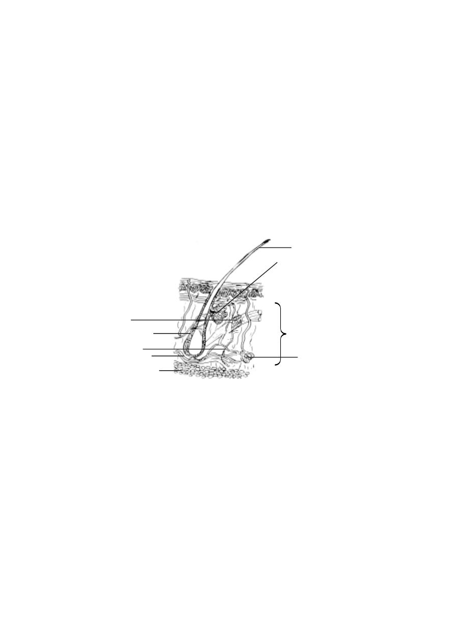
105
3. 1. 1 The Integumentary System
The word integument means covering. The integumentary system cov-
ers the outside of the body. It protects internal structures, prevents the entry
of infectious agents, reduces water loss, regulates body temperature, produces
vitamin D and detects stimuli such as touch, pain and temperature. Since the
integument performs several functions, it is commonly referred to as Jack of
all trades.
The skin or integument rests on layers of cells called hypodermis.
The hypodermis attaches the skin to underlying bones and muscles. It supplies
blood vessels and nerves to the skin.
The skin is composed of two major tissues, namely dermis and
epidermis. The dermis is mostly formed of connective tissue having fibro-
blasts, adipose cells and macrophages. It provides the structural strength to
the skin. The dermis accommodates nerve endings, hair follicles, smooth muscles
and glands.
It is divided into two layers, namely the superficial papillary layer
and deeper reticular layer. The papillary layer has projections called papil-
lae. The reticular layer is the major layer of the dermis. It is dense in nature.
It is continuous with the hypodermis.
Epidermis
:- The epidermis is made up of stratified squamous epithelium. It
is separated from the dermis by a basement membrane. It contains mel-
Fig.3.1.1. Skin and hypodermis
sebaceous gland
epidermis
dermis
hypodermis
sweat gland
fat
blood vessels
nerve
hair follicle
arector pili (smooth
muscle)
hair
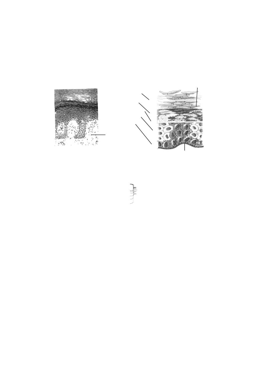
106
anocytes giving colour to the skin. Many of the cells of the epidermis produce
a protein substance called keratin. Hence they are called as keratinocytes.
The deepest layers of the epidermis produce nerve cells by mitosis.
As new cells are formed, the older cells are pushed to the surface. The sur-
face cells will protect the inner new cells. Gradually the shape and chemical
nature of the surface cells will get altered. Slowly they get filled with keratin.
This process is called keratinization. During this process the epidermis gets
divided into five distinct regions or strata. They are the stratum basale, stra-
tum spinosum, stratum granulosum, stratum lucidum and stratum cor-
neum.
Stratum basale is in the deeper region of the epidermis. It consists of
one layer of columnar cells. Keratinization of cells begins in this region. Above
this layer stratum spinosum is seen. It has 8-10 layers of polygonal cells.
The stratum granulosum is the next upper layer. It has 3-5 layers of flat-
tened cells. Above this layer stratum lucidum occurs. It is a thin zone having
several layers of dead cells. The top most layer is called the stratum cor-
neum. It consists of more than 20 layers of dead cells. These cells get filled
with keratin. They are said to be cornified. The cornified cells are surrounded
by a hard protective envelope.
The skin can be either thick or thin. All five epithelial layers are seen
in the thick skin. However stratum corneum contains more number of cells.
Thick skin is formed in the soles of the feet, the palms of hands and tips of
fingers. The general body surface has thin skin. In the thin skin each epithe-
lial layer inturn has few layers of cells. There are only one or two layers of
cells in stratum granulosum.
papilla
keratin
basement membrane
stratum basale
stratum spinosum
stratum lucidum
stratum granulosum
stratum corneum
Fig.3.1.2. Epidermis

107
Callus
:- The regions of skin subjected to constant friction or pressure are
thickened to form the callus. The callus has several layers of cells in the
stratum corneum.
Skin colour
:- The colour of the skin is due to pigments in the skin. The
thickness of the stratum corneum and blood circulation can also cause skin
colour. Normally the colour is caused by the pigment melanin. It provides
colour to skin, hair and eye. It protects the body from sun’s ultraviolet rays.
Melanin is produced by melanocytes. Melanin production is genetically deter-
mined. However, hormones and exposure to light can also alter the colour.
Skin dervatives
Hair :- The hairs are integumentary structures. A hair has a root and a shaft.
While the shaft projects above the skin, the root remains well below the sur-
face. The base of the root has a hair bulb. It is an expanded region. The shaft
and most of the root of the hair are formed of dead keratinized epithelial cells.
These are arranged in three concentric layers called the medulla, the cortex
and the cuticle. The central axis of the hair is formed of the medulla. Major
part of the hair is formed of a single layer of cells.
According to the amount and types of melanin, the hair colour may
vary. The colour of the hair is genetically determined. During old age the
amount of melanin decreases causing white hair. Grey hair has a mixture of
faded, unfaded and white hairs.
The hair growth is due to addition of cells at the base of the hair root.
The growth stops at specific stages. After a resting period, new hair replaces
old hair. The hairs on the head grow for a period of three years and rest for
1-2 years.
Fig.3.1.3. Cross section of a hair within a follicle
medulla
cortex
cuticle
internal epithelial root sheath
external epithelial root sheath
dermal root sheath
hair
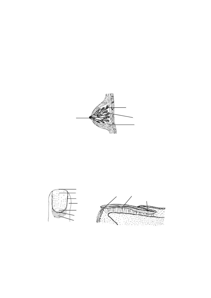
108
The muscle cells found associated with hair follicles are called the
arrector pili. Contraction of these muscles cause ‘goose flesh’ making the
hairs to ‘stand on end’.
The skin has sebaceous glands and the sweat glands. The seba-
ceous glands are located in the dermis. They produce an oily substance called
the sebum. These glands are connected by a duct to the upper part of the hair
follicles. The mammary glands are the modified sweat glands.
The most common type of sweat gland on the skin are the merocrine
glands. They are simple coiled tubular glands. They open directly on to the
skin through sweat pores. The gland has two parts. They are the deep coiled
portion and the duct which passes to the surface of the skin. The number of
sweat glands are more in the palms of the hands and soles of the feet.
Nails :- Each nail is made up of two parts. They are the nail root and the nail
body. The nail body is the visible part. The nail root is covered by the skin.
The proximal and lateral edges of the nail are covered by nail fold.
longitudinal section
Fig.3.1.5. Nail
nail root
nail bed
hyponchium
free edge
nail body
nail groove
nail fold
lunula
eponchium
dorsal view
nail root
Fig.3.1.4. The mammary gland
nipple
fat
lactiferous duct
glandular lobe
109
The stratum corneum of the nail fold grows onto the nail body as the
eponchium. The free edge of the nail body is the hyponchium. The nail is
found placed on the nail matrix and nail bed. A small white region seen at the
base of the nail is the lunula. It contains the nail matrix. The nails grow at an
average rate of 0.5-1.2 mm per day.
3.2. Skeletal system
The skeletal system is constituted by bones, cartilages and ligaments.
This system provides ‘the shape’ to the body. Further, bones remain as re-
gions for the attachment of muscles. It also helps to hold weight. Structures
like skull, protect inner organs. This system is also useful in locomotion. The
bones remain as reservoirs of fat and certain minerals. The bone marrow is
the site for the production of erythrocytes.
Fig.3.2.1. The complete skeleton
ulna
radius
carpals
metacarpals
phalanges
ribs
phalanges
vertebral column
sacrum
skull
mandible
clavicle
scapula
sternum
humerus
femur
patella
tibia
fibula
tarsals
metatarsals
coxa
110
The bones can be long, short, flat or irregular in shape. Hands and legs
have long bones. Short bones are broad in shape. Carpals (wrist bones)
and tarsals (antkle bones) are shorter. Flat bones are thin and flattened. Skull
bones, ribs, sternum and scapula (shoulder blade) are flat bones. Verterbral
and facial bones are irregular in shape.
Structure of a typical long bone
A bone is covered by a double layered sheath called the periosteum.
The outer layer of the periosteum is fibrous in nature. It is a dense collag-
enous layer having blood vessels and nerves.
A growing long bone has three regions. The long bony part is the dia-
physis or shaft. It is made up of compact bone.
The end of the bone consists of epiphysis. It is made up of spongy
bone. The outer surface of epiphysis is formed of compact bone. In between
the epiphysis and diaphysis epiphyseal or growth plate is found. It is made
up of hyaline cartilage. Growth in length of bone occurs at this plate.
The cavity inside the diaphysis is called the medullary cavity. This
cavity is lined by a membrane called the endosteum. The cavity inside the
diaphysis in adults contain yellow marrow. It is mostly adipose tissue. The
medullary cavity of the epiphysis contains red marrow concerned with blood
cell formation.
head
neck
body
periosteum
Fig.3.2.2. Long bone - Femur or thigh bone
articular cartilage
epiphysis
epiphyseal plate
cancellous bone
compact bone
diaphysis
medullary cavity
endosteum
patellar groove
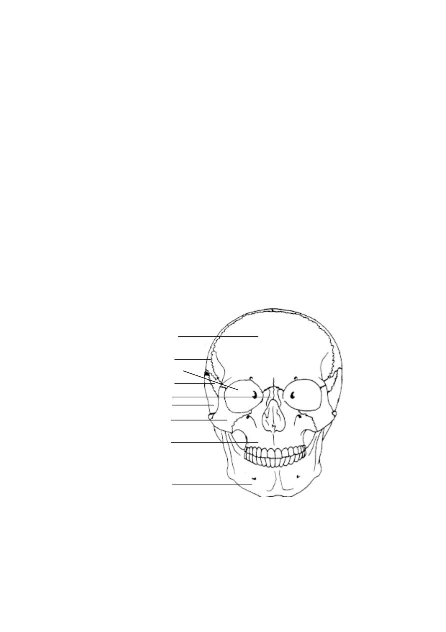
111
Dried, prepared bones are used to study skeletal anatomy. The bones
are named according to their position in the body. The named bones are di-
vided into two categories: (1) the axial skeleton and (2) the appendicular
skeleton. The axial skeleton consists of the skull, hyoid bone, vertebral col-
umn and thoracic cage. The appendicular skeleton consists of the limbs and
their girdles. In human body, there are 206 bones, of these 80 are in the axial
skeleton, 126 in the appendicular skeleton. Among the bones of the axial
skeleton 28 bones are in the skull, 26 bones in the vertebral column, 25
bones in the thoracic cage and one remains as the hyoid bone. (details as
found below)
Axial skeleton
- It forms the upright axis of the body. It protects the brain,
the spinal cord and the vital organs found within the thorax.
a) Skull - The human cranial capacity is about 1500 cm
3
. It consists of 22
bones. It protects the brain. It supports the organs of vision, hearing, smell
and taste. The lower jaw or mandible remains specially attached to the skull.
The skull or cranium is covered by eight bones. They are one pair each of
parietal and temporal, individual bones as frontal, sphenoid, occipital and
ethmoid. These bones are joined by sutures to form a compact box like struc-
ture. The sutures are immovable joints.
Fig.3.2.3. Skull
temporal bone
mandible
maxilla
zygomatic bone
nasal bone
sphenoid bone
parietal bone
frontal bone
eye orbit
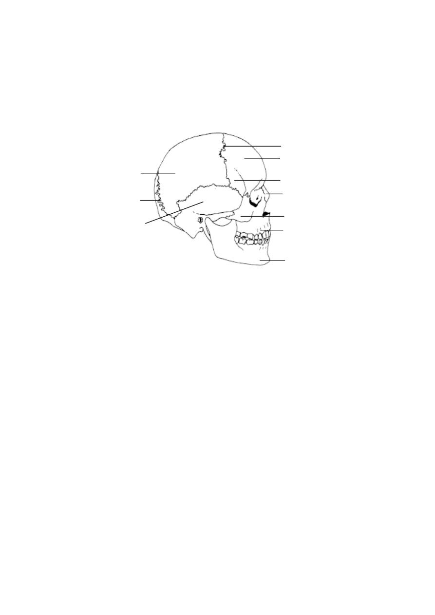
112
In the front there are 14 facial bones. Of these maxilla, zygomatic,
palatine, lacrymal, nasal and inferior nasal koncha remain as pairs.
Mandible or lower jaw and vomer are unpaired bones .
The parietal and occipital bones are major bones on the posterior
side of the skull. The parietal bones are joined to the occipital bone at the
back. The side of the head is formed of the parietal and the temporal bones.
The large hole in the temporal bone is the external auditory meatus. This
opening is meant for transmitting sound waves towards the eardrum. On the
lateral side immediately anterior to the temporal, the sphenoid bone is seen.
Anterior to the sphenoid bone is the zygomatic bone or cheek bone. It is a
prominent bone on the face. The upper jaw is formed of the maxilla. The
mandible constitutes the lower jaw.
The major bones seen from the frontal view are the frontal bone,
zygomatic bone the maxillae and the mandible. The most prominent open-
ings in the skull are the orbits and the nasal cavity. The two orbits are meant
for accommodating the eyes. The bones of the orbits provide protection for
the eyes and attachment points for the muscles that move the eyes. The bones
forming the oribits are the frontal, sphenoid, zygomatic, maxilla, lacrymal,
ethmoid and palatine. The head region also contains 6 ear ossicles. They
are Maleus (2), incus (2) and stapes (2).
parietal bone
temporal bone
occipital bone
Fig.3.2.4. Skull - Lateral view
suture
frontal bone
sphenoid bone
nasal bone
zygomatic bone
maxilla
mandible
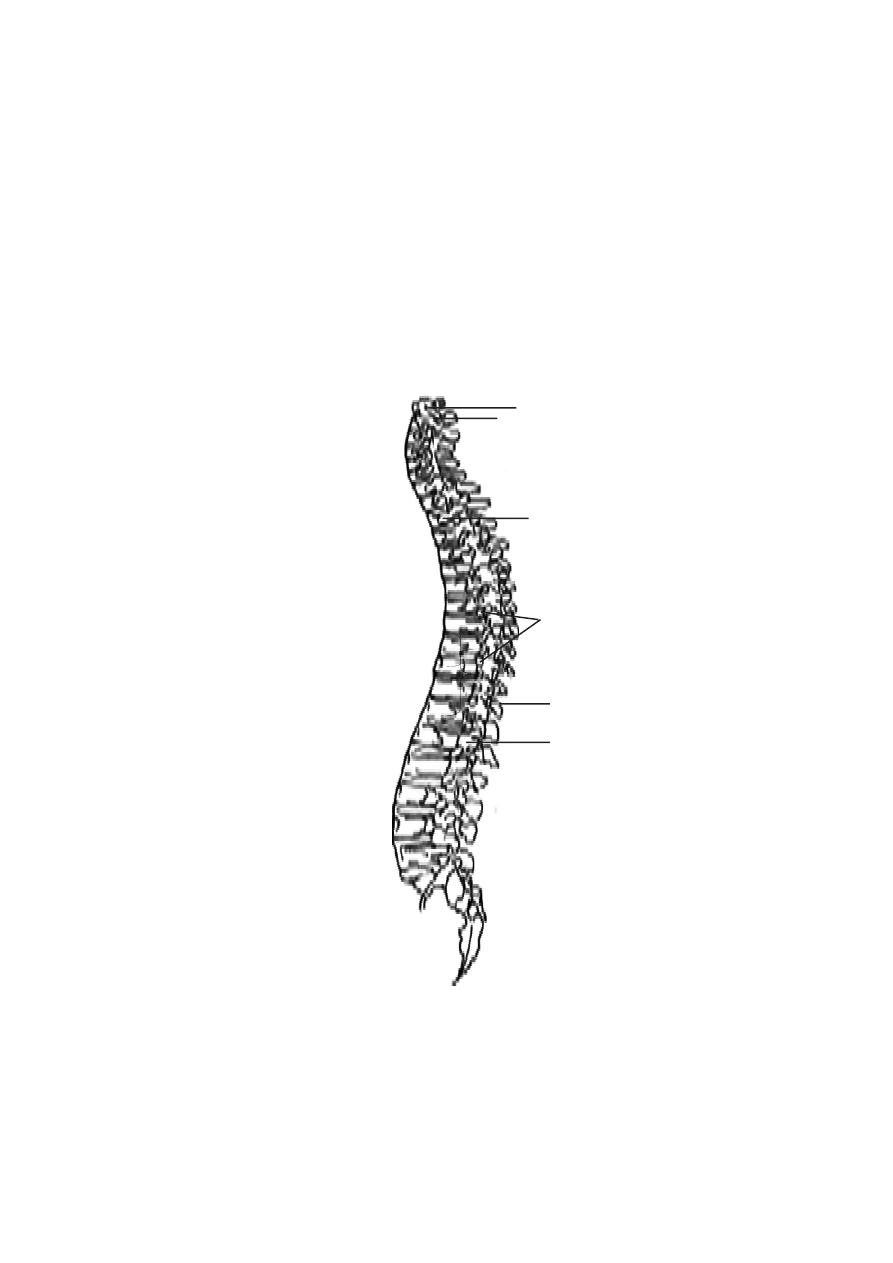
113
A large opening found at the base of the skull is the foramen
magnum. Through this opening the medulla oblongata of the brain descends
down as the spinal cord.
b). Vertebrae - The vertebrae make up the slighty S-shaped vertebral
column or backbone. The vertebral column consists of 26 bones. They are
divided into 5 regions. They are the cervical (7), thoracic (12), lumbar (5),
sacral (1) and coccygeal (1) vertebrae.
Fig.3.2.5. Vertebral column
Cervical region
Thoracic region
Lumbar region
Sacral and coccygeal region
first cervical vertebra (atlas)
intervertebral disc
intervertebral foramina
spinous process
sacrum
transverse process
coccyx
second cervical vertebra (axis)
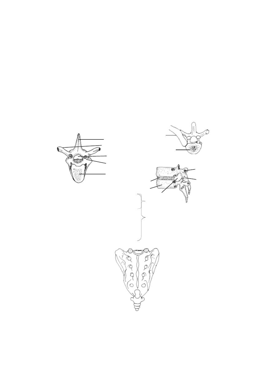
114
Vertrebra - Structure : The main load - bearing portion of a vertebra is a
solid disc of bone called the centrum. The centra of adjacent vertebrae are
separated by intervertebral discs of cartilage. Projecting from the centrum
dorsally is a vertebral arch. It encloses the neural canal. This canal contains
the spinal cord. Several bony projections are seen on the vertebral arch. On
each side of the centrum ther are two transverse processes. On the dorsal
side there is a neural spine. These bony projections are used for attachment
of muscles. Further, there are two superior and two inferior processes meant
for articulation with the neighbouring vertebra.
The first cervical vertebra is the atlas. It balances and supports the
head. It has no centrum. The second is the axis. The sacral vertebrae are
fused. They form a triangular structure called the sacrum. The coccygeal
vertelera has no function. It is a vestige. During development in the embryonic
stage there are nearly 34 vertebrae present. Of these, 5 sacral bones are
fused to form a single sacral bone. 4 or 5 coccygeal bones are fused to form
a single coccyx.
Fig.3.2.7. Sacrum and coccyx
coccyx
sacrum
Fig.3.2.6. Vertebra
body
rib
transverse
process
s p i n o u s
process
intervertebral foramen
intervertebral body
transverse process
vertebral foramen
vertebral arch
spinous process
body(centrum)
body(centrum)
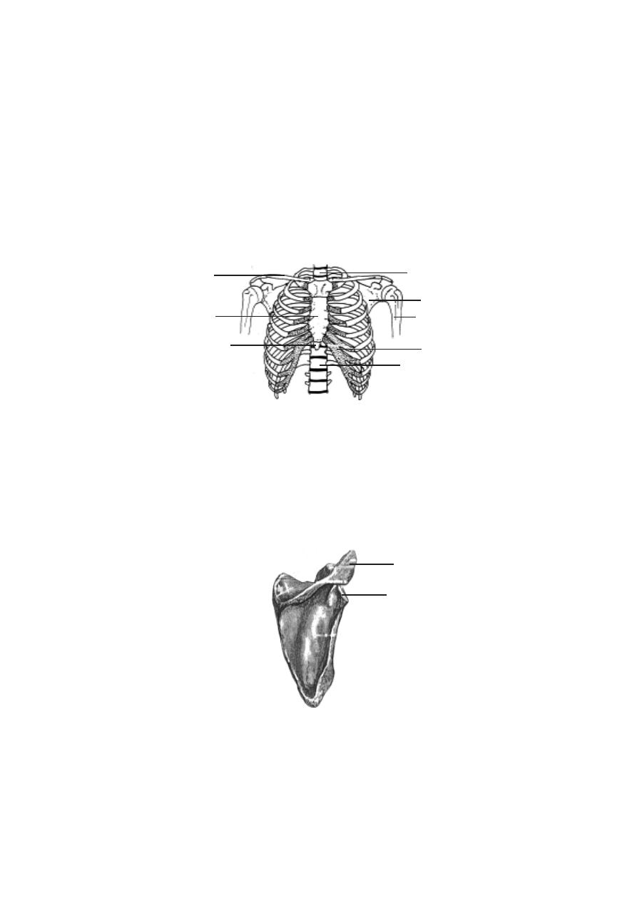
115
c). Rib cage - There are 12 pairs of ribs. Each articulates with a thoracic
vertebra. In the front, the first ten pairs are attached to the sternum (breast
bone) by costal cartilages. The first seven are attached directly to the ster-
num. They are called the true ribs. Cartilages of 8
th
, 9
th
and 10
th
ribs are
fused and attached to 7
th
. They are called the false ribs. 11
th
12
th
pairs are
not attached to the sternum. They are called floating ribs.
Appendicular skeleton
It consists of the bones of the upper and lower limbs and the girdles
by which they are attached to the body.
Pectoral girdle - The hands are attached to the pectoral girdle. Both of them
are attached loosely by muscles to the body. This arrangement facilitates free-
dom of movement. Hence it is possible to place the hand in a wide range of
positions.
Fig.3.2.9. Scapula
acromion process
glenoid fossa
Fig.3.2.8. Rib cage
xiphoid process
clavicle
1
st
thoracic vertebra
scapula
humerus
costal cartilage
12
th
thoracic vertebra
sternum
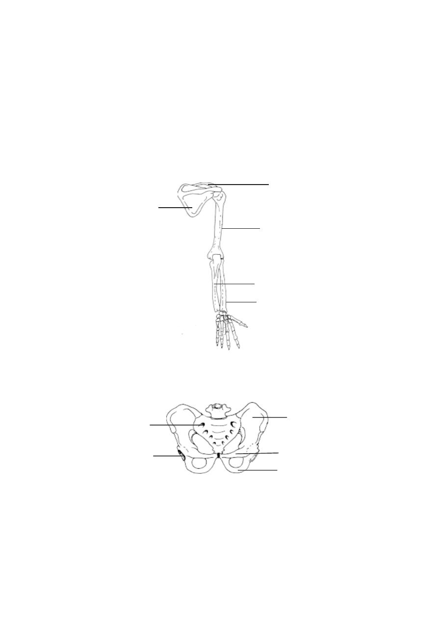
116
The pectoral or shoulder girdle consists of two pairs of bones. Each
pair has a scapula or shoulder blade and a clavicle or collarbone. The scapula
is a flat, triangular bone. A glenoid fossa is located in the superior lateral
region of the scapula. It articulates with the head of the humerus. The clavicle
is a long bone. It has a slight S-shaped curve. It can be easily seen and felt.
The clavicle holds the upper limb away from the body.
Pelvic girdle or pelvis
- It is a ring of bones formed by the sacrum and
paired bones called the coxae or hip bones.
ulna
radius
humerus
scapula
clavicle
carpals
metacarpals
phalanges
Fig.3.2.10. Skeleton of the arm
Fig.3.2.11. Pelvis
sacrum
acetabulum
ilium
pubis
ischium
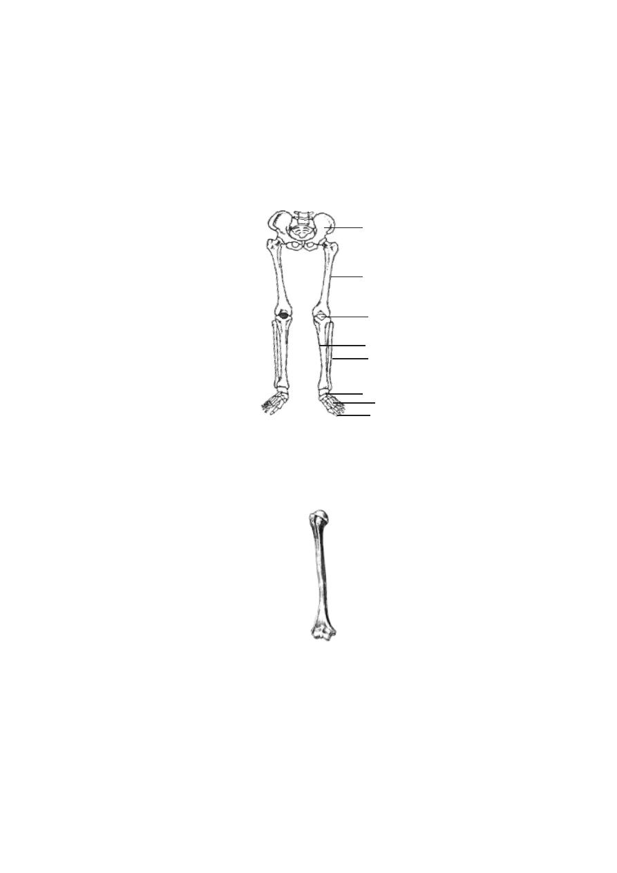
117
Each coxa is formed by the fusion of three bones, namely ilium,
ischium and pubis. A fossa called the acetabulum is located on the lateral
surface of each coxa. The acetabulum is meant for the articulation of the
lower limbs.
Upper limb or hand
- The part of the upper limb from shoulder to the elbow
is the arm. It contains one long bone called the humerus. The head of hu-
merus articulates with the glenoid fossa of the scapula. The distal end of the
bone articulates with the two forearm bones.
Forearm
- This part of the hand is in between the arm and the wrist. The
forearm has two bones. They are the ulna and the radius. While the ulna is
on the side of the little finger, the radius is on the lateral or thumb side of the
forearm.
Fig.3.2.12. Skeleton of the leg wih pelvic girdle
pelvis
femur
patella
tibia
fibula
tarsals
metatarsals
phalanges
Fig.3.2.13. Femur
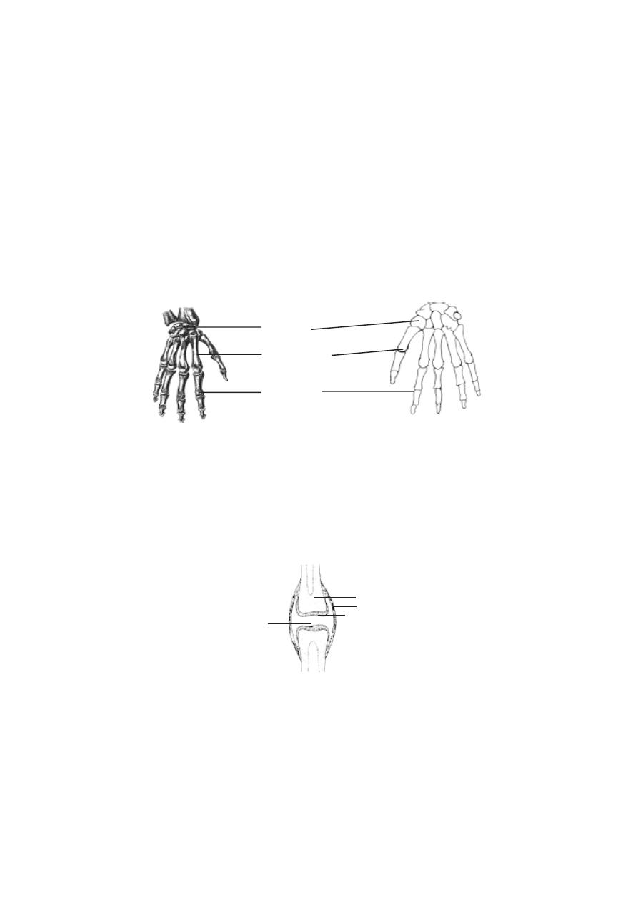
118
Wrist
- This short region is composed of eight carpal bones. These are
arranaged into two rows of four each. The carpals along with accompanying
ligaments are arranged in such a way that a tunnel on the anterior surface of
the wrist called the carpal tunnel has been formed. Tendons, nerves and
blood vessels pass through this tunnel to enter the hand.
Hand
- The bony framework of the hand is formed of five metacarpals.
They are attached to the carpals in the wrist. The concave nature of the palm
in the resting position is due to curved arrangement of metacarpals.
Each hand has five digits. They include one thumb and four fingers.
Each digit has small long bones called phalanges. While the thumb has two
phalanges other fingers have three each.
Lower limb
or Leg : The general pattern of the lower limb is similar to that of
the upper limb.
Fig.3.2.14. Hand
carpals
metacarpals
phalanges
Fig.3.2.15. A Synovial joint
bone
fibrous capsule
articular cartilage
synovial fluid

119
The upper region of the leg is the thigh. It contains a single longest
bone called the femur. It has a prominent rounded head for articulating with
the acetabulum of the pelvic girdle. The distal end of the femur has two condyles
for articulation with the tibia.
The knee region has a large, flat bone called the patella. It articulates
with the patellar groove of the femur.
Leg
- The leg is that part of the lower limb between the knee and the ankle. It
consists of two bones namely, the tibia and the fibula. The tibia is larger and
it supports most of the weight of the leg.
Ankle
: The ankle consists of seven tarsal bones. The ankle articulates with
the tibia and the fibula through the talus.
Foot
: It is formed of metatarsals and phalanges. They correspond to the
metacarpals and phalanges of the hand.
Joint
All bodily movements are caused by muscles. Our skeletal muscles
are firmly attached to bones. Movements involving such muscles cause pull
on our bones. Hence movements need movable bone joints.
A joint or an articulation is a place where two bones come together.
All joints are not movable. Many joints allow only limited movements.
The joints are named according to the bones that are united.
Kinds of joints
- There are three major kinds of joints. They are the fibrous,
cartilaginous and synovial joints.
Fibrous joints
- In this type, the joints are united by fibrous connective tis-
sue. There is no joint cavity. These joints show little or no movement. Sutures
formed between cranial bones, a syndesmosis (to bind) between radius and
ulna are examples for this type.
Cartilaginous joints
- These joints unite two bones by means of either hya-
line cartilage (synchondroses) or fibrocartilage (symphyses). The articula-
tion between the first rib and the sternum is an example for syncondrosis.
Symphysis pubis and intervertebral discs are examples for symphyses.
Synovial joints
- These joints contain a synovial fluid. This fluid is a com-
plex mixture of polysaccharides, proteins, fats and cells. It forms a thin lubri-
cating film covering the surfaces of a joint. Elbow and knee joints are of this
type.

120
3.3. Muscular System
Locomotion and bodily movements are characteristic features of the
animals. The movements are effected by various cell organelles such as cilia,
flagella and organs like muscles. Muscular movements are more powerful and
energetic. The skeletal muscles apart from their role in smarter movements,
provide beautiful shapes to the body. The inner smooth muscles of the visceral
organs make them work like machines all through the life period. The muscle
cells function like small motors to produce the forces responsible for the move-
ment of the arms, legs, heart and other part of the body. Thus the highly spe-
cialized muscle tissues are responsible for the mechanical processes in the
body.
Based on structure, functioning and occurrence three different types
of muscle tissues have been identified. They are the skeletal, visceral and
cardiac muscles.
1).
Skeletal muscles or striped muscles
: These muscles are attached to
the bones. The muscle cells are long and cylindrical. These voluntary muscles
cause body movements.
2).
Visceral muscles or Nonstriated muscles
: These are found in the walls
of the inner organs such as blood vessels, stomach and intestine. The muscle
cells are spindle shaped. These are involuntary in nature.
3).
Cardiac muscle
: These are found in the wall of the heart. The muscle
cells are cylindrical and branched. The muscles are involuntary in nature.
Skeletal muscles.
The skeletal muscles are attached to bones by tendons. The tendons
help to transfer the forces developed by skeletal muscles to the bones. These
muscles are covered by sheets of connective tissue called fascia.
Tendons
: These are connective tissue structures showing slight elasticity.
They are like cords or straps strongly attached to bones. The tensile strength
of tendons is nearly half that of steel. A tendon having 10 mm diameter can
support 600 - 1000 kg.
Fascia
: These are assemblages of connective tissues lining skeletal muscles
as membranous sheets. The fascia may be superficial or deep. The superficial
fascia is a layer of loose connective tissue found in between skin and muscles.
The deep fascia are collagen fibres found as a tough inelastic sheath around
the musculature. They run between groups of muscles and connect with the
bones.

121
Shapes of muscles.
There is a wide variety of shapes and sizes in muscles. Based on
general shape and the orientation or muscle fibres in relation to the direction
of pull, they can be grouped into two classes.
1. Parallel
: These muscle fibres are parallel to the line of pull. The muscles
may be flat, short, quadrilateral or long and strap like. The individual fibres run
the entire length of the muscle.
2. Oblique
: These muscle fibres are oblique to the line of pull. The muscles
may be triangular or pennate (feather-like). The pennate forms may be
unipennate, bipennate, multipennate or circumpennate. Some muscles have a
spiral or twisted arrangement.
Naming of muscles.
The muscles are named according to their size, shape, position and
action.
Shape
Size
deltoid
-
triangular
major
-
large
quadratus -
square
minor
-
small
gracilis
-
slender
longus -
long
lattismus - broadest
Number of Heads
Position
biceps - 2 heads
dorsi - of the back
triceps - 3 heads
pectoralis - of the chest
quandriceps - 4 heads
brachii - of the arm
anterior, posterior.
Depth
Action
superficialis - superficial
extensor
internus - internal, flexor
constrictor
profundus - deep
Flexor
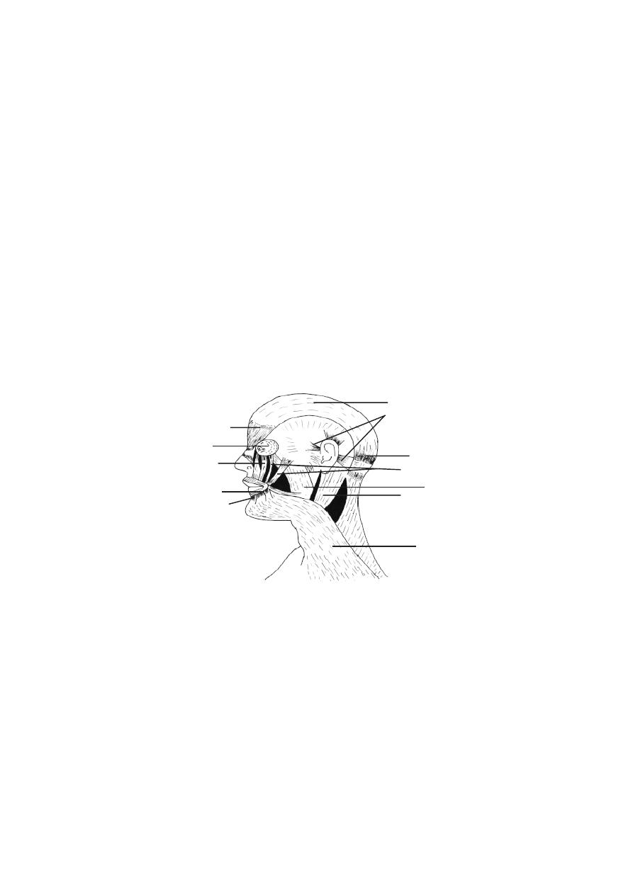
122
Distribution of muscles
I. Muscles of the head
There are two groups of muscles. They are craniofacial and masti-
catory muscles. The craniofacial muscles are related to eye orbital margins,
eyelids, nose, nostrils, lips, cheeks, mouth, pinna, and the scalp. These muscles
are also known as muscles of facial expression. Among these muscles those
that are related to the lip movement are significant. The facial expression is
mostly due to lip movement and positioning of the lips. Such thought related
movements are caused by several muscles associated with lips and the skin
around the mouth. Since orbicularis oris and buccinator muscles provide
lip movement for kissing posture they are known as “kissing muscles”. Smil-
ing is accomplished by zygomasticus major and minor, levator anguli oris
and risorius. The muscles of the lips can also provide expressions such as
sneering and frowning. The chin dimples are located between the mentalis
muscles
The masticatory (or speech) muscles move the mandible of the
lower jaw. The muscles responsible for this movement are masseter
temporalis and pterygoid. Tongue movements are caused by intrinsic and
extrinsic muscles. Swallowing of food is facilitated by several muscles re-
lated to the mouth, roof of pharynx, uvula and other regions.
II. Muscles of the Neck region.
The movements of the neck region are caused by cervical, suprahy-
oid, infrahyoid and vertebral muscles.
Fig.3.3.1. Muscles of the Head
orbicularis oculi
orbicularis oris
masseter
pars frontalis
levator labii
mentalis
aponeurosis
auricular muscles
pars occipitalis
zygomaticus muscles
sternocleidomastoid
platysma
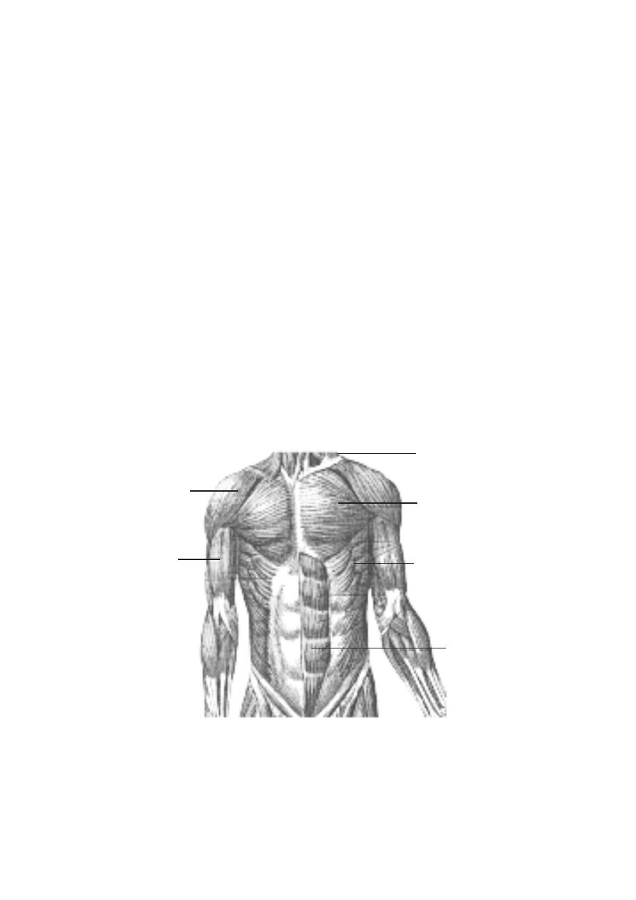
123
III. Muscles of the Trunk region.
The muscles of the vertebral column help to bend and rotate the body.
These are strong back muscles that help the trunk to maintain erect posture.
The most prominent muscles of this region are the erector spinae, longissi-
mus and spinalis.
Four important thoracic muscle groups are associated with the pro-
cess of breathing. While the process of inspiration is due to scalene and ex-
ternal intercostal muscles, the expiration is performed due to internal
intercostals and transverse thoracis. Major breathing movement is due to
diaphragm, a curved musculofibrous sheet that separates thoracic and ab-
dominal cavities.
Abdominal muscles can aid in forced expiration, vomitting, defaecation,
urination and childbirth.
The inferior opening of the pelvic bone is covered by pelvic dia-
phragm muscles. Below these muscles perineum is pressent. The perineum
and other “subfloor” muscles form the urogenital diaphragm. Pelvic and
urogenital diaphragm may get strecthed in pregnancy due to weight of the
foetus. However by specific exercises they can be strengthened.
Fig.3.3.2. Muscles of the Trunk
trapezius
pectoralis major
serratus anterior
deltoid
biceps
abdominis
brachii

124
IV. Muscles of the upper limb.
The hands are attached to the pectoral girdle and to the vertebral
column by large conspicuous muscles such as trapezius, rhomboid major
and minor, levator scapulae and lattissimus dorsi.
The trapezius is a flat, triangular muscle. It extends over the back of
the neck and upper thorax. It maintains the level and poise of the shoulder. It
helps to rotate the scapula forward, so that the arm can be raised above the
head. It helps to bend the neck backwards and laterally.
Latissimus dorsi is a large flat triangular muscle. It is a conspicu-
ous muscle stretching over the lumbar region and lower thorax. This muscle is
useful in adduction, extension and medial rotation of the humerus. It helps in
the backward swinging of the arm. By raising the arm above the head it helps
to pull the trunk upwards and forwards. It is useful in violent expiratory activi-
ties such as coughing or sneezing. It helps in deep inspiration.
Serratus anterior and pectoralis major connect the ribs to the
scapula. Pectoralis major extends from the upper thorax and abdomen to act
on the humerus. It is a fan shaped muscle. It spreads between the clavicle and
the 7th costal cartilage in the front of the chest. It helps to swing the extended
arm forward and medially. It helps in climbing. It is active in deep inspiration.
The muscles of the upper arm are the coracobrachialis, biceps,
triceps and brachialis. The coracobrachialis arises from the coracoid bone
in the shoulder and ends in the humerus of the upper arm. It helps to move the
arm forward and medially. The biceps brachii is a large fusiform muscle. It
has two proximal heads for attachement. They are connected to the coracoid
and shoulder joint. The lower head ends in the radius of the lower arm. It is a
powerful muscle causing flexing of the hand. The triceps arises by three
heads from scapula and upper part of humerus on the posterior side. The
wrist, hand and finger movements are caused by several extrinsic and intrin-
sic hand muscles. A detailed study of them could be made in higher classes.
V. Muscles of the lower limb.
Thigh movements are caused by anterior, postereolateral and deep
muscles. The anterior muscles are the iliacus and psoas major which help to
flex the thigh. The gluteus maximus form the mass of the buttocks region.
Leg movement is caused by the anterior thigh muscles, quadriceps femoris
and sartorius. The sartorius is the longest muscle of the body. It runs from
the hip to the knee. Muscle movement of ankle foot and toe are caused by
several groups of extrinsic and intrinsic muscles. A detailed study of them
could be made in higher classes.
rectus
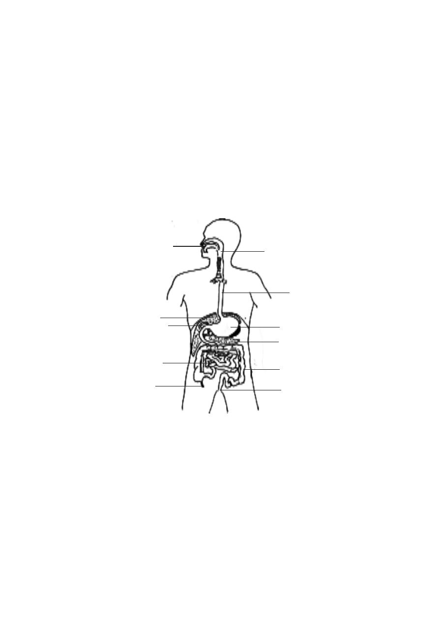
125
3. 4. Digestive system
The process of nutrition in man is holozoic. In this type of nutrition,
the nutrients are made available to the body through digestion of food. In
digestion, the macromolecules or biopolymers of food are hydrolysed to yield
their corresponding monomers. This process is facilitated by enzymes. Thus
the digestive system comprises the alimentary canal, associated glands and
regions of absorption of food. The digestive tract comprises various organs
from mouth to anus.
Mouth or Buccal cavity
The opening of the oral cavity is bounded by the lips. The lips are
muscular folds covered internally by mucosa. The lateral walls of the oral
cavity are the cheeks. The inner walls of the cheeks are lined by moist strati-
fied squamous epithelium. The lips and cheeks are useful in the process of
mastication and speech.
Fig.3.4.1. Digestive system
oesophagus
stomach
pancreas
large intestine
anus
small intestine
appendix
liver
pharynx
gall bladder
oral cavity (mouth)
126
Tongue
- It is a large muscular organ. It is attached to the floor of the oral
cavity. The anerior part of the tongue is free. A thin fold of tissue called the
frenulum attaches the free end to the floor of the mouth. The tongue is di-
vided into two parts by a groove called the terminal sulcus. About two thirds
of the anterior surface is covered by papillae. Some of them contain taste
buds.
Teeth
- There are 32 teeth in the mouth of a human adult. These are called as
permanent teeth. There are 4 different types of permanent teeth seen. This
nature is known as heterodontism. The types of teeth are incisors(8), ca-
nines(4), premolars(8) and molars(12). Since the teeth in the right and left
side of the mouth are mirror images of each other, the dental arrangement is
represented as follows.
i 2/2 : c 1/1 : pm 2/2 m 3/3 X 2 or
×
Each tooth consists of three regions. These are the upper crown,
middle neck and basal root regions. The crown region has one or more cusps.
The tooth is made up of a calcified tissue called dentine.
The dentine of the tooth crown is covered by an extremely hard sub-
stance called enamel. The surface of the dentine in the root is covered with a
bonelike substance called cementum. It helps to anchor the tooth in the jaw.
In the centre of the tooth there is a pulp cavity. This cavity is also called as
the root canal. This canal contains blood vessels and nerves. The canal opens
at the base through apical foramen.
Fig.3.4.2. Dentition
upper jaw
lower jaw
incisor
canine
I premolar
II premolar
I molar
II molar
III molar
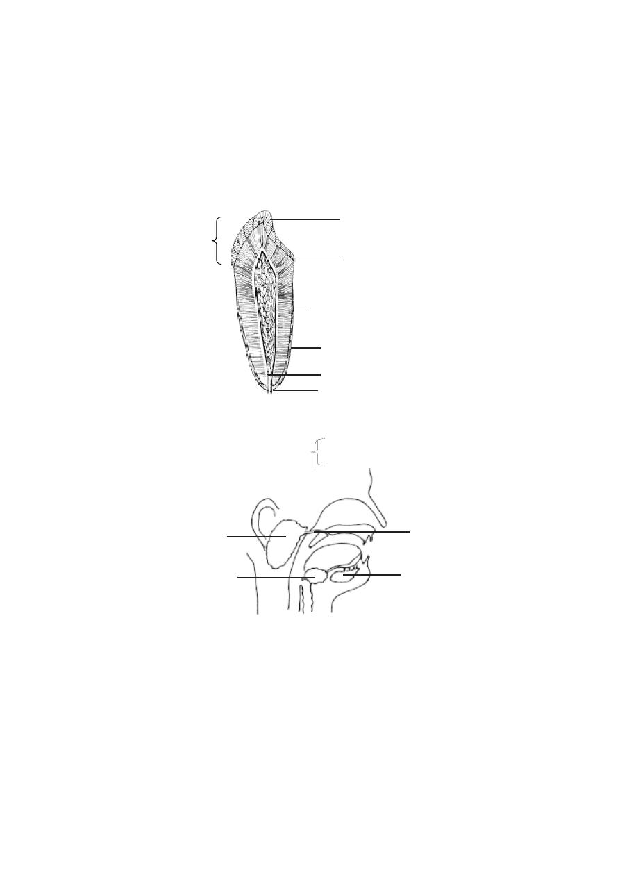
127
The teeth are set in sockets along the edges of the upper and lower
jaws. This region of the jaw is covered by dense fibrous connective tissue and
stratified squamous epithelium. It is called as the gingiva.
dentine
pulp cavity with nerve and
blood vessels
root canal
apical foramen
Fig.3.4.3. A tooth
crown
neck
root
enamel
cementum
Salivary glands
: These are scattered throughout the oral cavity. Three pairs
of glands are larger. They are the parotid, submandibular and sublingual
glands. The Parotid glands are the largest. They are located just anterior to
Fig.3.4.4. Salivary glands
parotid gland
submandibular gland
sublingual gland
parotid duct
the ear on each side of the head. The submandibular glands are found on
the inferior borders of the mandible. The sublingual glands are the smallest.
They lie immediatly below the mucous membrane in the floor of the mouth.
There are other numerous small, coiled, tubular glands in the mouth. They are
the lingual (tongue), palatine(palate), buccal and labial(lips) glands.
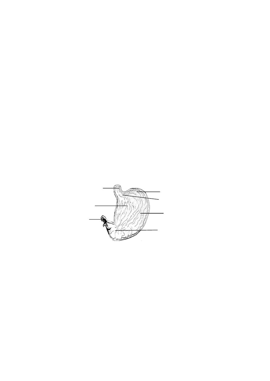
128
Pharynx
: A description of pharynx is provided under respiratory system.
Oesophagus
: This part of the digestive tube extends between the pharynx
and the stomach. It is about 25 cm long. It lies in the mediastinum of the
thorax, anterior to the vertebra and posterior to the trachea. It passes through
the diaphragm and ends at the stomach. The oesphagus has thick walls. The
inner wall is lined by a moist stratified squamous epithelium. The upper and
lower ends of this tube have sphincters to regulate the movements of mate-
rials.
Stomach
: It is an enlarged sac like structure. This sac found in the upper part
of the abdomen is horizontally placed. It is divisible into two regions namely
cardiac and pyloric stomachs. The cardiac stomach is towards the left of the
abdomen. The oesophagus opens into the cardiac stomach through the gas-
troesophageal or cardiac opening. A part of the stomach to the left of the
cardiac region is the fundus. The largest part of the stomach is the body. The
body narrows to form the pyloric region. The pyloric opening between the
pylorus and intestine is surrounded by a ring of muscles called the pyloric
sphincter.
Small intestine
: It consists of three parts: the duodenum, the jejunum and
the ileum. The entire small intestine is about 6m. long.
Duodenum
- It is about 25 cm. long. It curves within the abdominal cavity
and completes nearly 180 degree arc. Liver and pancreas are associated
with the duodenum.
Jejunum and ileum
- The jejunum and ileum are 2.5m. and 3.5m in length
respectively. These two are similar in structure to the duodenum. However
Fig.3.4.5. Section of the stomach
cardiac region
pyloric opening
fundus
oesophagus
gastroesophageal opening
pyloric region
body
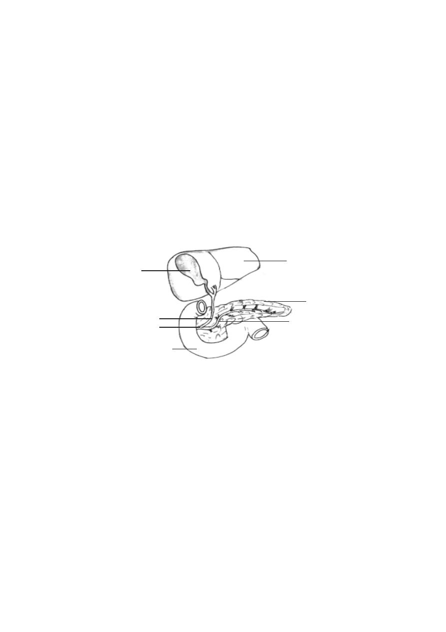
129
there is a gradual decrease in the diameter of the small intestine. The junction
between the ileum and the large intestine is the ileocaecal junction. It has a
ring of smooth muscles forming a sphincter, and a one way ileocaecal valve.
Liver
: It is the largest visceral organ. It weighs about 1.36 Kg. The liver
consists of two major left and right lobes, and two minor lobes caudate and
quadrate. The bile secreted by the liver gets collected in the gall bladder.
There are two hepatic ducts and they unite to form a single duct. The com-
mon hepatic duct is joined by the cystic duct from the gall bladder to form the
common bile duct. It empties into the duodenum.
Gall bladder :
It is a sac like structure on the inferior surface of the liver. It
is about 8 cm long and 4 cm wide.
Pancreas :
It is a compelx organ. It is composed of both endocrine and exo-
crine tissues. The endocrine part of the pancreas consists of pancreatic is-
lets. They produce insulin and glucagon. The exocrine part of the pancreas
consists of acini. They produce digestive enzymes. The pancreas consists of
several lobules. The ducts from the lobules unite to form the pancreatic
duct. It joins the common bile duct at the hepatopancreatic ampulla.
Large intestine.
Caecum :
It is the proximal end of the large intestine. At this region large, and
small intestines meet. The caecum is about 6 cm in length. Attached to the
caecum is a small blind tube about 9 cm long. It is called the vermiform
appendix.
Fig.3.4.6. Duodenal region
liver
gall bladder
pancreas
common bile duct
pancreatic duct
hepatopancreatic
ampulla
duodenum
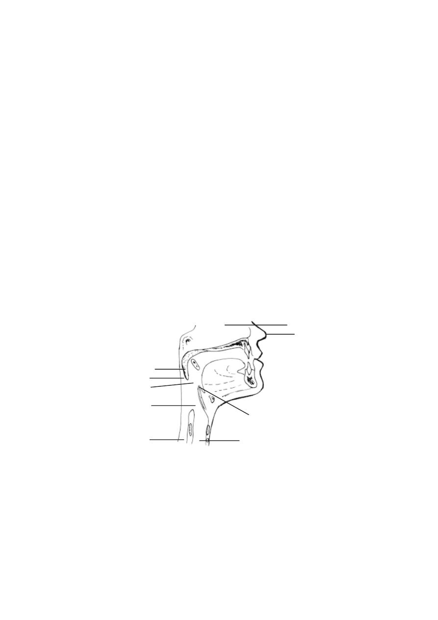
130
Colon :
It is about 1.5 - 1.8 m long. It consists of four parts namely the
ascending colon, transverse colon, descending colon and sigmoid co-
lon. The sigmoid colon extends into the pelvis and ends at the rectum.
Rectum
: It is a straight, muscular tube that extends from the sigmoid colon to
the anal canal.
Anal canal
: It is the last 2cm of the digestive tract. It ends at the anus. The
canal has two sphincters, namely the internal anal sphincter and the external
anal sphincter.
3. 5 Respiratory system
The process of respiration involves movement of air in and out of the
lungs, gas exchange between air in lungs and the blood, transport of O
2
and
CO
2
. These processes are facilitated by working together of well developed
respiratory organs and the circulatory system.
The respiratory organs include nasal cavity, pharynx, larynx, tra-
chea, bronchi and lungs. These organs are organised into upper and lower
respiratory tracts.
Upper respiratory tract.
1. Nasal cavity
: The nasal cavity follows the external nose. The nose is a
visible prominent structure. Internally it is supported by cartilage plates. The
bridge of the nose is formed of the nasal bones and extension of the skull
Fig.3.5.1. Upper Respiratory tract
nasal cavity
external nose
uvula
nasopharnyx
epiglottis
oropharynx
oesophagus
laryngopharynx
trachea
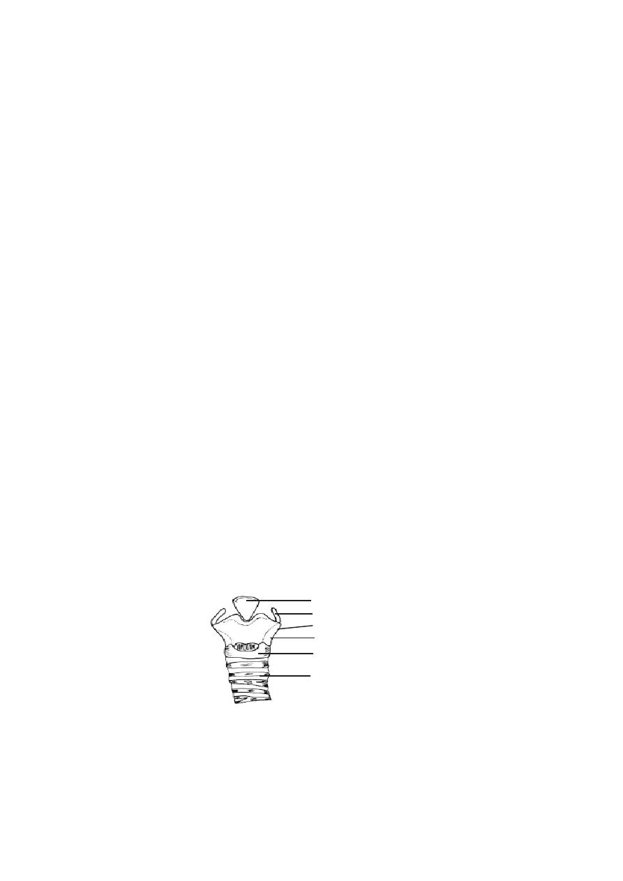
131
bones(frontal and maxillary). The respiratary passage is divided into two cham-
bers by a median partition. The nasal passage opens to the outside through
external nostrils. It opens inside by internal nostrils at the pharynx.
2. Pharynx
: The buccal cavity and the nasal passage open into the pharynx.
It is a common pathway that opens into the oesophagus of the alimentary
canal and larynx of the respiratory system.
The pharynx is divided into three regions, namely the nasopharynx,
the oropharynx and the laryngopharynx.
The nasopharynx extends from the internal nostril to the region of
the uvula. The uvula is a soft outgrowth hanging in between the posterior part
of the oral cavity and the pharynx. It prevents the entry of food into the nasal
cavity. The wall of the nasopharynx is lined by ciliated columnar epithilium.
The middle ear opens into the nasopharynx through two auditory tubes. This
arrangement is meant for equalizing the air pressure between the atmosphere
and the middle ear. The inner surface of the nasopharynx also contains the
pharyngeal tonsil or adenoid meant for defence against infections. An
enlargement of the tonsil can interfere with breathing.
The oropharynx remains between the uvula and the epiglottis. The
oral cavity opens into the oropharynx. Near the opening of the oral cavity 2
sets of palatine tonsils and lingual tonsils are present.
The laryngopharynx extends in between the epiglottis and the
oesophagus.
3. Larynx
: The larynx is seen just behind the pharynx and the buccal cavity.
This region is surrounded by cartilages(3 unpaired and 6 paired). These are
interconnected by muscles and ligaments.
The unpaired cartilages are the thyroid, cricoid and epiglottis. The
thyroid cartilage is the largest. It is also known as the Adam’s apple.
epiglottis
cricoid cartilage
cartilage rings
hyoid bone
thyrohyoid membrane
thyroid cartilage
Fig.3.5.2. Larynx

132
The cricoid cartilage forms the base of the larynx. The other cartilages are
placed above the cricoid. The epiglottis is attached to the thyroid. It projects
as a free flap over the opening of the larynx. It prevents food particles from
saentering into the tracheal tube.
The ligaments inside the larynx form the vocal folds or vocal cords.
The vocal cords and the openings between them are called the glottis. The
vocal cords are involved with sound production. The air moving past the vocal
cords make them to vibrate. Louder sounds are made by increasing the ampli-
tude of vibrations. Frequency of the vibrations can be altered by changing the
length of the vibrating segments of the vocal cords. The length is altered by
muscles attached to the cartilage. Males usually have longer vocal cords than
females. The sound made by the vocal cords can be altered by the tongue, lips
and teeth to form words.
4. Trachea (or wind pipe)
: It is a membranous tube. The wall is made up of
connective tissue and smooth muscles. The wall is provided support by 15-20
‘C’ shaped cartilage rings. They protect the trachea and keep it open all the
time.
The inner wall of the trachea is lined by mucous membrane. It con-
sists of ciliated columnar epithelium. The cilia of this epithelium help to propel
mucus and foreign particles towards the larynx.
The length of the trachea is 10-12 cm. Its inner diameter is 12 mm.
The trachea extends from the larynx to the level of the 5th thoracic vertebra.
The basal part of the trachea divides to form 2 smaller tubes called the pri-
mary bronchi (sing : bronchus). The cartilage ring found at the basal region is
called the carina. Foreign objects reaching carina stimulate a powerful cough
reflex.
4. Lungs
: The pair of lungs are the actual organs of respiration. Each lung is
conical in shape. The base of the lung rests on the diaphragm. The right lung
is larger than the left and it weighs around 620g. The left lung weighs 560g.
The right lung has three lobes and the left lung has two.
The lungs are placed within the thoracic cavity. Each lung is sur-
rounded by separate pleural membrane. The region inside the pleural mem-
brane is named as the pleural cavity. This cavity is filled with pleural fluid.
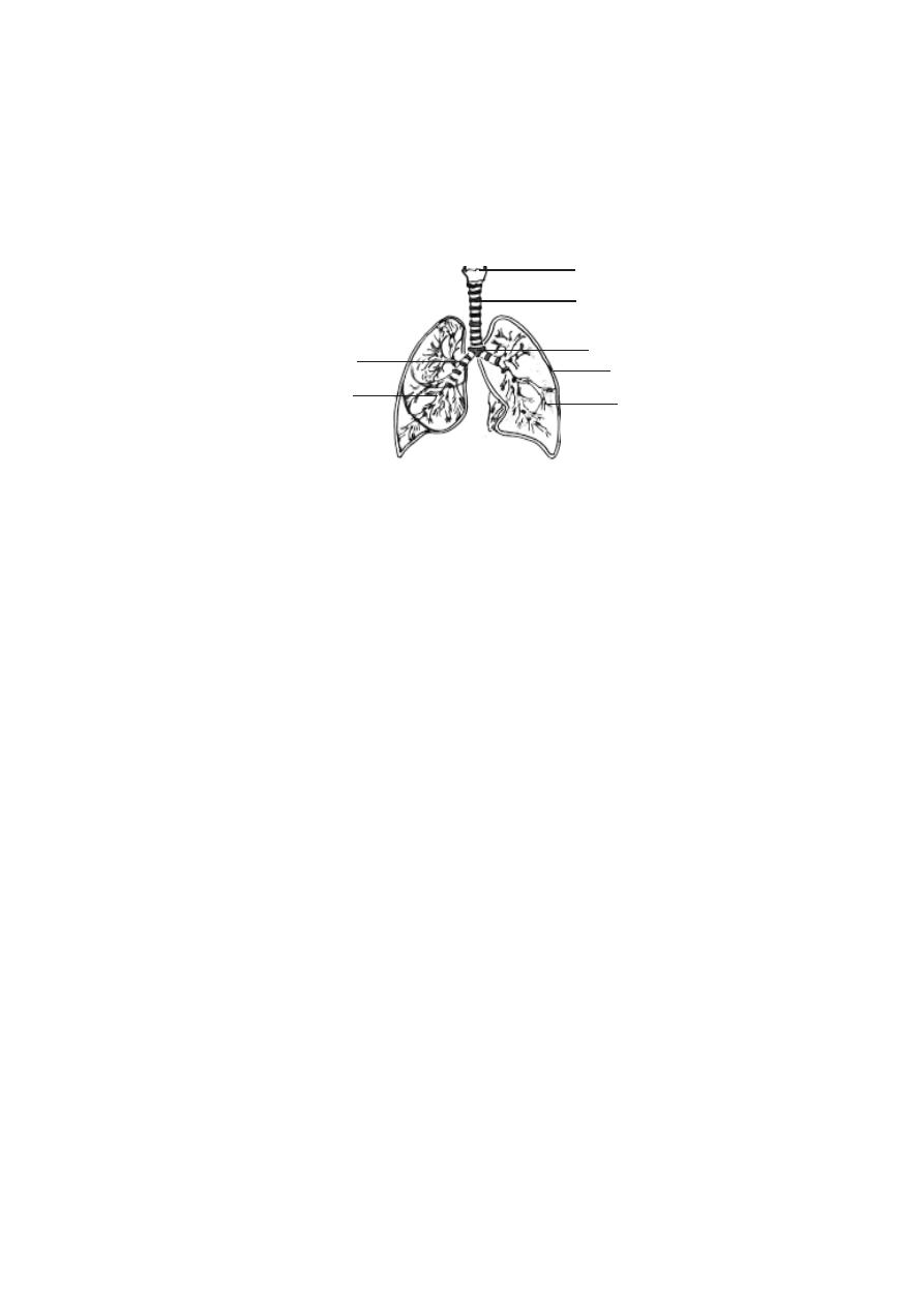
133
The region in between the two lungs is named as the mediastinum.
It is a midline partition, being occupied by the heart, trachea and oesophagus.
Structures such as the primary bronchi, blood vessels, nerves and
lympatic vessels enter or exit the lungs at a specific region on the inner margin
of the lungs. This region is known as the hilum. All structures passing through
the hilum are referred to as the root of the lung.
The primary bronchi on entering into each lung divide further into
secondary bronchi. There are two secondary bronchi in the left lung and
three in the right lung. The secondary bronchi inturn give rise to tertiary bronchi.
They divide still further and finally give rise to bronchioles. The diameter of
the bronchioles is less than 1 mm. These bronchioles divide several times to
become still smaller terminal bronchioles.
Like the trachea, the primary bronchi are supported by ‘C’. shaped
cartilages and smooth muscles. As the bronchi become smaller the cartilages
are replaced by smooth muscles.
The terminal branchioles end in small air filled chambers called al-
veoli. The alveoli are thin walled pouches. They collectivelly provide the res-
piratory surface for gaseous exchange. The wall of the alveolus is very thin
providing a minimal barrier to gaseous exchange between air and blood. The
thickness of the wall of the alveolus is as little as 0.05m. Studies have shown
that in human lungs there are about 300 million alveoli. They provide a mean
total alveolar surface area value of 143 m
2
.
Fig.3.5.3. Lungs
Larynx
trachea
carina
pleura
branchioles
secondary bronchi
primary bronchus
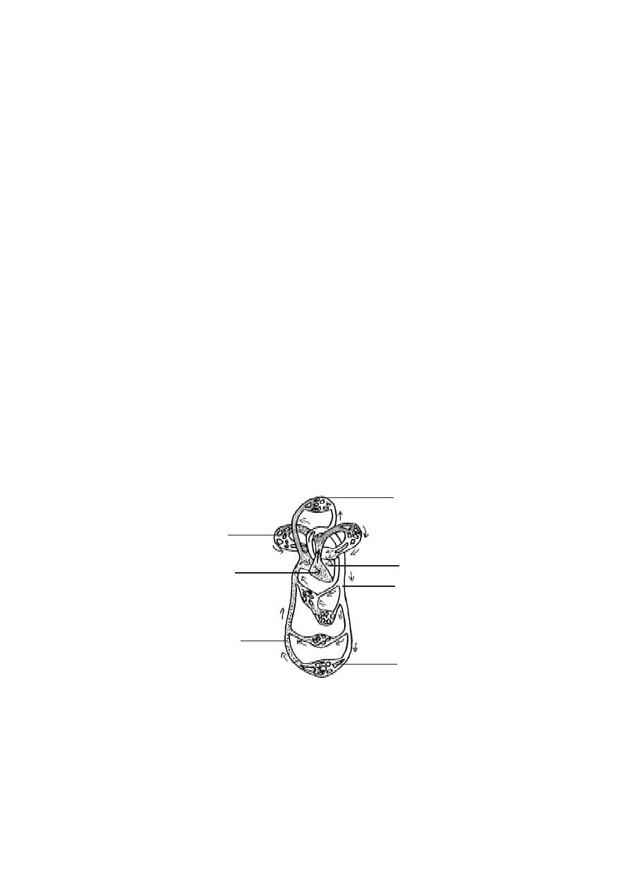
134
5.Thoracic wall and muscles of respirations.
Eventhough the lungs are the principal organs of respiration, the pro-
cess of ventilation happens by an indirect method. Air pressure gradients be-
tween thoracic chamber and lung cavity due to thoracic enlargement and re-
duction cause ventilation of lungs. Thoracic modifications during respiration
happen due to several muscles. These are called the muscles of inspiration
and expiration. These muscles are the diaphragm, external and internal
intercostal muscles between the ribs, pectorals and scalene (Ref. Muscu-
lar system).
3. 6. The Circulatory System
The multicellular organisation in animal world has resulted in the ori-
gin and evolution of circulatary system in animals. This arrangement facili-
tates internal transport of various substances to all organs and organ systems.
Among majority of multicellular animals this system remains as a closed type.
It has blood running inside closed blood vessels, the blood being pumped by
heart. In man, as in all mammals there is a double circulation of blood. The
primary circulation through pumping action of heart, supplies blood to all re-
gions of the body. The blood later returns to the heart. It is called the
systemic circulation or body circulation. A similar circulation carries blood
to lungs for oxygenation and returns it back to the heart. It is called the pul-
monary circulation.
Fig.3.6.1. Systemic and Pulmonary circulations
tissue capillaries
lung capillary
left side of heart
arteries
veins
tissue capillaries
right side of heart

135
Systemic and Pulmonary circulations
The most important component of this system is the heart. It is a
large, muscular, valved structure having four chambers. The chambers are
the right atrium, left atrium, right ventricle and left ventricle. Each atrium
opens into a corresponding ventricle. The right and left chambers are sepa-
rated by septa.
Systemic circulation
:- The left atrium receives oxygenated blood from the
lungs, through the pulmonary vein. When the atria contract, blood from the
left atrium is forced into the left ventricle. Later by a contraction of the ven-
tricle, the blood leaves the heart through the aorta. The aorta is the single
systemic artery emerging from the heart. By successive branchings, the aorta
gives rise to hundreds of arteries taking blood to all regions of the body. As the
branchings happen, the arteries divide into numerous (4 × 10
6
) arterioles. In
the target organs they produce four times as many capillaries. A similar num-
ber of venules converge into each other forming veins of increasingly larger
size. Finally, only two veins, the superior and inferior vena cavae
return the
blood to the right atrium. Thus the course of blood from left ventricles through
the body organs and back to the atrium forms the systemic circulation.
Pulmonary circulation
:- The venous blood from right atrium is conducted to
the right ventricle. The ventricle expels the blood via the pulmonary trunk to
the lungs. The oxygenated blood later returns by the pulmonary veins to the
left atrium. This circulation from right ventricle to the left atrium via the lungs
is termed the pulmonary circulation.
Portal circulation
:- In the systemic circulation the venous blood passing
through spleen, pancreas, stomach and interstine is not carried back directly
to the heart. It passes through the hepatic portal vein to the liver. This vein
begins as capilaries from the visceral organs and ends in the liver again as
capillaries. These capillaries converge to form the hepatic vein which joins
the inferior vena cava, conveying blood to right atrium. This route is the portal
circulation.

136
Arterial system
The main arterial trunk carrying blood from the heart to the tissues is
the aorta. At its commencement it lies in the thorax, and it is known as the
thoracic aorta. Its terminal portion is in the abdomen, where it is called as
the abdominal aorta. The thoracic aorta comprises the short ascending
aorta, the arch of the aorta and the longer descending aorta. The de-
scending aorta passes downward through the diaphragm. It continues up to
the level of the fourth lumbar vertebra, where it terminates by dividing into
two common iliac arteries. The main branches of the various portions of
the aorta are as follows.
Ascending
-
Coronary
Right common carotid
Arch of aorta -
Brachiocephalic (Innominate) Right sub clavian
Left common carotid
Left sub clavian
Visceral branches
Parietal branches
Bronchial
Inter costal
Esophageal
Sub costal
Thoracic aorta
Pericardial
Superior phrenic
Mediastinal
Visceral branches
Parietal branches
Coeliac
Superior mesenteric
Inferior phrenic
Abdominal aorta
Gonadial
Lumbar
Middle suprarenal
Renal
Interior mesenteric
Terminal branches
Common iliac
External iliac
Middle sacral
Internal iliac
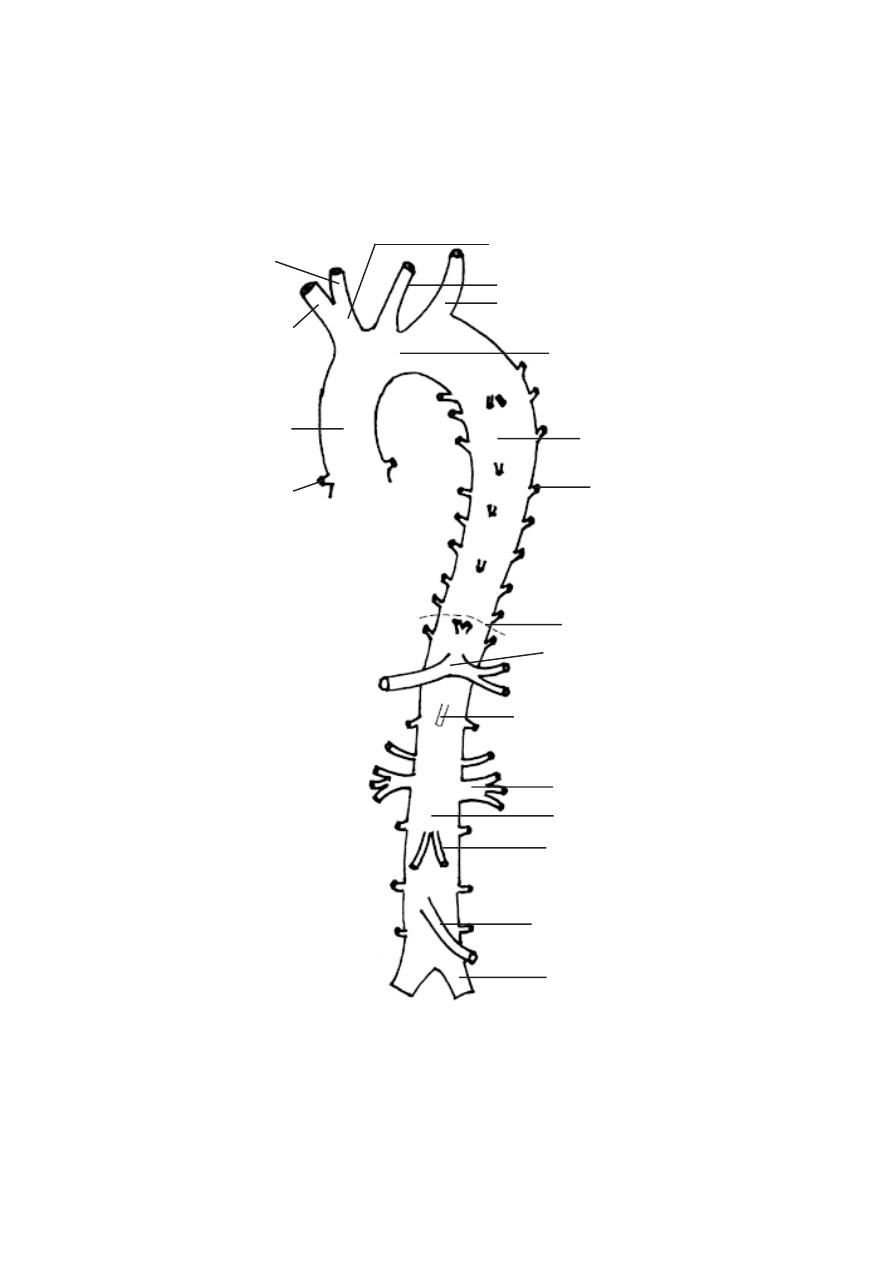
137
brachiocephalic (innominate) artery
left common carotid artery
left suclavian artery
arch of aorta
descending aorta
inter costal artery
diaphragm
coeliac artery
superior mesenteric artery
renal artery
abdominal aorta
testicular artery
inferior mesenteric artery
left common ilac artery
Fig. 3.6.2.Human aorta and its branches
right common carotid artery
right subclavian
artery
ascending aorta
right coronary artery

138
Ascending aorta:
The branches of the ascending aorta are the left and right coronary
arteries. They arise close to the origin of the aorta. They supply the heart
muscles with blood.
Arch of the aorta:
The branches of the aortic arch are the innominate, left common
carotid and left sub clavian arteries.
Innominate: (Brachiocephalic) this is the first branch of the aortic arch. It
passes upward and diagonally to the right and divides into two branches, the
right common carotid and the right subclavian. The common carotid
artery passes up to the neck and divides into the external carotid artery
and internal carotid artery. The external carotid artery supplies blood to
muscles, glands, skin and other structures of the face and scalp. The internal
carotid artery goes to the brain.
The right subclavian artery passes laterally to the arm. In the arm, the
subclavian artery forms the axillary arteries. The right axillary continues as
the brachial artery, which in the region of the elbow, divides into radial and
ulnar arteries. These continue in the distal portion of the arm, giving off
branches to wrist and hand.
The second branch of the aortic arch is the left common carotid
artery. It arises close to the innominate. It passes up the left side of the neck,
following a course and possessing branches similar to that of the right
common carotid. The left sub clavian artery arises from the aortic arch
independently of the left common carotid and supplies the left arum.
Thoracic aorta:
The branches of the thoracic portion of the descending aorta supply
both the internal organs and the body wall. The internal organs include
bronchi, esophagus, pericardium, lungs, rib cage and intercostal muscles.
Superior phrenic arteries (one pair) supply blood to the upper surface of
the diaphragm.

139
Abdominal aorta. This portion of the aorta has many branches. It serves
the entire lower region of the body. The following branches are given off
from the abdominal aorta.
1. The coeliac artery arises shortly behind the diaphragm, and sends
branches to the liver, gall bladder, stomach and duodenum and digestive glands
such as pancreas and liver.
2. The superior mesenteric artery, supplies the major portion of the small
intestine and a part of the large intestine.
3. The middle suprarenal arteries, supply the suprarenal glands,
4. The renal arteries supply the kidneys.
5. The internal testicular arteries in the male and ovarian arteries in the
female supply the testis and ovary respectively.
6. The inferior mesenteric artery supplies, the large intestine and rectum.
The common iliac arteries from the dorsal aorta enter into the legs.
Each iliac artery further divides into sciatic and femoral arteries supplying
blood to the leg muscles.
Pulmonary circulatory structures:
The pulmonary artery emerges from the superior surface of the right
ventricle, passes diagonally upward to the left, and crosses the root of the
aorta. It divides into right and left pulmonary arteries, branches of which
enter the right and left lungs, respectively. Pulmonary veins are four in
number, two from each Lang. They carry oxygenated blood from the lungs
to the left atrium.
Venous system:
Systemic veins are the vessels that collect blood from the tissues to
the heart. The principal systemic veins are:
1). The coronary sinus.
2). The superior vena cava.
3). The inferior vena cava and their branches and
4). The portal vein which drains the abdominal viscera.
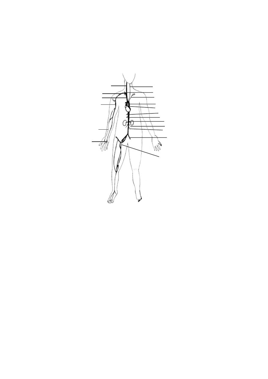
140
internal thoracic artery
left common carotid artery
left subclavian artery
descending portion of aorta
ascending portion of aorta
coeliac artery
renal artery
superior mesenteric artery
gonadial artery
common iliac artery
inferior mesenteric artery
femoral artery
brachio cephalic artery
right subclavian artery
brachial artery
ulnar artery
right common carotid artery
radial artery
Fig. 3.6.3 Human - Arterial system
Coronary sinus:
The coronary sinus is a short vein lying on the posterior side of the
heart. It receives most of the blood from heart tissues.
Superior vena cava and its branches:
This vessel is a large venous trunk, which empties blood to the heart
from the head, neck, upper extremities and thorax. It is sounded by the union
up the right and left
Innominate (brachiocephalic) veins. It opens into the right atrium.
The principal veins draining the head and neck are the external and
internal jugular veins. The right and left subclavian veins drain the upper
extremities, each terminating at its junction with the internal jugular vein to
form the innominate vein. Near their termination form each subclavian vein
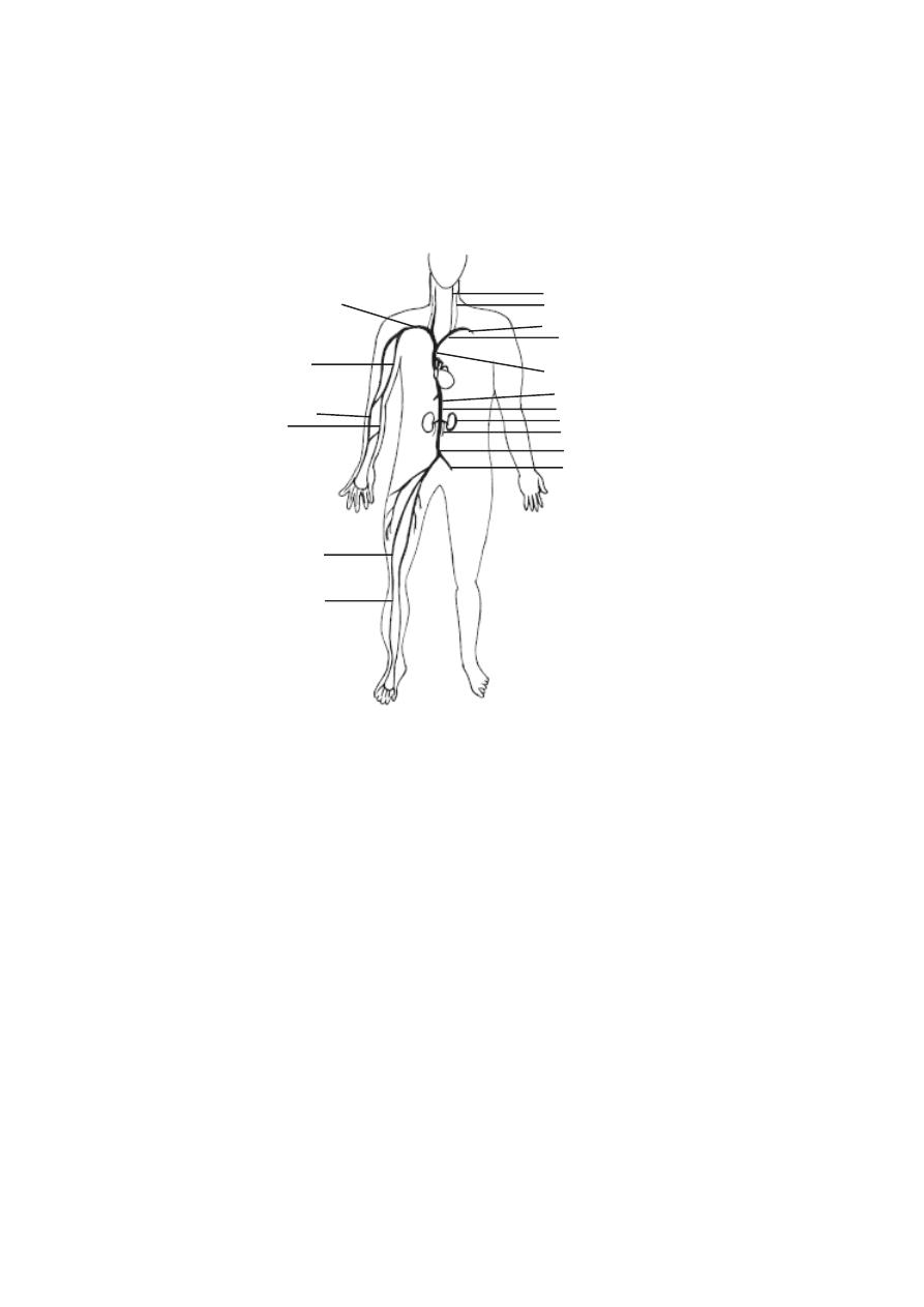
141
Innominate vein
hepatic vein
inferior vena cava
ouarian (testicular) vein
renal vein
common iliac vein
internal iliac vein
R. subclavian vein
internal jugular vein
external jugular vein
L. subclavian
superior vena cava
femoral vein
brachial vein
radial vein
ulnar vein
anterior tibial vein
(Brachio cephalic vein)
Fig. 3.6.4.Human - Venous system
receives an external jugular vein. Each subclavian vein inturn is formed by
the union of the cephalic and axillary vein.
Each innominate vein receives the deep cervical, vertebral, internal
mammary and inferior thyroid veins. In addition, the left innominate receives
the left superior intercostals vein and veins from thymus, trachea, esophagus
and pericardium. The subclavian veins also drain blood from the veins of the
hand (brachial)
Inferior vena cava and its branches:
This is the venous trunk which receives most of the blood from
regions of the body below the level of the diaphragm. It is the largest vein in
the body. The inferior vena cava is formed by the union of the two
common iliac veins. It extends forwards to the right of the aorta, passes
through the diaphragm and opens into the right atrium.

142
The inferior vena cava receives blood from following veins, inferior
phrenic (from the diaphragm), hepatic (from the liver), right suprarenal (from
the adrenal gland), renal (from the kidneys), right spermatic or ovarian
(from the right testis or ovary), lumbar (from the body wall) and common
iliac (from the pelvic region) and the veins of the lower extremities.
Pulmonary circulation
:- The venous blood from right atrium is conducted to
the right ventricle. The ventricle expels the blood via the pulmonary trunk to
the lungs. The oxygenated blood later returns by the pulmonary veins to the
left atrium. This circulation from right ventricle to the left atrium via the lungs
is termed the pulmonary circulation.
Portal circulation
:- In the systemic circulation the venous blood passing
through spleen, pancreas, stomach and interstine is not carried back directly
to the heart. It passes through the hepatic portal vein to the liver. This vein
begins as capilaries from the visceral organs and ends in the liver again as
capillaries. These capillaries converge to form the hepatic vein which joins
the inferior vena cava, conveying blood to right atrium. This route is the portal
circulation.
Components of Circulatory system
Blood vessels
The blood vessels carrying blood away from the heart are the arter-
ies. The Veins carry blood towards the heart. The arteries and veins are
named and classified according to their anatomical position. They can also be
classified according to their size and wall structure. Functionally, arteries are
subdivided into conducting, distributing and resistance vessels.
1. Conducting vessels
:- These are large arteries from the heart and their
main branches. the walls of these vessels are elastic in nature.
2. Distributing vessels
:- These are smaller arteries reaching individual or-
gans. They branch into the organs. They have muscular walls.
3. Resistance vessels
:- These are mostly arterioles. While these vessels
are smaller, their walls are highly muscular. Hence these vessels can reduce
pressure of blood due to peripheral resistance.
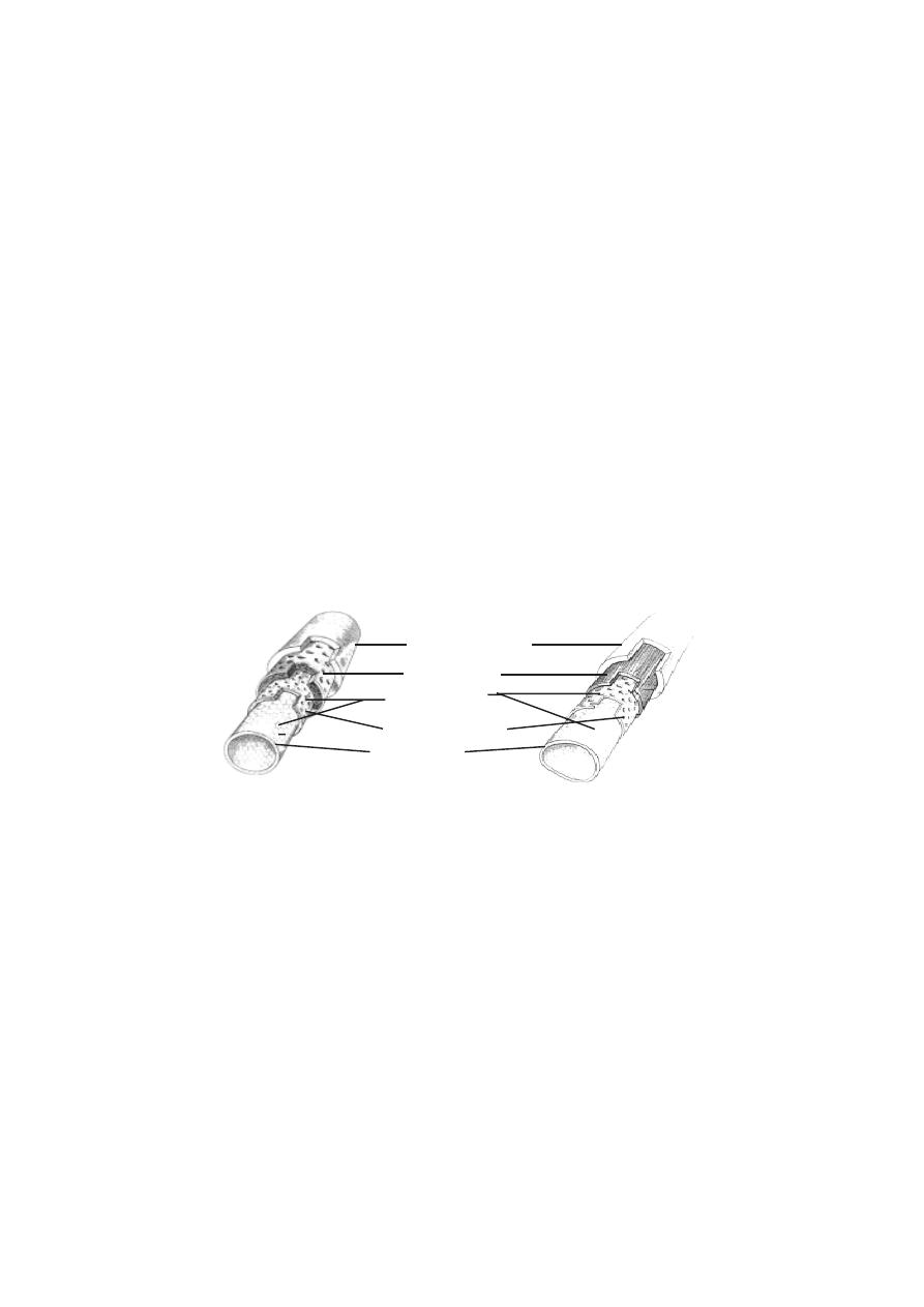
143
4. Exchange vessels
:- These are the capillaries. The walls of these vessels
allow exchanges between blood and the tissue fluid surrounding the cells. The
substances commonly exchanged are oxygen, carbon-di-oxide, nutrients, wa-
ter, inorganic ions, vitamins, hormones, metabolic products and antibodies.
5. Capacitance or reservoir vessels
:- These are the larger vessels and
veins. These are of varying sizes. They collect and convey blood back to the
heart. The higher capacitance of these vessels is due to their distensibility.
Hence their blood content is more, even at low pressure. The number of such
veins is also enormous.. Thus the veins are called as the “blood reservoirs”
Structure of blood vessels
The blood vessels show a vast range of structural modifications.
However a few basic patterns can be studied.
A blood vessel consists of a wall and a lumen or cavity. The wall of
the blood vessels is made up of 3 distinct layers or tunica. They are the
tunica intima, tunica media and tunica externa or tunica adventitia.
The tunica intima is formed of an endothelium, a delicate connective
tissue and elastic fibres. The tunica media contains smooth muscle cells. It
causes vasoconstriction and vasodilation. The tunica externa is composed
of connective tissue. The composition and thickness of layers varies with the
diameter of the blood vessels and the type.
Types of blood vessels
1. Large elastic arteries
:- The walls of these arteries contain elastic fi-
bres. The smooth wall measures about 1micron in thickness. It gets stretched
under the effect of pulse and recoils elastically.
Fig.3.6.5. (a) Artery (b) Vein - Structural comparision
(a)
(b)
tunica adventitia
tunica interna
tunica media
elastic membrane
endothelium
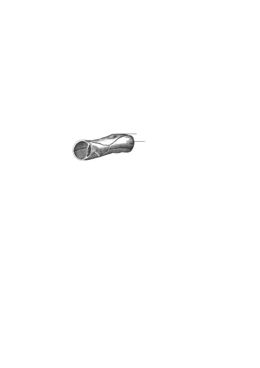
144
2. Muscular arteries
:- There are larger and smaller muscular arteries. The
larger muscular arteries are inelastic and they have thick walls. The wall has
30-40microns in diameter in the
layers of smooth muscles. Since they regulate
blood supply, they are called distributing arteries. The small muscular
arteries are capable of vasodilation and vasoconstriction.
3. Arterioles
:- They conduct blood from the arteries to the capillary bed.
These are small vessels capable of vasodilation and vasoconstriction.
4. Capillaries
:- These are fine vessels found between arterioles and venules.
They measure 5-8micron in diameter.
5. Venules
:- These are tubes of flat, oval or polygonal endothelial cells.
Each venule is formed by the convergence of two or more capillaries. Its
diameter ranges upto 30micron.
6. Veins
:- Veins seen in anatomy are medium veins. They run in between
venules and large veins. Large veins transport blood to the heart.
Veins with diameter above 2 mm have valves. They are of semilunar
type. They allow movement of blood towards the heart. There are several
valves in the medium veins.
Branching of blood vessels
:- When an artery divides into two equal branches,
the original artery ceases to exist. Hence the branches are called terminal
branches. The smaller branching vessels formed on the sides are called the
collateral branches. When arteries are joined to each other it is named as
anastomosis.
Blood supply to blood vessels
:- As any other region, the cells and tissue
on the wall of the blood vessel require nourishment. Some amount can diffuse
from blood in the lumen. For vessels having diameter greater than 1 mm, dif-
fusion of nutrients may not be possible. Such vessels have very minute ves-
sels called vasa vasorum spread over them. They penetrate into the wall of
the blood vessels.
Fig.3.6.6. A Capillary tube
capillary endothelial cell
nucleus

145
Fig.3.6.7. Location and External structure of the Heart
Innervation of blood vessels
:- The walls of the blood vessels are inner-
vated by sympathetic nerve fibres. They regulate the contraction of the mus-
culature. They effect vasoconstriction.
The Heart
The heart is a hollow, fibromuscular organ. It is somewhat conical or
pyramidal in form. It is roughly the size of a closed fist. An average heart
measures 12 cm from base to the apex. Transverse diameter at its broadest
region is 8-9 cm. It is 6 cm thick antero-posteriorly. While in adult male the
heart weighs 280-340 g, in female it weighs 230-280 g.
Fig.3.6.8. Internal structure of the Heart
The thoracic organs such as heart, trachea and oesophagus form a
midline partition called the mediastinum. The heart lies obliquely in the medi-
astinum.
The heart is surrounded by a double layered membrane called the peri-
cardium. The outer layer is called the fibrous pericardium. The inner mem-
brane is called the serous pericardium. In between heart and pericardium,
there is a pericardial space. This space is filled with a fluid called the pericar-
dial fluid.
pulmonary artery
aorta
left atrium
mediastinum
inferior vena cava
lungs
diaphragm
right atrium
superior vena cava
brachio cephalic artery
left common carotid artery
right subclavian artery
right common carotid artery
left subclavian
artery
pulmonary artery
semilunar valves
bicuspid valve
chordae tendineae
inter ventricular septum
aorta
pulmonary veins
left atrium
left ventricle
superior vena cava
right atrium
tricuspid valve
right ventricle
inferior vena cava

146
The wall of the heart is made up of three tissue layers. They are the
epicardium, myocardium and endocardium. The epicardium forms the
smooth outer surface of the heart. The middle myocardium is composed of
cardiac muscle. This layer plays an important role in the functioning of the
heart. The endocardium forms the smooth inner surface. It is formed of
squamous epithelium.
3.7. Lymphatic system
Lymphatic circulation along with blood circulation plays a key role in
maintaining the fluidity in all regions of the body. It helps to maintain fluid
balance in tissues and it absorbs fat from the digestive tract. It also functions
as body’s defence system against micro organisms and other harmful
substances. This system includes lymph, lymphocytes, lymphatic vessels, lymph
nodules, lymph nodes, tonsils, the spleen and the thymus gland.
Lymphoid cells and tissues
- Lymphatic organs contain lymphatic tissues.
These tissues primarily consist of lymphocytes. They also contain macroph-
ages, dendritic cells and reticular cells. Lymphocytes are a type of white
blood cells. They originate from red bone marrow and are carried by blood to
lymphatic organs and other tissues. There are several classes of lympho-
cytes. The B-lymphocytes or B cells synthesize antibodies for recognizing
and neutralising alien macromolecules. T- lymphocytes can recognize and se-
lectively kill cells infected with viruses. B and T lymphocytes are
produced from stem cells present in the bone marrow. The T lymphocytes get
matured only after entering into Thymus, a lymphoid organ through circula-
tion. Maturation and differentiation of B cells will occur in the bone marrow
itself. Thus the thymus and bone marrow are described as central or primary
lymphoid organs.
Thymus
- It is a roughly triangular, bilobed gland. It is located in the mediasti-
num (ie., between the lungs). It lies between the sternum and the pericardium.
Its size varies with age. It is largest in the early part of life (upto 15 years). At
birth it weighs 10 - 15 g. After puberty it greatly decreases in size.
Each thymus lobe is surrounded by a thin capsule made of the connec-
tive tissue. It has 2 layers. The inner layer is the medulla, the outer layer is
cortex. The lymphocytes are found only in cortex layer.
Lymph nodes
- These are small round structures. Their size ranges from 1-
25 mm. They are distributed throughout the course of the lymphatic vessels.
These nodes are found all over the body. However they are concentrated as
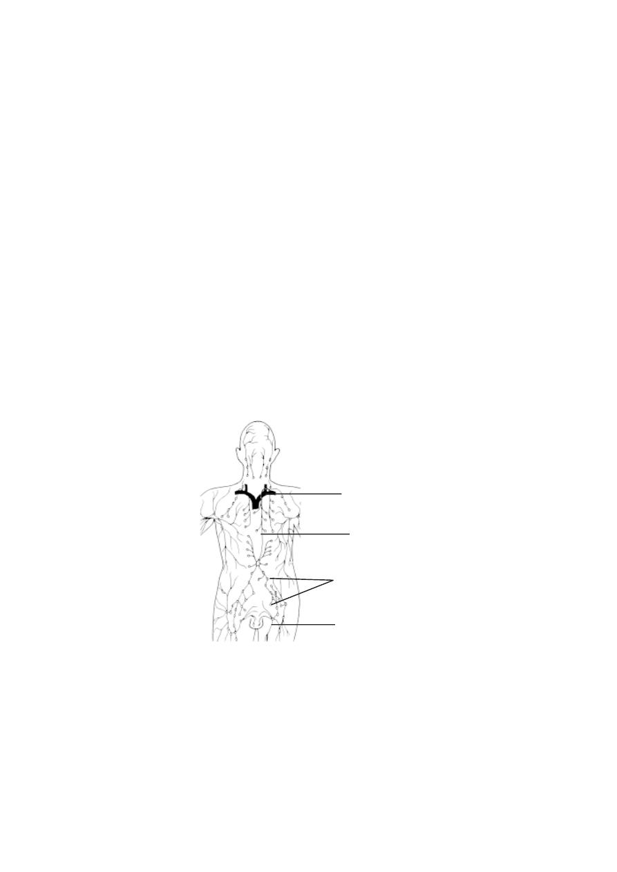
147
aggregations in 3 regions of the body. These are the inguinal nodes in the
groin, the axillary nodes in the axillary region and the cervical nodes of the
neck.
The lymph enters the lymph nodes through afferent lymphatic vessels
and exits through efferent vessels. The nodes contain open spaces called si-
nuses. The sinuses are lined with phagocytic cells.
Spleen
- It is roughly the size of a clenched fist. It is located on the left side
of the abdominal cavity. It has a fibrous capsule. The spleen contains two
types of lymphatic tissues, namely the red pulp and the white pulp.
Tonsils
- These are the largest lymph nodules. They provide protection against
bacteria and other harmful materials. In adults the tonsils decrease in size and
may disappear. There are 3 groups of tonsils in the pharyngeal walls. Of the
three, the palatine tonsils are usually refered to as “the tonsils”. These are
larger lymphoid masses on each side of the junction between the oral cavity
and the pharynx. The pharyngeal tonsil or adenoid are found near the junc-
tion between the nasal cavity and the pharynx. The lingual tonsil is a loosely
associated collection of lymph nodules on the posterior surface of the tongue.
The lymphatic circulation
- The lymph fluid from the tissues is drained by
lymphatic capillaries. These capillaries though present in many tissues are
absent in epidermis, hairs, nails, cornea, cartilages, CNS and bone
Fig.3.7.1. Lymphatic system
subclavian vein
thoracic duct
lymph nodes
lymphatic vessel

148
marrow. The lymphatic capillaries join into larger vessels. The larger vessels
pass to local or remote lymph nodes. These vessels and associated lymph
nodes are arranged in regional groups. Each group has its region of drainage.
Nodes within a group are interconnected. Such regional groups with nodes
and vessels are organised in (1) Head and neck (2) Upper limbs (3) Lower
limbs (4) Abdomen and pelvis (5) thorax.
The regional vessels return to the venous blood circulation via the
right and left lympho venous portals. Nearly eight lymphatic trunks con-
verge at the site of the vertebral column and open into the venous portals
nearer to the neck
3. 8. The Nervous System
A complete understanding of the human nervous system remains a
challenge. Several billion cells remain associated with this system. The vary-
ing functions of these cells and the nervous system are responsible for human
behaviour and activities. Hence, scientists from different fields collectively
are interested in understanding the functioning of this system. Studies on brain
and other related structures began several years ago. Even to-day thousands
of scientists are involved in researches for knowing the structure and func-
tioning of the brain. For a thorough knowledge of this system, further works in
anatomy, physiology, molecular,biology, psychology, medicine and other related
fields are needed.
Basically the nervous system is formed of nerve cells or neurons.
Neurons are responsible for transmission of impulses. They also help in realising,
analysing and storing messages. They can stimulate muscles to work. The
network of interconnected neurons in the nerves, brain and spinal cord have
highly complicated methods of functioning.
A neuron has a basic cell structure called the cyton. The projections
of the cyton are the dendrites and the dendrons. The inter communicating
long projection is the axon. There are variations in the shape of the cyton,
number of dendrons and nature of axon.
A neuron is interconnected with the dendrite of the neighbouring neu-
ron through the endplate of the axon. Such specialized connections are called
as synapses. In the terminal regions of the effector nerves the axon of the
nerve cells are in contact with the muscle tissue. These joints are named as
neuro - muscular junctions.

149
The structure of a peripheral nerve
A nerve is made up of several nerve fibres. A nerve fibre contains
axons with their coverings called schwann cells. The fibres are grouped into
fasciculi. The number and pattern of fasciculi vary in different nerves. Thus
a nerve trunk possesses many such fasciculi. Such a trunk is surrounded by an
epineuruium. The individual fasciculi are enclosed by a multilayered perineu-
rium. The perineurium surrounds the endoneurium or intra fascicular con-
nective tissue.
In a peripheral nerve the epineurium constitutes 30 -70 % of the total
cross sectional area of the nerve bundle. The thickness is more when there
are more fasciculi. A layer of fat in the epineurium provides a ‘cushion’ effect
to the nerve.
The perineurium contains alternating layers of flattened polygonal cells.
The endoneurium remains condensed around axons. The components of the
endoneurium remain bathed in endoneurial fluid.
The fasciculi of the nerve are supplied blood by vasa nervosum. These
minute blood vessels radiate upto the endoneurium.
Nervous system
The organs of the nervous system are continuous in nature. However,
for study purposes it can be divided into systems and organs.
A. Central nervous system (C N S)
This system includes the brain and the spinal cord or medulla spina-
lis. They are protected by surrounding bones. While the brain is located within
the cranium, the spinal cord is placed within the vertebral canal of the ver-
tebrae. Through an opening called foramen magnum, the spinal cord de-
scends down from the brain.
B. Peripheral nervous system.
It consists of nerves and ganglia. The nerves that are formed from the
brain are called the cranial nerves. There are 12 pairs of cranial nerves and
31 pairs of spinal nerves.
C. Autonomous nervous system.
The nerves in this system transmit impulses from the C N S to smooth
muscles, cardiac muscles and glands. It is also called the involuntary ner-
vous system. It is subdivided into sympathetic and parasympathetic divisions.
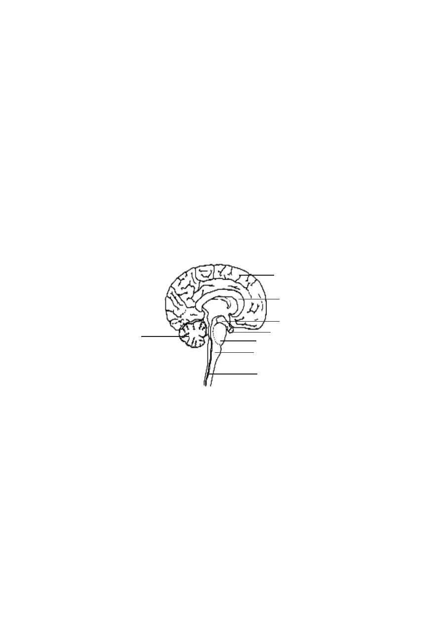
150
1. Brain
The brain is safely kept inside the cranial vault. Inside the skull the
brain is surrounded by three protective coverings. They may be grouped un-
der two divisions.
1.Pachymenix -It includes the duramater.
2.Leptomeninges - In includes the arachnoid mater and pia mater.
The duramater is the outermost membrane. It is thick and inelastic in
nature. The arachnoid mater is the middle covering over the brain. In between
arachnoid and piamater there is a space called the subarachnoid space. It
contains cerebro-spinal fluid and blood vessels. The piamater is a delicate
membrane closely applied to the brain. This membrane contains blood capil-
laries supplying blood to the brain cells.
The human brain weighs about 1.3 Kg. It contains more than a billion
neurons. Based on embryological development the brain can be divided as
follows.
1. Prosencephalon (Fore brain)
- It consists of the cerebrum and the dien-
cephalon. The cerebrum is the largest part of the brain. It is divided into right
and left hemispheres by a longitudinal fissure. However, at the base the
two hemispheres are connected by a sheet of nerve fibres called the corpus
callossum.
infundibulum
Fig.3.8.1 L.S. of the human brain
cerebrum
pituitary gland
cerebellum
corpus callossum
pons
medulla oblongata
spinal cord

151
The outer surface of the cerebrum is called the cortex or grey mater.
It is 2 to 4 mm thick. The inner content of the cerebrum is the white mater.
The surface of the cerebrum has several folds called the gyri. They greatly
increase the surface area of the cortex. The shallow grooves in between the
gyri are called the sulci. A central sulcus runs in the lateral surface of the
cerebrum from superior to inferior region.
Each cerebral hemisphere is divided into four lobes. They are the fron-
tal at the front, the parietal towards the top of the head, the temporal on the
side and the occipital at the rear.
The diencephalon contains the thalamus and hypothalamus. This
region is found between the cerebrum and the brain stem.
The thalamus has a cluster of nuclei which act as the relays for par-
ticular sensory pathways. Just beneath the thalamus, the hypothalamus is
present. It contains reflex centres linked to the autonomic system. A funnel
shaped stalk called the infundibulum extends from its floor. It is connected to
the neurohypophysis of the pituitary gland.
2. Mesencephalon (mid brain)
- It is the smallest region of the brainstem.
On its dorsal surface there are four rounded bodies called the carpora
quadrigemina.
3. Rhombencephalon (hind brain)
- The three main regions of the rhomben-
cephalon are the medulla oblongata, the pons varoli and the
cerebellum.
The cerebellum consists of two hemispheres. Its surface has many
ridges called folia. The cerebellum consists of three parts. They are the small
anterior flocconodular lobe, a narrow central vermis and two large lateral
hemispheres.
The pons is just superior to the medulla oblongata. It contains
ascending and descending nerve tracts.
The medulla oblongata is about 3 cm long. It is continuous with the
spinal cord. It remains as a bridge between the brain and the spinal cord.
Brain stem
- The medulla oblongata, pons and mid brain form the brain stem.
It connects the spinal cord to the brain. Ten of the twelve cranial nerves enter
or exit the brain through the brain stem.
Spinal cord
- The spinal cord extends from the foramen magnum to the level
of the second lumbar vertebra. It is considerably shorter than the vertebral
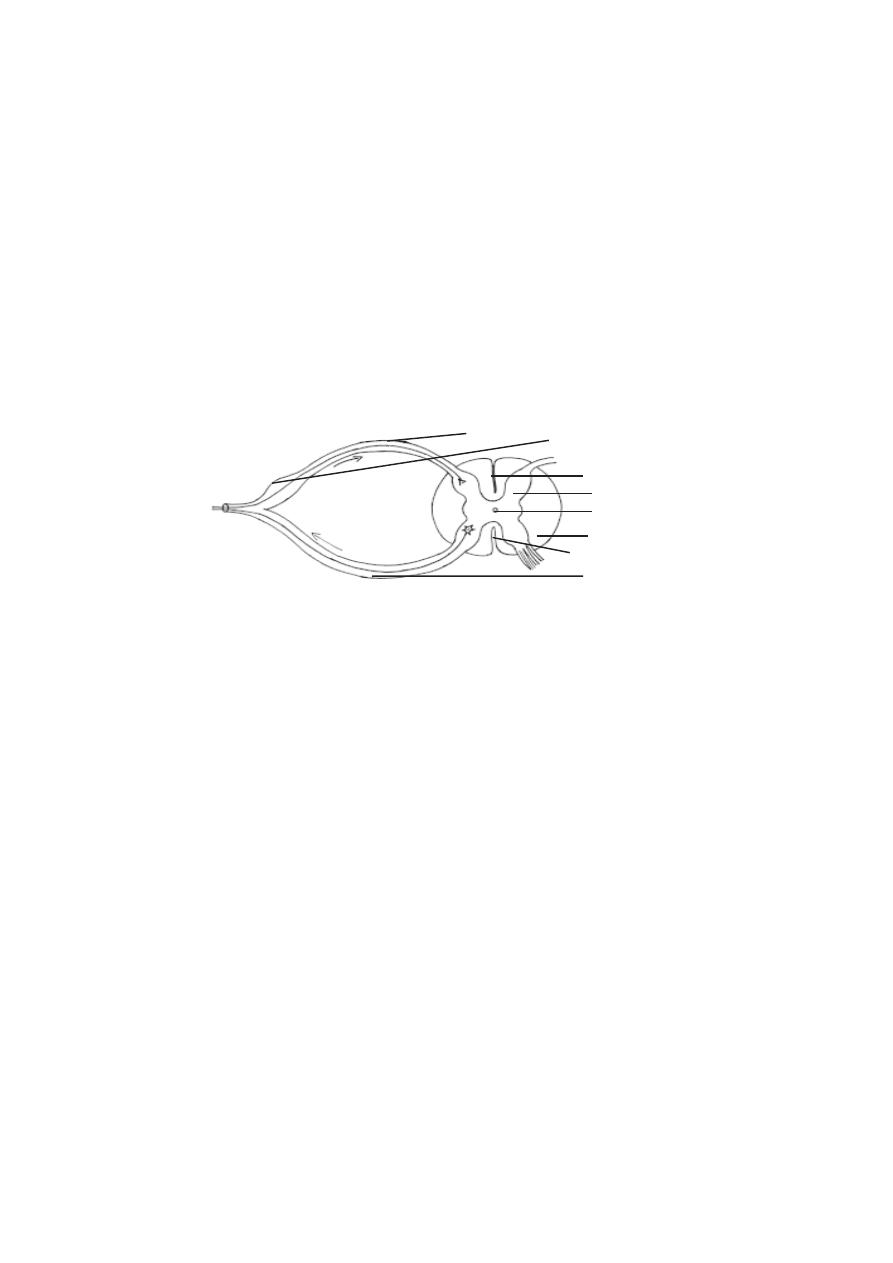
152
column. There are two enlargements in the spinal cord. They are the cervical
and lumbar enlargements. Below the lumbar enlargement the spinal cord tapers
to form a cone like region called the conus medullaris. A connective tissue
filament called the filum terminale extends inferiorly from conus medullaris
to the coccyx. The conus medullaris and the nerves extending below resemble
a horse’s tail. Hence it is called cauda equina.
A cross section of the spinal cord reveals a central grey portion and a
peripheral white portion. The white matter consists of nerve tracts and the
grey matter consists of neuron cell bodies and dendrites.
The dorsal and ventral sides have long fissures. There are 31 pairs of
spinal nerves arising from the spinal cord. Each nerve has a dorsal root and a
ventral root from the spinal cord. The dorsal roots have dorsal root ganglia.
Ventricles
The entire CNS remains as a hollow tube. The tube inside the adult
brain forms ventricles.
Each cerebral hemisphere contains a large cavity called the lateral
ventricle. It corresponds to the hypothetical first and second ventricles. The
two lateral ventricles communicate with the third ventricle located in the
centre of the diencephalon. This connection is made through two
interventricular foramina (foramen of Monro). The third ventricle inturn
opens into the fourth ventricle found inside the medulla oblongata. This
communication happens through a narrow canal called the cerebral
aqueduct (aqueduct of sylvius). The fourth ventricle is continuous with the
central canal of the spinal cord. The central canal extends nearly to the full
length of the cord.
Cerebro-spinal fluid (CSF)
This fluid fills the ventricles of the brain and the central canal of the
spinal cord. About 80-90 % of CSF is produced by specialized cells called
Fig.3.8.2. Cross section of the spinal cord
grey mater
white mater
central canal
dorsal fissure
dorsal root
ventral root
dorsal ganglion
ventral fissure
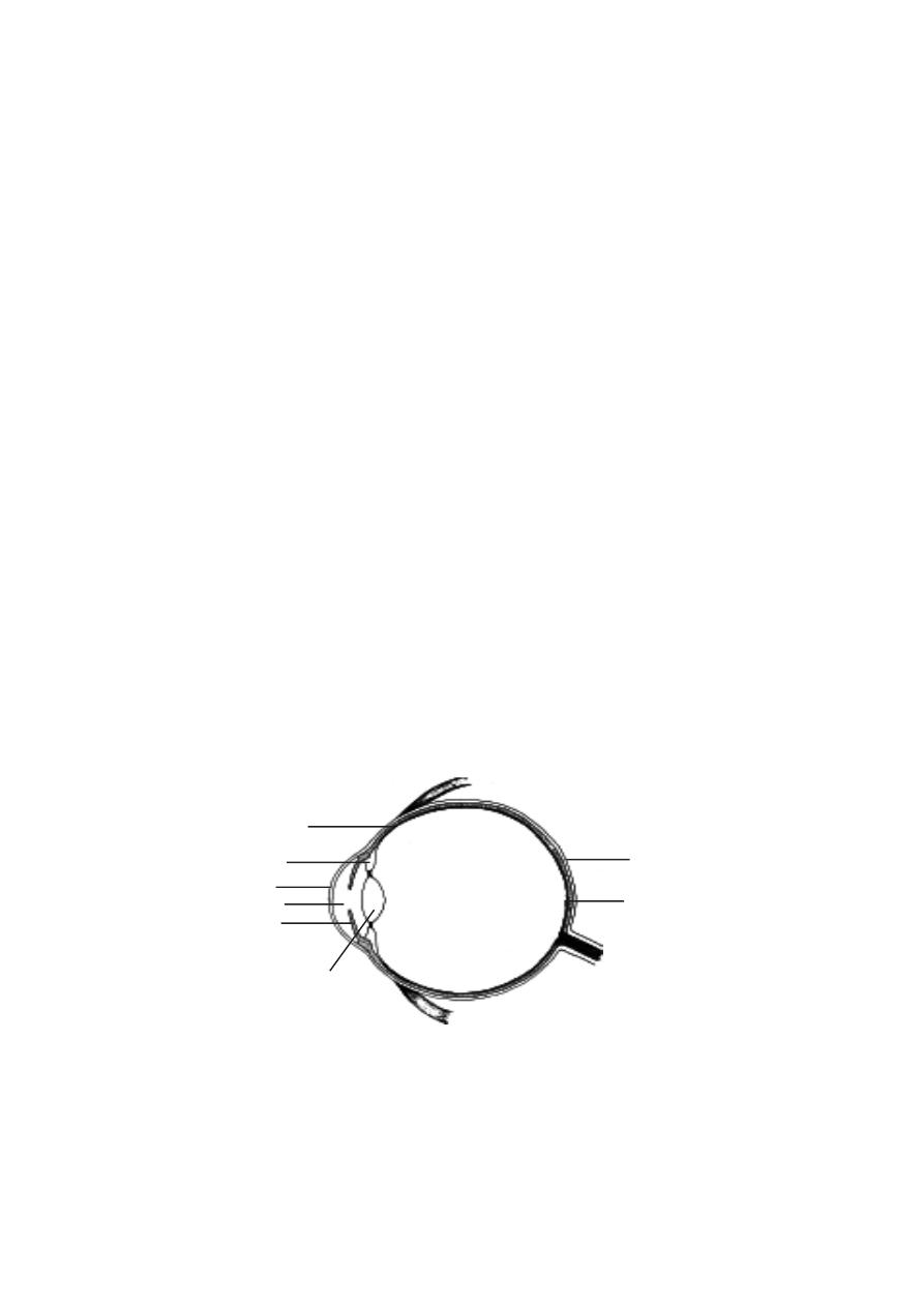
153
ependymal cells within the lateral ventricles. Remaining 10-12 % is pro-
duced by similar cells in the 3
rd
and 4
th
ventricles. These ependymal cells, their
supportive tissue and the associated blood vessels together are called choroid
plexuses. The plexuses are formed by invagination of the vascular piamater
into the ventricles.
3. 9. The Sensory Organs.
Living organism respond to several stimuli such as light, heat, sound,
chemicals, pressure, touch, stretch and orientation. These stimuli are felt by
specific receptors. The receptors convert the stimuli into impulses in the ner-
vous systems.
The touch receptors in the skin are the simplest receptors. Such
receptors are single nerve cells responding directly to the stimulus. Other re-
ceptors are complex sense organs. On these organs the stimulus is chan-
nelled into a receptive region of the organ. Among the several organs, the
most important are the eyes and ears.
The eye
The eye is formed of 3 coats or tunics.
Coats or tunic
Regions
1. Outer or fibrous
- sclera & cornea
2. middle or vascular
- choroid, ciliary body & iris
3. inner or nervous
- retina
lens
sclera
retina
choroid
ciliary body
cornea
pupil
iris
Fig.3.9.1. C.S. of human eye
Optic Nerve
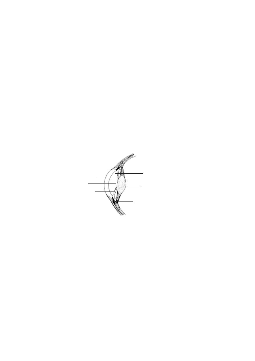
154
The sclera is the white outer layer of the eye. It covers posterior five-
sixths of the eye. This firm layer provides shape and protects the internal
structures. A small region of the sclera can be seen as the “white of the eye”.
In the front, the outer layer forms a transparent region called the cor-
nea. It permits entry of light. The cornea is made up of a connective tissue
having collagen, elastic fibres and proteoglycans.
The middle tunic of the eyeball is the vascular tunic. It contains most
of the blood vessels. The vascular tunic contains melanin containing pigment
cells. It appears black in colour. A major part of the vascular tunic is found in
association with the sclera and called the choroid. Anteriorly this layer forms
the ciliary body and iris.
The ciliary body consists of smooth muscles called the ciliary
muscles. Contraction of the ciliary muscles can change the shape of the lens.
The iris is the coloured part of the eye. It may be black, brown or
blue. It is a contractile structure surrounding an opening called the pupil.
Light enters the eye through the pupil. The iris regulates such entry by con-
trolling the size of the pupil.
The inner most tunic of the eye is the retina. It consists of an outer
pigmented retina and an inner sensory retina. The sensory retina is light
sensitive. It contains nearly 120 million photoreceptor cells called rods and
another 7 million cones.
Fig.3.9.2. Front of the Eye
ciliary body
lens
pupil
iris
cornea
aqueous humour
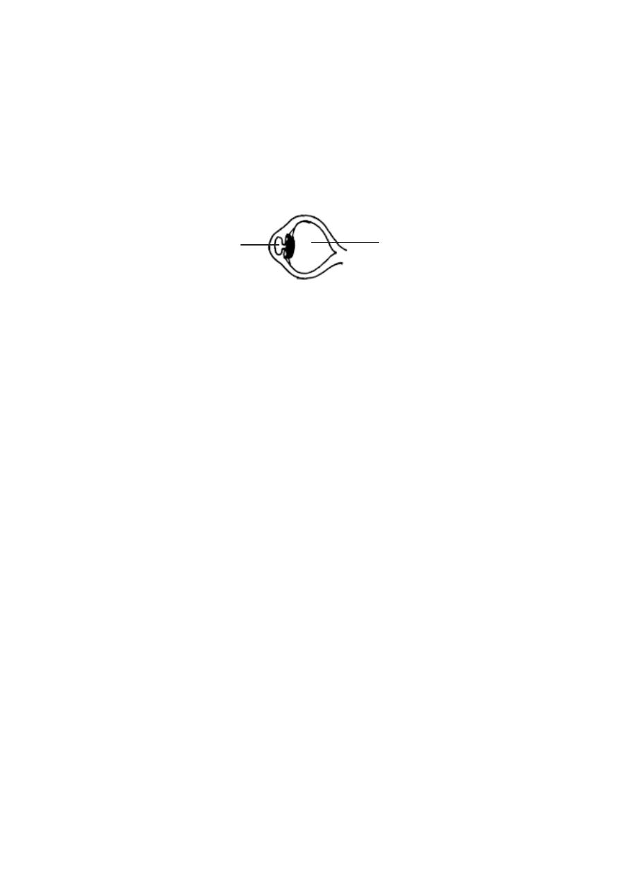
155
Compartments of the eye
: The eye has 2 major compartments. There is a
smaller compartment anterior to the lens. Behind the lens there is a larger
compartment.
The anterior compartment is divided into two chambers. There is
an anterior chamber found between the cornea and iris. A smaller
posterior chamber lies between the iris and lens. These two chambers are
filled with a substance called the aqueous humor. It helps to maintain
intraocular pressure.
The posterior compartment of the eye is much larger and it
contains a transparent jellylike substance called vitreous humor.
The eye lens is an unique biological structure. It is transparent and
biconvex. It is made up of long columnar epithelial cells called lens fibres.
These fibres have an accumulation of proteins called crystallines. The lens is
placed between the two eye compartments by suspensory ligaments.
The functioning of the eye is aided by accessory structures. They
include the eyebrows, eyelids, conjunctiva and lacrimal apparatus.
The eyebrows prevent the sweat during perspiration from running
down into the eye. They help to shade the eyes from direct sunlight.
The eyelids and associated lashes protect the eyes from foreign ob-
jects. The medial region where the eyelids join has a small reddish-pink mound
called the caruncle. It contains modified sebaceous and sweat glands. There
are two or three rows of hairs attached to the free edges of eyelids. Modified
sweat glands called the ciliary glands open into the follicles of the
eyelashes. It keeps them lubricated. The inner margin of the eyelids contain
Melbomian glands. These glands produce sebum for lubricating the eyelids.
The inner surface of the eyelids and the anterior surfae of the eye are
covered by a thin, transparent mucous membrane called the conjunctiva.
The lacrymal glands or tear glands are situated in the superolateral
corner of the eye orbit. They produce tear at the rate of about 1 ml / day. It
Fig.3.9.3. Compartments of the eye
aqueous humour
vitreous humour
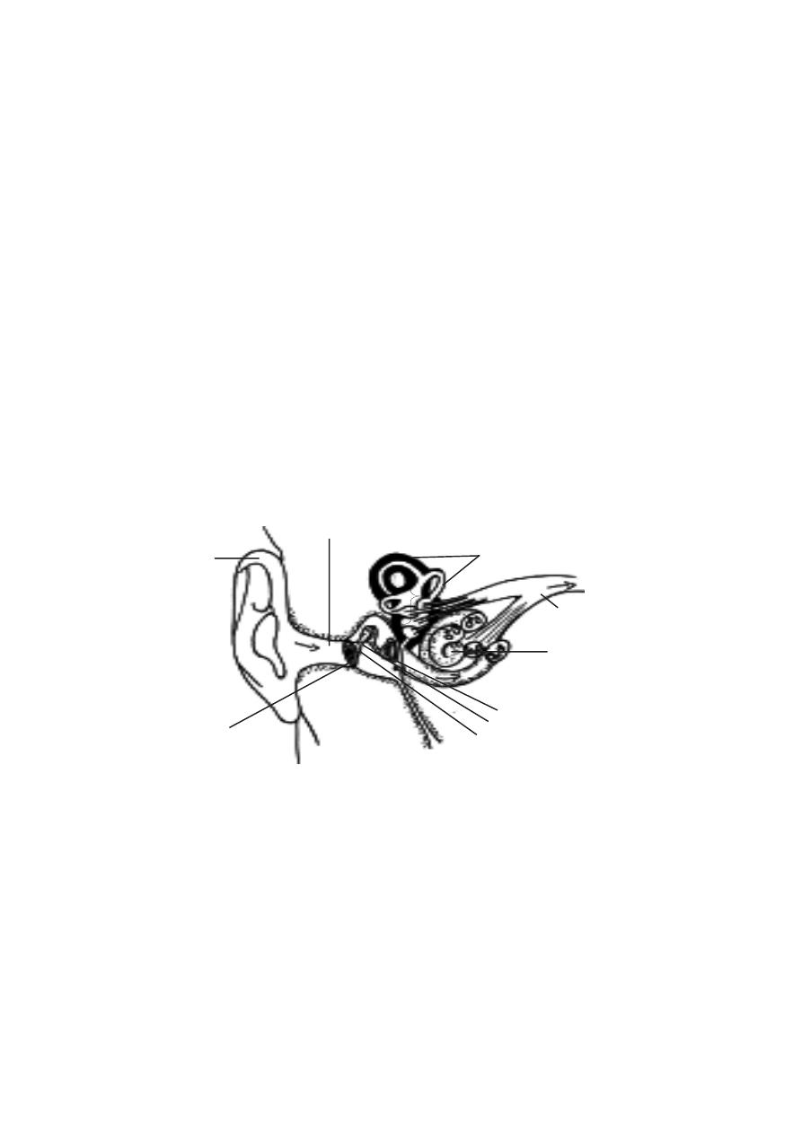
156
helps to moisten the eye surface and wash away foreign substances. At the
corners of the eye there are small openings called the puncta. Each punctum
inturn opens into a lacrymal canaliculus. The lacrimal canaliculi open into a
lacrimal sac. This sac enters into a nasolacrimal duct which opens into the
inferior nasal concha. These ducts help to drain the excess tear. The entire
organization related to ‘tear’ is called the lacrymal apparatus.
Ears (The organs of hearing)
The ears are the organs of hearing and balance. They have three parts,
namely external, middle and inner ears.
External ear
- The fleshy part outside the head is called the pinna. It is
made up of elastic cartilage and skin. It is followed by the external auditory
meatus. This passage is lined with hairs and ceruminous glands. These
glands produce cerumen or earwax. The hair and wax prevent foreign ob-
jects from reaching the ear drum. The ear drum or tympanic membrane is
a oval, three layered structure. It separates outer and middle ears.
Middle ear
- It is an air filled cavity. It contains three auditory ossicles called
the malleus, incus and stapes. The handle of malleus is in contact with the
inner surface of the ear drum. The head of the malleus is attached to the
incus. While the stapes on one side is attached to the incus, its other side fits
into the oval window. The oval window leads to the inner ear.
Inner ear
- This region has tunnels and chambers inside the temporal bone
malleus
pinna
semi circular canals
cochlea
stapes
incus
external auditory meatus
auditory nerve
ear ossicles
tympanic membrane
Fig.3.9.4. Ear - Inner structure
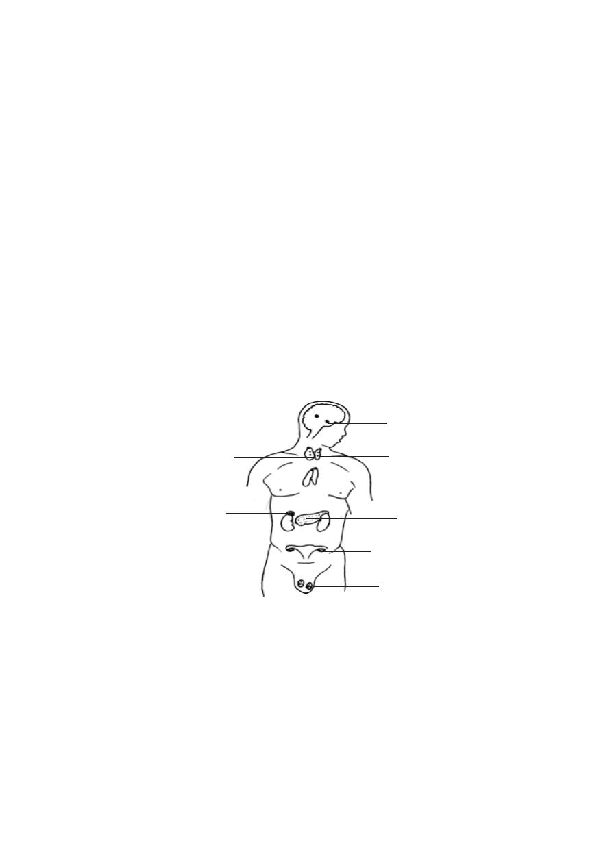
157
called the bony labyrinth. The bony labyrinth contains three regions called
the cochlea, vestibule and semicircular canals. The oval window found in
between the middle and inner ears communicates with the vestibule of the
inner ear. The organs of the inner ear perceive the sound.
3.10 Endocrine system
Our body has two major regulatory systems. They are the nervous
and endocrine systems. Together they regulate and co-ordinate the activi-
ties of all other body structures. The endocrine system sends
information to the tissues it controls in the form of chemical signals. These
signals, in the form of hormones are released into the circulatory system. They
are carried to all parts of the body . Body cells are able to recognise the
chemical signals and respond to them. The hormones of the endocrine glands
regulate and control the functioning of several organs in the body. Thus, this
system in general helps to maintain homeostasis. There are several endocrine
glands in our body. The major glands are the pituitary, thyroid, parathy-
roids, pancreas, adrenals, testes and ovaries.
An understanding of the structure and form of each endocrine gland,
its secretory products and the means by which its activity is regulated is abso-
lutely essential. This knowledge will help us to treat several diseases related
to these glands.
pituitary
thyroid
parathyroids
pancreas
adrenal
testes
ovaries
Fig.3.10.1. The Major Endocrine glands
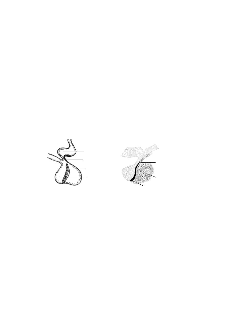
158
Pituitary gland (or) Hypophysis
It is an organ, that secretes eight major hormones. These hormones
regulate numerous body functions and controls the secretory activities of sev-
eral other endocrine glands. The hypothalamus of the brain is connected to
the pituitary. The posterior pituitary is an extension of the hypothalamus.
Structure of the pituitary gland.
This gland is approximately 1 cm in diameter. It weighs 0.5-1g. It is
placed in a region called the sella turcica of the sphenoid bone in the floor of
the skull. It is placed inferior to the hypothalamus. It is connected to it by a
stalk of tissue called the infundibulum.
Based on origin and function the pituitary is divided into two parts.
They are the posterior pituitary or neurohypophysis and anterior pitu-
itary or adenohypophysis.
Posterior pituitary or Neurohypophysis.
The posterior pituitary is continuous with the brain. Hence it is called
the neurohypophysis. During embryonic development, it is formed as an
outgrowth of the inferior part of the brain in the area of the hypothalamus.
The outgrowth of the brain, forms the infundibulum. The distal end of the
infundibulum enlarges to form the posterior pituitary. Since this part of the
pituitary is an extension of the nervous system, its secretions are known as
neurohormones.
Anterior Pituitary or Adenohypophysis
During embryonic development an outpocketing of the roof of the
oral cavity arises. It is called as the Rathke’s pouch. This pouch grows
Fig.3.10.2. (a) Entire (b) L.S. of the pituitary gland
hypothalamus
infundibulum
a d e n o h y p o p h y s i s
(ant. pituitary)
neurohypophysis
(post pituitary)
(a)
pars tuberalis
pars distalis
pars intermedia
(b)
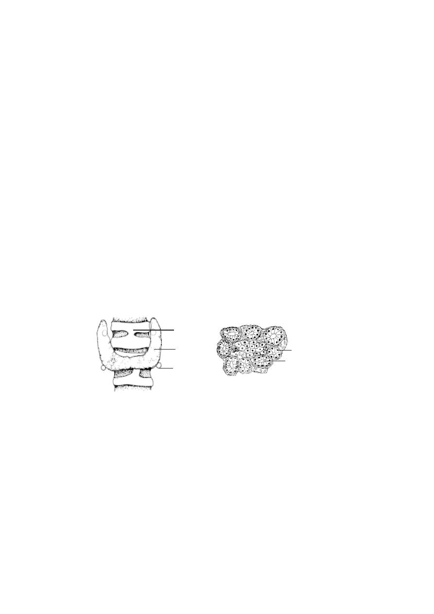
159
towards the posterior pituitary. Later, the pouch loses its connection with the
oral cavity and becomes the anterior pituitary. The anterior pituitary is subdi-
vided into three areas. They are, the pars tuberalis, pars distalis and pars
intermedia.
Relationship of the pituitary to the brain.
There is a network of blood vessels on the hypothalamus. It is called
the primary capillar y nerwork. A portal system called the
hypothalamohypophyseal portal system extends from a part of the hypo-
thalamus to the anterior pituitary (a portal blood vessel begins and ends as
capillaries). The portal system in turn opens into the secondary capillary net-
work of the anterior pituitary. The neurohormones produced by the hypothala-
mus are collected by the primary capillary network. Through the portal sys-
tem they enter into the secondary network of the anterior pituitary.
Thyroid gland
The thyroid gland is composed of two lobes. They are placed on the
lateral sides of the upper portion of the trachea. These lobes are connected by
a narrow band of thyroid tissue called the isthmus. The isthmus extends across
the anterior aspect of the trachea.
The thyroid is one of the largest endocrine glands. It weighs approxi-
mately 20g. It is richly supplied with blood capillaries. It is more red than its
neighbouring tissues.
The gland is composed of numerous follicles. They are small spheres.
Their walls are made up of cuboidal epithelial cells. The central cavity or
lumen of each follicle is filled with a protein called the thyroglobulin. It stores
large amount of thyroid hormone. The thyroid secretes thyroxine and calci-
tonin.
Fig.3.10.3. Thyroid and Parathyroid glands
Fig.3.10.4. Thyroid C.S
trachea
thyroid
parathyroid
glandular cells
acinus

160
Parathyroid glands.
The parathyroid glands are found in association with the thyroid glands.
They are found embedded in the posterior part of each lobe of the thyroid
gland. There are four parathyroid glands. Inside the glands the cells are
organised in densely packed masses. The cells of the glands secrete parathy-
roid hormone.
Adrenal glands or Suprarenal glands.
These glands are found near the superior pole of each kidney. They
are surrounded by adipose tissue. The glands are enclosed by a connective
tissue capsule.
The adrenal glands are composed of an inner medulla and outer cor-
tex. These regions are formed from two separate embryonic tissues. The
medulla consists of closely packed polyhedral cells. They are centrally lo-
cated in the gland. The cortex is composed of smaller cells. These cells form
three distinct layers, namely the zona glomerulosa, the zona fasciculata
and the zona reticularis. These layers are structurally and functionally spe-
cialized.
The adrenal medulla secretes two major hormones. They are epi-
nephrine (or adrenaline) and norepinephrine (or noradrenaline). The hormones
of the adrenal cortex are the cortisol and aldosterone.
Pancreas.
The pancreas lies between the greater curvature of the stomach and
the duodenum. It is an enlarged structure. It is approximately 15 cm long. It
weighs 85 -100g.
The pancreas is both an exocrine and an endocrine gland. The endo-
crine part consists of pancreatic islets (islets of Langerhans). They are
approximalety 500,000 to 1,000,000 in number. The islets are distributed in the
pancreas. The islets are composed of alpha (
α
) cells (20%) and beta (
β
)cells
(75%). While the œ cells secrete glucagon the
β
cells secrete insulin. A
third type of cells called the delta (
δ
) cells (5%) have been identified. These
cells secrete somatostatin.
3.11 Urinary system
It is customary to link the organs of urinary excretion and reproduc-
tion as urino-genital system. The suitability of this concept is questionable.
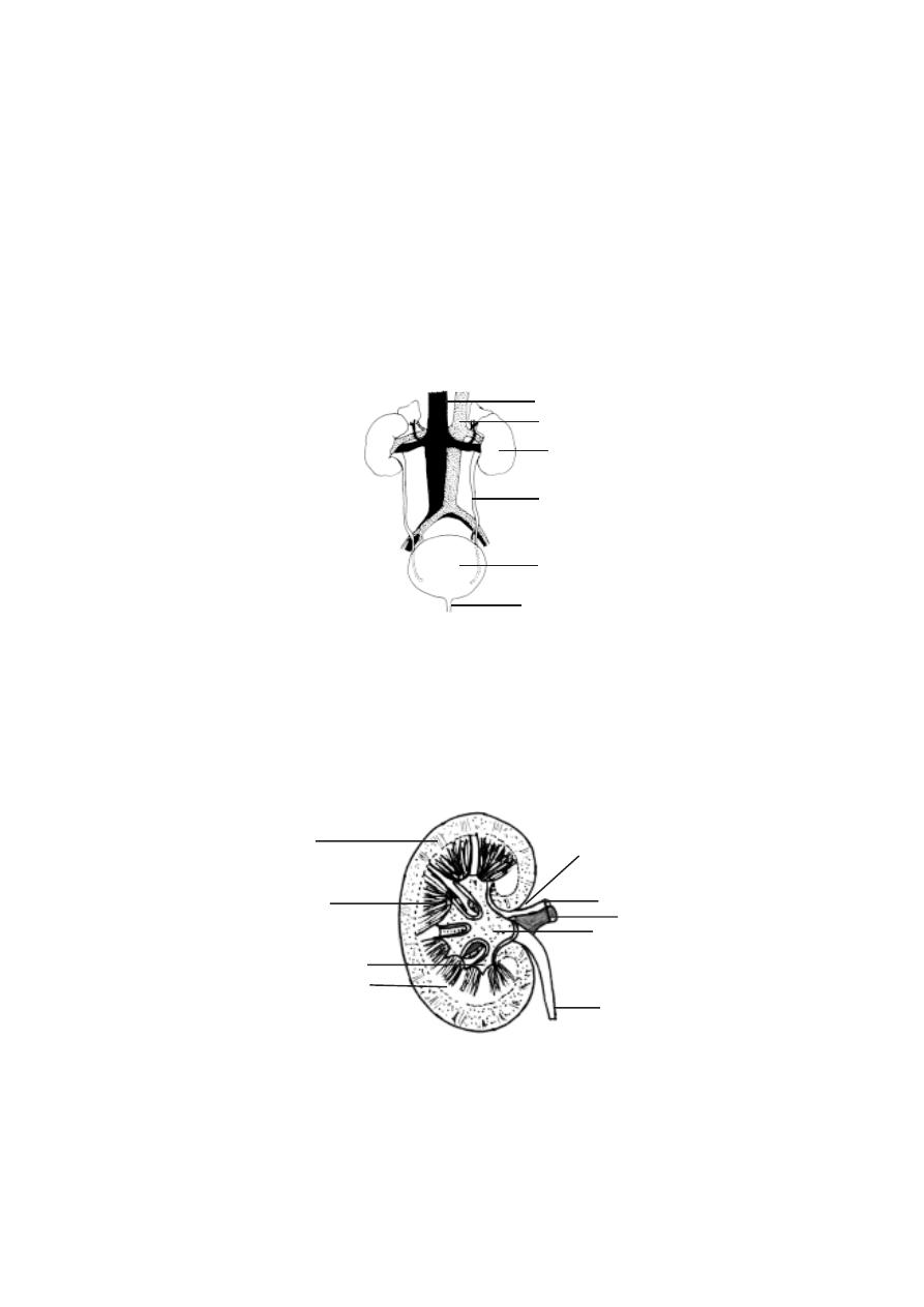
161
The urinary and reproductive organs differ in their embryological origin and
development. In postnatal human beings, the association between the compo-
nents of the urinary and the reproductive systems is very much limited. Hence
the urinary and reproductive systems are considered separately.
The urinary organs comprise, two kidneys (renes), ureters, the uri-
nary bladder (vesica urinaria) and the urethra.
The kidneys.
The kidneys are bean shaped organs. In fresh state the kidneys are
reddish brown in colour. They lie on the posterior abdominal wall. In the
abdomen, the right kidney is slightly lower than the left. It is because of the
presence of liver superior to it. The kidneys are surrounded by adipose tissue.
Each kidney is about 11 cm in length, 6cm in breadth and 3cm in antero-
posterior dimensions. In adult males the average weight of kidney is about
150g (in adult female 135g).
Fig.3.11.1. Human Urinary system
renal vein
renal artery
kidney
ureter
urinary bladder
urethra
Fig.3.11.2. L.S. of Kidney
renal artery
cortex
renal pyramids
renal hilum
renal pelvis
renel vein
medulla
calyx
ureter
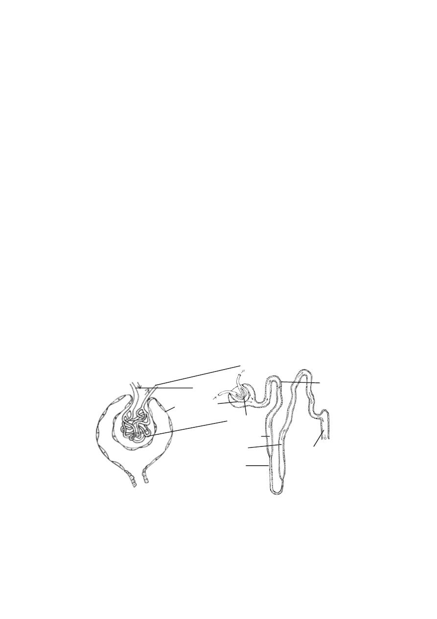
162
The inner margin of each kidney has a small depression called the
hilum. The renal artery and nerves enter and the renal vein and the ureter
exit at this region. The hilum opens into a cavity called the renal sinus.
Each kidney is enclosed by a fibrous connective tissue layer, called
the renal capsule. Internally the kidney is divided into an outer cortex and an
inner medulla. The medulla consists of several cone-shaped
renal pyramids. Extensions of the pyramids called the medullary rays, project
from the pyramids into the cortex. Extension of the cortex called renal col-
umns, project between the pyramids. The tips of the pyramids are called the
renal papillae. They are pointed toward the renal sinus. The renal papillae
are surrounded by funnel shaped structures called the minor calyces. The
minor calyces of several pyramids join together to form larger funnels called
major calyces. There are 8-20 minor calyces and 2 or 3 major calyces per
kidney. The major calyces converge to form an enlarged channel called the
renal pelvis. The renal pelvis then narrows to form the ureter. The ureter
leaves the kidney and gets connected to the urinary bladder.
Nephron.
The basic functional unit of each kidney is the nephron. There are
approximately 1.3 million nephrons in each kidney. Atleast 450,000 of them
must remain functional to ensure survival. Each nephron consists of an en-
larged terminal end called the renal corpuscle, a proximal tubule, a loop
of Henle and a distal tubule. The distal tubule opens into a collecting duct.
The renal corpuscle, proximal tubule and distal tubules are in the renal cortex.
The collecting tubules and parts of the loops of Henle enter the renal medulla.
u r i n i f e r o u s
B o w m a n ’ s c a p s u l e
afferent blood
g l o m e r u l u s
efferent blood vessels
loop of Henle
c o l l e c t i n g t u b u l e
Fig.3.11.3. Nephron
v e s s e l s
t u b u l e
Malpighian capsule
p r o x i m a l t u b u l e
d i s t a l t u b u l e

163
Most nephrons measure 50-55 mm in length. 15% of the nephrons
are larger and they remain near the medulla. These are called the
juxtamedullary nephrons. They have larger loops of Henle.
The renal corpuscle of the nephron consists of a Bowman’s capsule
and a bunch of capillaries called the glomerulus.
In the Bowman’s capsule the outer and inner layers are called pari-
etal and visceral layers respectively. The outer parietal layer is composed of
simple squamous epithelium. The inner visceral layer surrounds the glomeru-
lus. It consists of specialized cells called podocytes. The walls of the glom-
erular capillaries are lined with endothelial cells. There is a basement
membrane between the endothelial cells of the glomerular capillaries and the
podocytes of Bowman’s capsule. The capillary endothelium, the basement
membrane and the podocytes of Bowman’s capsule make up the filtration
membrane.
The glomerulus is supplied with blood by an afferent arteriole. It is
drained by an efferent arteriole.
The cavity of Bowman’s capsule opens into the proximal tubule. The
proximal tubule is also called the proximal convoluted tubule. It is approxi-
mately 14mm long and 60 µm in diameter.
Posteriorly the proximal tubule continues as the loop of Henle. Each
loop has a descending limb and an ascending limb. The first part of the de-
scending limb is similar in structure to the proximal tubule. The loops of Henle
that extend into the medulla become very thin near the end of the loop. The
first part of the ascending limb is also very thin and it consists of simple squa-
mous epithelium, but it soon becoms thick. The distal tubules, also called the
distal convoluted tubules are not as long as the proximal tubules.
Ureters and Urinary bladder
The ureters extend inferiorly from the renal pelvis. They arise medi-
ally at the renal hilum to reach the urinary bladder. The bladder is meant for
temporarily storing the urine. The urinary bladder is a hollow muscular bag. It
lies in the pelvic cavity. The size of the bladder depends on the presence or
absence of urine. The bladder capacity varies from 120-320ml. Filling upto
500 ml is tolerated. Micturition will occur at 280ml. The ureters enter the
bladder inferiorly on its posterolateral surface. The urethra exits the bladder
inferiorly and anteriorly. At the junction of the urethra with the urinary bladder
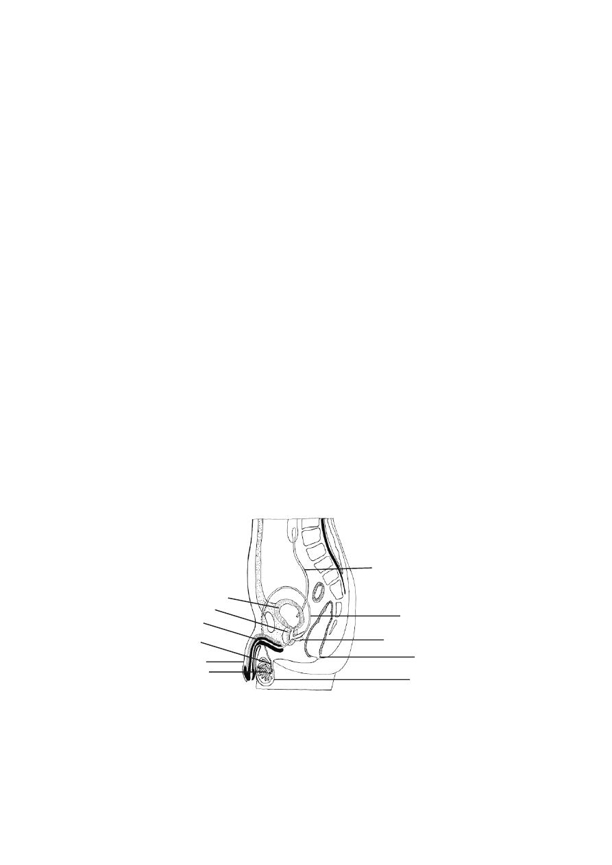
164
smooth muscles of the bladder form the internal urinary sphincter. Around
the urethra there is another external urinary sphincter. The sphincters control
the flow of urine through the urethra.
In the male the urethra extends to the end of the penis where it opens
to the outside. In male the urethra is 18-20cm long. In the female the urethra
is shorter. It is about 4 cm long and 6 mm in diameter.
3.12. Reproductive system
The process of sexual reproduction is a wonderful act in nature. This
process, apart from ensuring a healthy progeny provides an oppurtunity to
produce enormous range of genetically varied offsprings. Organisms have
adopted several strategies for sexual reproductive processes. Such
adaptations have resulted in suitable morphological, anatomical and behav-
ioral modifications. Human reproductive organs as internal and external geni-
talia are highly sophisticated yet simple in their functioning. The functioning is
in accordance with psychological and endocrinological thresholds. An
academic approach towards an understanding of the human male and
female reproductive organs and their functions will go a long way in
avoidance of unethical, unhealthy and unhygenic practices encoun-
tered at specific periods in life.
Male reproductive organs
The male reproductive system consists of the testes (singular : tes-
tis), epididymides (sing: epididymis), ductus deferentia or vasa deferentia
(sing : ductus deferens, vas deferens), urethra, seminal vesicles, prostate
gland, bulbourethral glands, scrotum and penis.
Fig. 3.12.1. Human male reproductive organs
seminal vesicle
scrotum
anus
vas deferens
ureter
urinary bladder
prostate gland
urethra
epididymis
testis
penis
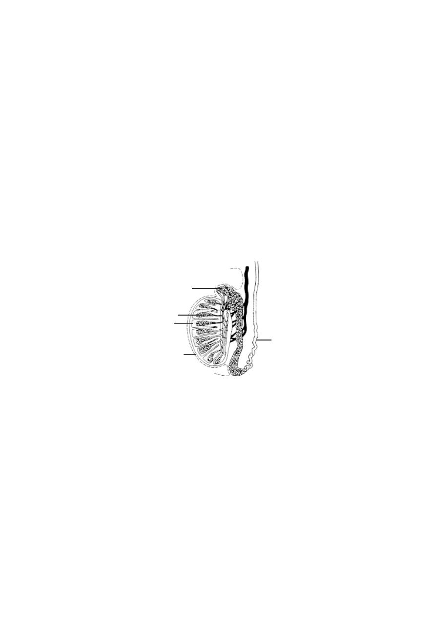
165
Testes :
The testes are the primary reproductive organs or gonads in the
male. These are suspended in the scrotum by scrotal tissues.
The sperm cells are temperature sensitive. They do not develop nor-
mally at usual body temperatures. Hence the testes and epididymides in which
the sperm cells develop, are located outside body cavity in the scrotum, where
the temperature is low.
The left testis usually is 1 cm lower than the right. An average testis
is 4-5 cm in length, 2-5cm in breadth. Its weight varies from 10.5-14g.
The outer part of each testis is a thick, white capsule called tunica
albuginea. Internally the testis contains several incomplete septa. The septa
divide each testis into about 300-400 cone shaped lobules. The lobules contain
seminiferous tubules and interstitial cells or Leydig cells. Sperm cells de-
velop within the seminiferous tubules.
The seminiferous tubules are extensive. The combined length of the
tubules in both testes is nearly 800 metres. These tubules through a set of
short, straight tubules open into tubular network called the rete
testis. The rete testis in turn open into efferent ductules. Internally the tu-
bules and ductules are lined by ciliated columnar epithelium. These cells help
to move the sperm cells out of the testis.
Epididymis
: It is formed of extremely convoluted ductules coming out of the
testis. It occurs on the posterior side of the testis. The maturation of sperm
cells occurs within the ductules of the epididymis.
Fig. 3.12.2. L. S. of the Testis
tunica albuginea
testis
epididymis
coiled seminiferous tubule
vas deferens

166
Vas deferens or ductus deferens
: It emerges from the tail end of the
epididymis and ascends along the posterior side of the testis. It becomes asso-
ciated with the blood vessels and nerves that supply the testis. Collectively
these structures constitute the spermatic cord. Thus the spermatic cord con-
sists of (1) Vas deferens (2) testicular artery and venus plexus (3) lymph
vessels (4) nerves (5) fibrous processes and muscles. This cord enters into
the pelvic region. The end of the vas deferns enlarges to form the ampulla. At
this region the vas deferens is surrounded by smooth muscles capable of peri-
staltic contraction. They help to propel the sperm cells through the ductus
deferens.
Ejaculatory duct : Nearer to the ampulla of each vas deferens there is a sac
like seminal vesicle. It joins the ductus deferens to form the ejaculatory
duct. These ducts are about 2.5 cm long. They project into the prostate gland
and end by opening into the urethra.
Urethra :
The male urethra extends from the urinary bladder to the distal end
of the penis. It is about 20 cm long. It is a passageway for both urine and
reproductive fluids. The urethra is divided into three parts. They are
1. The prostatic Urethra - It is closest to the bladder and passes through the
prostate gland.
2. The membranous urethra
- It is the shortest part of the urethra and it
extends from the prostatic urethra.
3. The spongy urethra or penile urethra
- It is the longest part of the
urethra. It extends from the membranous urethra, through the length of the
penis. There are several minute mucus secreting urethral glands opening into
the urethral passage.
Penis
- It is the male copulatory organ. It consists of two parts namely the
radix or root and the corpus or body. The radix attaches the penis to the
lower abdomen. The corpus is normally pendulous. It is covered by a loose
skin.
Fig. 3.12.3. C. S of Penis
corpus cavernosum
corpus spongiosum
spongy urethra

167
The corpus of the penis consists of three masses of erectile tissue.
Flooding these tissues with blood causes the penis to enlarge and become
firm. These tissues are the right and left corpora cavernosa and the median
corpus spongiosum penis. Most of the corpus is formed of the corpora
cavernoas. The corpus sporgiosum penis surrounds the urethra and near the
end of the penis it expands into a conical, glans penis. Its
swollen base is the corona glandis.
The skin over the penis is thin. It is loosely connected to the tunica
albuginea. At the tip of the penis it is folded to form the prepuce or the
foreskin. It overlaps the glans penis. The corona glandis and penile neck
have numerous preputial glands.
Seminal vesicles
- These are two sac-like structures located between the
bladder and rectum. Each vesicle is about 5 cm long. Their secretions contrib-
ute about 70% of the seminal fluid.
Prostate
- It is a firm structure. It is partly glandular and partly fibromuscu-
lar. It is found around the beginning of the male urethra. It is about 3 cm in
diameter. It weighs about 8g.
The muscular part of the prostate may help in dilating the urethra to
hold the seminal fluid (3-5ml) during the period of sexual excitement prior to
ejaculation.
After the middle age the prostate often enlarges. It may project into
the bladder and interrupt urination.
Bulbo-urethral gland
- These are two glands. They are small round masses
about 1 cm in diameter. They lie lateral to the membranous urethra. Its secre-
tion may control genito-urinary diseases.
Scrotum
- It is a fibromuscular sac. It contains the testes and their associated
ducts. It is divided into right and left by cutaneous raphe. Its left side is usually
lower. The external appearence varies according to age and body tempera-
ture. The scrotal skin is thin and pigmented. It has numerous sweat glands and
nerve endings.
Female reproductive organs
In human female the internal reproductive organs are the ovaries,
uterus, uterine tubes and vagina. Externally the organs are the mons pu-
bis, labia majora and labia minora, clitoris and vestibular glands.
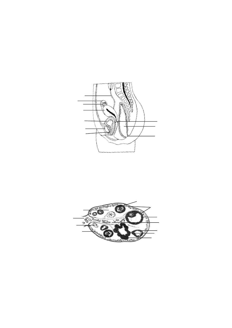
168
Ovaries
- These are paired structures. The two ovaries are placed on each
side of the uterus in the pelvic region. They are greyish pink in colour. Each
ovary is almond shaped. They are about 3cm long,1.5cm wide and 1cm thick.
The ovary is attached to the posterior surface of the inner body wall
by a membranous fold called the mesovarium. The ovary is further supported
by suspensory and ovarian ligaments.
Ovarian structure
- In young female the surface of the ovary is covered by
a layer of ovarian surface epithelium. It consists of a single layer of cuboi-
dal cells. Beneath the epithelium the ovary is surrounded by a tough coat
named tunica albuginea. It is made of collagenous tissue.
The overy proper is divisible into two regions, namely the cortex and
the medulla. The cortex region contains the ovarian follicles. The medulla is
interior. It receives blood vessels and nerves at the hilum.
Fig. 3.12.5. Histology of the Ovary
mesovarium
tunica albuginea
cortex
medulla
corpus luteum
theca interna
primary follicles
granulosa cells
secondary follicle
cumulus mass
ureter
ovary
uterus
urinary bladder
urethra
pubic bone
anus
rectum
cervix
Fig. 3.12.4. Human female reproductive organs
fallopian tube or
uterine tube

169
After puberty the cortex forms the major part of the ovary. It con-
tains ovarian follicles and corpora lutea of various sizes. Their size de-
pends on the stage of menstrual cycle or age. The cortex is filled with stroma
composed of collagen. The follicles are embedded in the stroma.
Ovarian follicles
The formation of the female gamete has many different phases and it
is complex. At birth, the primordial follicles are found in the superficial zone of
the cortex. They contain primary oocytes (about 25mm in
diameter). Each one of them is surrounded by a single layer of flat follicular
cells. The follicles undergo changes as the female attains puberty. The vari-
ous follicular stages are :
1. Primary follicle
- The follicle cells are converted from squamous to cuboidal
cells. The follicular membrane or membrana granulosa becomes multilay-
ered. The oocyte increases in size. It has an outer thick layer called the zona
pellucida. The follicular cells divide and form granulosa cells.
2.Secondary follicle
- It is about 20µm thick. The granulosa cells surround
the oocyte and form a mound of cells called the cumulus ovaricus. The inner
and outer theca become prominent. The theca interna is well established.
3. Tertiary follicle
- Only one follicle reaches the tertiary stage. It increases
in size (2mm diameter). Now it is called the graffian follicle. The oocyte and
ring of cells surrounding the oocyte (corona radiata) break away and float
freely in the follicular fluid. Finally the wall of the follicle ruptures and the
contents are released into the peritoneum.
The ovary of the foetus at 5 months gestation has 7 million oocytes.
At birth the ovary of the child contains about 1 million oocytes. Due to further
degeneration at the time of puberty only about 40,000 oocytes remain. Of the
40,000 oocytes only about 400 undergo ovulation during the reproductive years.
Corpus luteum
- It is formed after ovulation. The walls of the empty follicle
collapses and fold extensively. The granulosa cells of the theca externa get
enlarged.
They are now termed as luteal cells. They secrete hormones. In
pregnancy the corpus luteum persists. Otherwise, it degenerates after 10-12
days. The connective tissue cells get enlarged. It becomes white in colour and
is now called as the corpus albicans. In course of time it shrinks and
disappears.
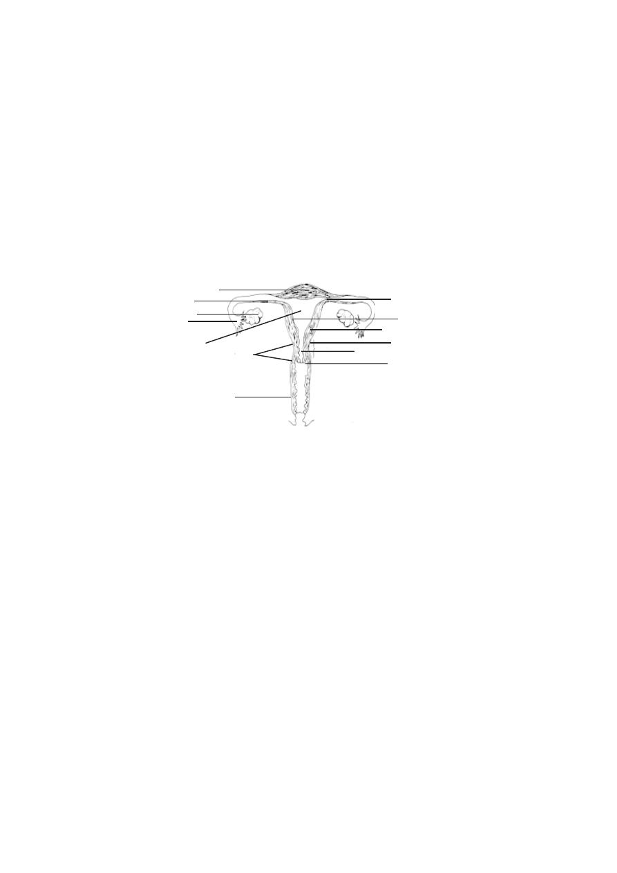
170
Uterine tubes (Fallopian tubes)
- There are two uterine tubes or oviducts,
one on each side of the uterus. Each one is associated with a ovary. Each
tube is about 10 cm length. The terminal part of the tube is enlarged to form
the infundibulum. It opens into the peritoneal cavity. The opening is called the
ostium. The uterine tube consists of three parts. The part nearer to the in-
fundibulum is called the ampulla. It is the longest part. That part of the tube
nearer to the uterus is called the isthmus. It is narrow. The tubular part enter-
ing into the uterus is called the uterine or intramural part.
Uterus
It is a hollow thick walled muscular organ. It is pear shaped. It is
about 7.5cm long and 5 cm wide. It weighs about 50g.
During pregnancy its weight may go upto 1kg. Its larger rounded part
is called as the fundus. The narrower part is called as the cervix. The cervix
is directed inferiorly. The middle part is the body . The uterus
continues as the cervical canal and opens into the vagina through a opening
called the ostium.
The wall of the uterus is three layered. The outermost layer is the
perimetrium or serous layer. The major part of the wall is made up of the
next layer called the myometrium or muscular coat. The
innermost layer is the endometrium or mucous membrane. The endometrium
is a functional layer. It undergoes menstrual changes and sloughing during
female sex cycle.
Vagina
- It is the female copulatory organ. It is a fibromuscular tube. It is
about 10 cm long. It extends from the uterus to the outside. The vaginal pas-
sage is used during intercourse and it allows menstrual flow and child birth.
Fig. 3.12.6. Human female reproductive system
ovary
isthmus
uterine part
fundus
cervix
cervical canal
ostium
perimetrium
endometrium
myometrium
Uterus
vagina

171
External Genitalia
Vestibule
- The external female genitalia is known as the vulva or puden-
dum. It consists of the vestibule and its surrounding structures. The vestibular
region remains in between the two labia majora. It contains the vaginal open-
ing and the urethral opening. The vestibular region is surrounded by the
mons pubis anteriorly and labia majora and labia minora on the lateral
sides.
Mons pubis
- It is a rounded eminence situated anteriorly. It is made up of
subcutaneous adipose connective tissue. It is covered by coarse hair at the
time of puberty. It corresponds to similar structure in the male.
Labia majora
- These are two longitudinal folds of skin. They form the outer
boundary for the vestibule.
Labia minora
- These two small skinfolds lie between the labia majora. They
remain nearer to the vaginal opening.
Clitoris
- It is homologus with male penis. It is an erectile structure. It is
found in the anterior margin of the vestibule. It is a sensitive region having
sensory receptors.
Hymen vaginae
- It is a thin mucous membrane. It is found within the vagi-
nal orifice or opening. If the membrane completely closes the vaginal opening,
it should be removed to allow menstrual flow. In young women the hymen
may normally get torn during physical exercise. In some women it may be
absent. It has no established function.
External urethral opening
- This opening is about 2.5 cm below the clitoris.
It is anterior to the vaginal opening. It remains as a small cleft.

172
Self evaluation
Choose the correct answer
1. The cornified region of the skin is formed of
a) stratum lucidum
b) stratum basale
c) stratum spinosum
d) stratum corneum
2. The goose flesh is formed due to the contraction of
a) diaphragm
b) errector pili
c) trapezius muscle
d) gluteus maximus
3. The skin colour is due to the pigment.
a) melanin
b)haemoglobin
c) neutral red
d) Janus green
4. The number of facial bones are
a) 26
b) 25
c) 14
d) 22
5. The cervical vertebra supporting the head is
a) axis
b) atlas
c) sacral
d) lumbar
6. Choose the flat bone in our body.
a) skull bone
b)ribs
c) sternum
d) all the above
7. The number of thoracic bones are
a) 5
b)12
c) 7
d) 1
8. The bone that holds the upper limb away from the body is
a) scapula
b)pelvic bone
c) clavicle
d) femur

173
9. The broadest muscles are named as
a) deltoid
b) gracilis
c) longus
d) lattismus
10. In shape the deltoid muscle is a
a) square
b) slender
c) circular
d) triangular
11. The major breathing movement is due to
a) scalene
b) thoracic
c) diaphragm
d) intercostals
12. The largest salivary glands are
a) parotid
b)submandibular glands
c) sublingual glands
d) labial glands
13. The human dentition is
a) heterodont
b) thecodont
c) diphyodont
d) all the above
14. The
length of the duodenum in human alimentary canal is
a) 8 cm
b) 1.8 m
c) 9 cm
d) 25 cm
15. The lung is surrounded by
a) duramater
b) pericardial membrane
c) pleura
d) meninges
16. Breathing process can be interfered with due to
a) closure of the ileo-colic valve
b) enlargement of the tonsil
c) closure of pyloric sphinctes
d) vibration of the vocal cord
17. The reduction in blood pressure may be caused due to
a) distributing vessels
b) resistance vessel
c) exchange vessels
d) reservoir vessels

174
18. Vaso-dilation and vaso-constriction are caused by
a) tunica intima
b) exchange vessels
c) tunica media
d) tunica adventitia
19. The wall of the blood vessels are supplied with blood by
a) vasa nervosum
b) exchange vessels
c) vasa vasorum
d) capacitance vessels
20. The exchange vessels are
a) arteries
b) arterioles
c) Venules
d) Capillaries
21. Spleen is located on the left side of
a) the abdominal cavity
b) the thoracic cavity
c) the lung
d) the kidney
22. The synapses are formed between
a) nerves and muscles
b) nerve tissues
c) capillaries
d) organs
23. The cerebral hemispheres are connected by a sheet of nerve fibres called
a) corpora quadrigemina
b) choroid plexus
c) corpus callossum
d) cauda equina
24. The number of human cranial nerves is
a) 12 pairs
b) 31 pairs
c) 10 pairs
d) 11 pairs
25. The Melbomian glands produce
a) sebum
b) tear
c) wax
d) oil
26. The interoccular pressure is maintained by
a) aqueous humor
b) vitreous humor
c) cerebro spinal fluid
d) lymph fluid

175
27. The hypophysis is connected with the brain through
a) pars tuberalis
b) adeno hypophysis
c) hypothalamus
d) pars distalis
28. The average weight of human thyroid gland is
a) 10 gms
b) 20 gms
c) 500 gms
d) 20 kg
29. The average weight of kidney in adult female is
a) 150 g
b) 135 g
c) 75 g
d) 250 g
30. The bunch of capillaries in the Bowman’s capsule forms the
a) medullary rays
b) calyces
c) glomerulus
d) capillary endothelium
31. The interstitial cells are otherwise called
a) Leydig cells
b) Sperm cells
c) Glandular cells
d) Secretory cells
32. The ovary of the foetus at 5 months gestation has
a) 40,000 Oocytes
b) 7 million Oocytes
c) 400 Oocytes
d) No Oocytes
33. The inner layer of membrane lining the uterus is
a) perimetrium
b) myometrium
c) endo metrium
d) serous layer
34. The uterine tubes are otherwise called as
a) ureters
b) spermiducts
c) fallopian tubes
d) birth canal

176
Part - II
1. What is keratinization ?
2. What is callus ?
3. What are floating ribs ?
4. What is a synovial joint ?
5. Name the two catagories of bones.
6. What is foramen magnum ?
7. What is acetabulm ?
8.Differentiate skeletal and visceral muscles.
9. Name the kissing muscles
10. Write a short note on the structure of the cardiac muscle.
11. Name the muscles which are essential for the movement of the neck.
12. Provide the human dental formula
13. What is gingiva.
14.Name the three pairs of Salivary glands.
15. Name the four parts of the Colon.
16. What is Adam’s apple ?
17. What is carina ?
18. What is acinus ?
19. What is double circulation ?
20. Name the layers found in the wall of the heart.
21. Provide the root of systemic circulation
22. What is the role of B-lymphocytes ?
23. What are sulci and gyri ?
24. What is a neuromuscular junction ?
25. What is choroid plexuses ?

177
26. What is melbomian gland ?
27. What is Rathke’s pouch ?
28. What are podocytes ?
29. What is the function of the ciliary muscles ?
30. What is an eardrum ?
31. Name the parts of the anterior pituitary.
32. Name the layers of the adrenal cortex.
33. What is the role of prostate glands ?
34. What is corpus luteum ?
35. What is graffian follicle ?
Part - III
1. Draw the different layers of the skin and label the parts.
2. Give an account of the nail and its structure
3. Describe the structure of typical human vertebra
4. Describe the structure of a typical long bone.
5. Describe the various types of joints.
6. Give an account of the muscles of the lower limb
7. Draw a neat labelled sketch of a tooth.
8. Give a description of the human liver
9. Describe the structure of stomach with diagram.
10. Explain the structure of the larynx with diagram.
11. Describe the paired and unpaired cartitages of larynx
12. Write notes on portal circulation
13. Describe the types of the blood vessels.
14. Draw a neat labelled sketch showing the internal structure of the heart
15. Give an account of the thymus
16. Explain
a) Lymph nodes
b) Tonsils

178
17. Describe the structure of a Neuron with diagram.
18. Explain the cross section of spinal cord with diagram
19. What is the structure of a peripheral nerve ?
20. Explain the organization of the lacrimal apparatus in human eye
21. Draw a neat labelled sketch of the C.S. of human eye.
22. Explain the structure of the inner ear.
23. Describe the structure of the thyroid gland with diagram.
24. Describe the endocrine nature of the pancreas
25. Provide the structure of human kidney
26. Explain the various stages of the Ovarian follicles.
27. Describe the Ovarian structure with diagram.
28. Give an account of corpus luteum
Part - IV
1. Explain the structure of the various skin-dervatives with suitable diagrams.
2. Name bones in the skull of man. Explain the same with a neat diagram.
3. Write an essay on the structure and organization of the axial skeleton in
human beings.
4. Explain the appendicular skeleton with diagram.
5. Name the muscles of the upper limb and explain how they work.
6. Give an account of the organs of digestion in the buccal cavity.
7. Describe the structure of digestive system with diagram.
8. Explain the structure of respiratory system with diagram.
9. Write an essay on structure and types of blood vessels
10. Describe the systemic and pulmonary blood circulations in man with
diagram.
11. Explain the structure of human heart.
12. Provide a detailed description of the anatomy of human brain
13. Explain the structure of human urinary system with diagram.
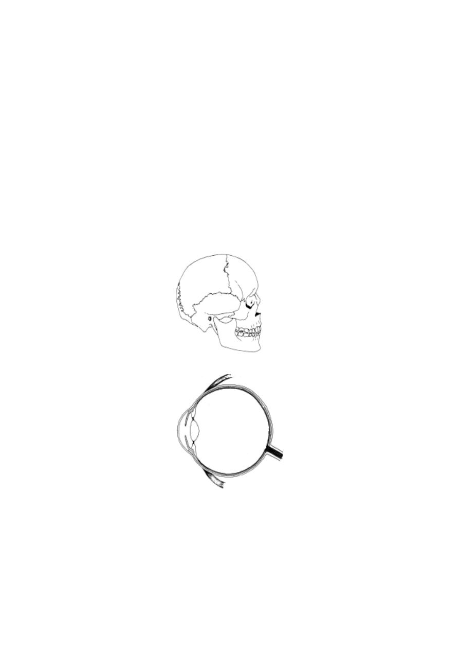
179
14. Describe the structure of a nephron with diagram.
15. Give an account of the structural organization of the hypophysis
16. Write an essay on the primary sex organs and associated structures of a
human male.
17. Explain the structure of female reproductive organs in human with suitable
diagrams.
18. Name the muscles in the human trunk region and explain how they work.
Label the diagrams

180
4. GENETICS
4.0. Introduction
Genetics is the study of inheritance or heredity. It deals with the trans-
mission of characters, specific for that particular type of plant or animal, from
the parent to the offspring of the next generation.
The history of Genetics is closely linked with the ancient cultural his-
tory of man. It began ten thousand years ago, when human beings changed
from a nomadic life to the life of settlers. As settlers in early civilization they
started living as groups. A need for food forced them to explore nature and
adopt agriculture. The agricultural revolution in early human civilization opened
the doors for very rapid improvements in human culture. They learnt to iden-
tify plants that could be grown and cultivated. Several cultivation processes
evolved. They started selecting suitable varieties for agriculture.
Simultaneously they started domestication of animals. Many animals
were brought under human control. Gradually they understood their reproduc-
tive methods. Hybridisation and generation of new varieties of animals like
horses, dogs, cats and cattle happened. The idea of Genetics was adopted in
everyday practice.
A scientific approach to understand inheritance was initially made by
Gregor Mendel. He did hybridization experiments in Pisum sativum more
out of curiosity and his devotion to science. He applied statistical methods to
biological research.
Since the ‘rediscovery’ of Mendelism in 1900, Genetics has made rapid
developments. The advancement in cell biology, microscopy, biochemistry and
other disciplines provided the necessary infrastructure for this science to reach
its climax. As of to-day all processes and causes for inheritance are well
understood. Through the developments in Genetic Engineering, Biotechnol-
ogy, Tissue culture and other methods we are trying to exploit nature. How-
ever, the steady processes of dominance of human beings on earth through
their knowledge of science and skill in technology will also ensure the safety
of earth in the times to come. In such a safeguarding the science of Genetics
with all its related fields will play a key role.
4.1 Multiple Alleles
According to Mendelism, a genetical character is controlled by the
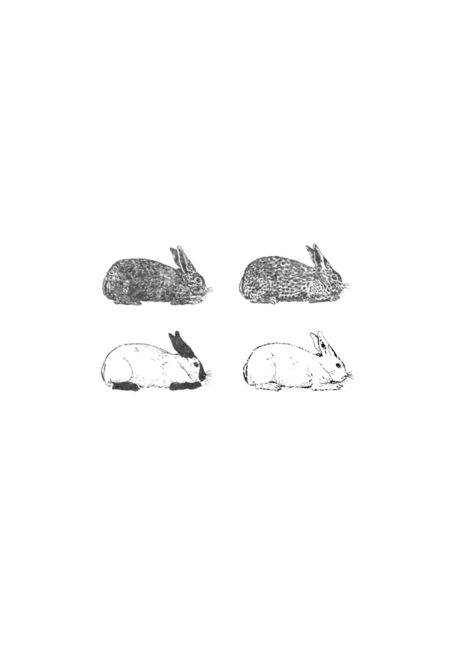
181
combined action of two genes or factors in the same loci of two homologus
chromosomes. It means that each gene has two alternative forms or
alleles and their expression are knwon as allelomorphs. Of these two, one is
dominant and the other recessive. However, some genetical characters are
determined by several forms of an allele known as multiple alleles. There
are many examples for multiple allelism.
1.
Skin colour in rabbits
Rabbits have five kinds of skin colour, coloured (agouti), chinchilla,
himalayan albino and albino, light grey. Of these, the chinchilla variety is
lighter in colour. Himalayan albino will have pink eyes, white coat colour and
black colour in tips like nose, tail and feet.
A crossing of a homozygous coloured rabbit with an albino resulted in
following F
1
and F
2
generation.
Parents
CC
x
c
a
c
a
(coloured)
(albino)
F
1
Cc
a
x
Cc
a
(coloured)
F
2
CC
Cc
a
c
a
c
a
25%
50%
25%
(coloured) (coloured)
(albino).
Fig. 4.1.1. Different colours in Rabbit
Coloured(agouti)
Chinchilla
Himalayan albino
Albino

182
This result shows that coloured condition is dominant over albino.
Other possible crossings are
Parents
CC
x
c
h
c
h
(coloured)
(Himalayan albino)
F
1
Cc
h
x
Cc
h
(coloured)
F
2
CC
Cc
h
c
h
c
h
25%
50%
25%
(coloured)
(coloured)
(Himalayan albino)
Parents
c
h
c
h
x
c
a
c
a
(Himalayan albino)
(albino)
F1
c
h
c
a
x
c
h
c
a
(Himalayan albino)
F2
c
h
c
h
c
h
c
a
c
a
c
a
25%
50%
25%
Himalayan albinos Himalayan albinos
albino
The above crossings show that the genes C, c
h
and c
a
representing
coloured, himalayan albino and albino are alleles of each other.
Parents
c
ch
c
ch
x
c
a
c
a
(chinchilla)
(albino)
F
1
c
ch
c
a
x
c
ch
c
a
(light grey)
F
2
c
ch
c
ch
c
ch
c
a
c
a
c
a
25%
50%
25%
(chinchilla)
(light grey) (albino)
In chinchilla, coat colour is lighter than the coloured (agouti). The
coloured character is dominant over chinchilla. However F
1
hybrids between
chinchilla and himalayan albino (C
ch
c
h
) or between chinchilla and albino (c
ch
c
a
)
show light grey skin colour. The occurrence of light grey colour is due to
partial expression of the gene for chinchilla in a heterozygous state.
Genotype
Phenotype
CC, Cc
ch
, Cc
a
,Cc
h
coloured (wild)

183
c
ch
c
ch
chinchilla
c
ch
c
h
,c
ch
c
a
light grey
c
h
c
h
,c
h
c
a
himalayan albino
c
a
c
a
albino
2.
ABO blood groups in human beings
The ABO blood group system in human beings was established by K.
Landsteiner. It is based on the presence or absence of certain antigens. There
can be two anitigens A or B in the blood, resulting in four blood groups, namely
A,B, AB and O. These are called ABO blood groups or Landsteiner blood
groups.
The inheritance of ABO system illustrates a new principle in genetic
control of phenotypes.
The blood of a person having A group will have the antigen A and a
person having B group will have the antigen B. With these antigens A and B
there are certain naturally occurrring antibodies in the serum of the blood. The
antibodies in a particular individual will be found only for those antigens which
are absent in blood of this individual. The presence of antigens and antibodies
occur as follows
Blood groups
Antigen
Antibody in the serum
A
A
anti B
B
B
anti A
AB
A and B
None
O
None
anti A and B
Antibodies in the blood of ‘group A’ will agglutinize red blood corpuscles
of the blood group B. Similarly the antibody in blood ‘group B’ will agglutinize
red blood corpuscles of the blood group A. Since no antibody is found in ‘group
AB’ blood, it will not agglutinize any other group . ‘Group O’ will have anti-
bodies for ‘group A’ and B. Hence ‘group O’ will agglitinize group A and B.
Compatibility of donor blood to that of the recipient will occur as fol-
lows.
Blood group of the donor
Blood group of the recipient
A
A and AB
B
B and AB

184
A B
A B
O
O, A, B, AB
From the table provided it is obvious that ‘group AB’ is universal
recipient. ‘Group O’ is universal donor.
The gene for ABO system is conventionally represented by the sym
bol ‘I’. Alleles I
A
and I
B
code for the enzymes involved in the formation of the
antigens A and B respectively and the allele ‘I
o
’ for no known protein. Thus
the genes can result in 6 possible genotypes but four possible phenotypes as
found in the table
Blood group
Possible genotype
O
I
o
I
o
A
I
A
I
A
or I
A
I
O
B
I
B
I
B
or I
B
I
O
A B
I
A
I
B
The alleles I
A
and I
B
jointly express themselves in the individual, they
are codminant. Both I
A
and I
B
are dominant to the recessive allele I
o
.
Disputed parentage and blood groups
The identification of blood group may help to decide in cases con-
cerned with parentage issues. By knowing the blood groups of parents, it is
possible to determine the possible blood groups in the children. The impossibil-
ity of a particular blood group in the progeny can also be pointed out.
Parents
Blood group in progeny
Impossible blood groups
O x O
O
A, B, AB
O x A
O, A
B, AB
O x B
O, B
A, AB
O x AB
A, B
O, AB
A x A
A, O
B, AB
A x B
A, B,AB, O
None
B x B
B, O
A, AB
A x AB
A, B, AB
O
B x AB
A, B, AB
O
AB x AB
A, B, AB
O

185
Rh blood group
Rh factor in blood was discovered by Landsteiner and Wiener in
1940. It was initially discovered in rabbits, immunized with the blood of Rhesus
monkey. The human beings whose blood will get agglutinated with rabbit se-
rum were designated as Rh
+
, and whose blood if not agglutinated were desig-
nated as Rh
-
. Wrong transfusion can cause agglutination of blood in the
recipient Hence before transfusion of blood, along with identification of ABO
blood group, it is necessary to test compatibility of Rh factor.
The presence of Rh
+
child in the uterus of the Rh
-
mother can cause
agglutination in the blood of the fetus. Even though such an unfortunate inci-
dent may not happen in the first pregnancy, it could occur in successive preg-
nancies. The death of the foetus in such cases is due to haemolytic anemia.
This disease is called erythroblastosis fetalis.
4.2. Quantitive inheritance :-
Charles Darwin while working on his theory of Natural selection,
recognised two principal types of variations, namely continuous and discon-
tinuous. The continuous variations show the whole range of variations in a
particular character. The discontinuous variations would appear all of a sud-
den and show no gradation.
Mendel in his work depended on sharp or alternate characters com-
prising discontinuous variations. For example, when tall and dwarf plants were
crossed, in F
1
only tall plants appeared. Crossing F
1
plants, produced only tall
and dwarf plants in the F
2
generation. Mendel discarded intermediate charac-
ters if any, seen.
However, by the end of the 19
th
century Galton, a British geneticist
and statistician was interested in the study of continuous variations. He tried
to find an answer for its origin. He called these characters as metrical char-
acters and found them inherited.
Thus in the begnning of the 20
th
century two groups of geneticists
emerged. They were the mendelians and the biometricians. While the
mendelians considered that all hereditary differences are discontinuous and
qualitative, the biometricians belived that hereditary variations are basically
continuous and quantitative. These two views remained contradictory. Later,
Johansen (1903), through his work on bean seeds proved that both the views
of mendelians and biometricians were only partly correct.
Yule (1906) suggested that quantitative variations may be controlled

186
by large number of individual genes, with each gene having a small effect.
Later on, such genes were called as polygenic systems. The hereditary pro-
cesses operating through such system was explained through multiple
factor hypothesis.
Multiple factors
Skin colour in human beings
The inheritance of skin colour in black and white populations in United
States of America was worked out and reported by C. B. Davenport in
1913. In USA, marriages between black and white individuals has resulted in
a population known as mulattoes. They have intermediate skin colour in the
first generation. When the mulattoes marry among themselves, all shades of
skin colours are obtained.
If the skin colour is due to genes in two loci A and B, the genotype of
black and white persons will be AABB and aabb respectively. The genotypes
of mulattoes will be AaBb. However the F
2
generation has yielded five or
more shades of skin colour. The observed results on number of
individuals with differing shades indicate the involvement of four or five gene
pairs in the control of skin colour. The effect of these genes may be further
modified due to modifying genes.
Studies have also shown that, the control of most characteristics are
multifactorial. For example, in human beings several different gene loci are
involved in determining characteristics such as body height and body mass.
An understanding of the polygenic influence on a specific genetic trait
requires enormous data. The data are to be analysed employing several bio-
statistical and biomathematical tools.
From such studies it becomes clear that while at individual level the
basic inheritance remains mendelian, at the population level it appears much
more complicated.
4.3 Sex Determination
Differentiation as male and female sex and reproduction through sexual
processes are basic phenomena in the living world. Sexual reproduction is a
strategy for the production of diversity in nature. It is achieved through effecient
functional anatomy of male and female individuals. In this natural system, the
sex cells and the basic reproductive organs are the primary sexual charac-
ters. Morphological, Physiological and behavioural characters exhibited by

187
male and female of a speices are called the secondary sexual characters.
Thus the characteristic differences between male and female animals consti-
tutes sexual dimorphism.
The fundamental mechanism concerned with sex determination are
genitical in nature. These genetical methods vary in different animals.
A. Sex Chromosome mechanism
The chromosomal basis for the determination of sex was first pro-
posed by Clarance Mc clung in 1902. He observed gametogenesis in grass-
hopper (Xiphidium fasciatum).He reported that while the somatic cells of
the females had 24 chromosomes the males had 23 chromosomes. Similar
observations were made in bugs and beetles by several other workers.
By further studies it was concluded that in dioecious organisms there
are two types of chromosomes. Among them the chromosomes concerned
with body or somatic characters were named as autosomes (A). The other
type of chromosomes concerned with sex determination were called sex chro-
mosomes (X and Y).
Later it was found that sex chromosomes (X and Y) had structural
differences. The cytological studies have shown that X-chromosomes in most
cases are straight, rod-like and slightly longer. The Y-chromosome was found
to be smaller with one end curved or bent to one side (as in Drosophila).
Types
Sub types
Organisms
chromosomes
Sex
Heterogametic
males
Heterogametic
females
XX - XO
Females-2X
Males - 1X
XX - XY
ZO -ZZ
ZW - ZZ
Females-
2
X
Males - XY
Females-
1
Z
Males -
2
Z
Females-ZW
Males - 2Z
Moths and butterfiles
Gypsy moth fishes,
reptiles, birds, mam-
mals
Man, other mammals
angiosperms
Drosophila certain
Plants like
Vallisneria spiralis
Bugs and grasshoppers
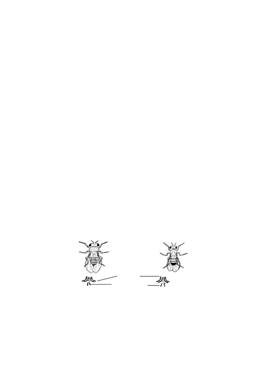
188
Based on sex chromosomal determination of sex, there may be two
types of organisms, namely 1. Heterogametic males 2. Heterogametic females.
Heterogametic males :- In this type of sex determination the female has
two x-chromosomes. The males have only one x-chromosome. During game-
togenesis the males could produce two types of gametes ie., one type having
‘X-chromosomes’ and the other without ‘X-chromosome’. Hence the males
could be called the heterogametic sex. The females can produce only one
type of gametes ie., all eggs having one ‘X’-chromosome each. Hence the
female could be called the homogametic sex. Each type of sex determina-
tion has subtypes as shown in the table.
B. Genic balance mechanism
Further studies on sex determination showed that sex determination
was not the inheritance of genes by the sex chromosomes alone. Studies on
intersex and supersex indicated the operation of still more a complex mecha-
nism for the determination of sex. Thus the genic balance mechanism of
sex was discovered. It was first described by C.B Bridges in 1921.
In Drosophila it was discovered that the sex of an individual depends
upon the ratio of x chromosomes to the autosomes. For sex determination,
each haploid set of autosomes carry factors with a male determining value
equal to one (1). Each x chromosome carries factors with a female determin-
ing value of one and a half (1 .5 ). Hence in a normal male (AAXY), the male
female determinants are in the ratio of 2 : 1.5(‘A’ represents a haploid set of
autosomes). Thus the genic balance is in favour of maleness. A normal female
(AAXX) has the male female determination ratio of 2:3. Therefore the bal-
ance is in favour of femaleness.
Fig.4.3.1. Drosophila flies
Male
Female
Chromosomes
autosomes
sex chromosomes
Chromosomes
189
Sex determination in Human beings
The human sex determination mechanism to a larger extent re-
sembles XX - XY type of Drosophila. However, the Y chromosome contains
male determining genes and it is the determiner of fertility and sex of male
individual.
Thus in human beings, the presence of Y chromosome deter-
mines maleness and its absence determines femaleness. Evidences to estab-
lish this viewpoint had been provided by certain abnormal conditions called
syndromes.
Sex anomalies in Human beings
1. Turner’s syndrome (XO Females) : In this abnormality the females are
sterile and have short stature. They have webbed neck, broad shield-shaped
chest, low intelligence, under developed breasts and poorly developed ova-
ries. These conditions result due to the presence of 44 autosomes and only
one X chromosome in her body cells. This abnormality is known as Turner’s
syndrome.
2. Klinefelter’s syndrome :- This syndrome is caused due to the presence
of an extra X chromosome in males. This happens when an abnormal egg with
XX chromosome is fertilized by a sperm carrying Y chromosome. The zygote
will have three sex chromosomes (XXY). The resulting young one is an ab-
normal sterile male. The symptoms of this syndrome are the presence of small
testicles, mental retardation, longer arms and high pitched voice.
X/A ratio
Phenotype
Number of X
chromosomes
Sets
Autosomes
Super male
Super female
Normal
female
Normal male
Intersex
Tetraploid
Triploid
Diploid
Haploid
3
2
1
1
1
4
3
4
2
3
2
2
3
1.0
1.0
3
1
0.67
0.5
2
1.0
1.0
1.5
0.33
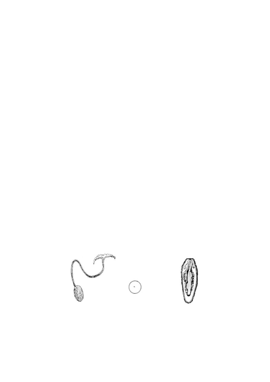
190
3. Super females :- These females are also known as Poly X females. They
possess an extra X-chromosome (44 autosomes +3 x chromosomes). The poly
X females are mentally retarded and sterile.
4. XYY males :- Such males will have an extra Y chromosome (XYY). This
condition results in mental retardation and criminal attitudes.
5. Hermaphroditism :- A hermaphroditic person will have one extra X and Y
chromosome. The person will have both ovarian and testicular tissues. The
external genitalia will not be well defined.
C. Male haploidy or Haplo-diploidy mechanism.
This mechanism is also known as arrhenotokus parthenogen-
esis. It is a common mechanism in several insects such as ants, bees and
wasps. In these insects, fertilized eggs develop into diploid females and unfer-
tilized egg into haploid males.
In a honey bee colony a queen bee can lay two types of eggs.
They are the fertilized and unfertilized eggs. It happens by controlling the
sphincter in the sperm receptacle of the female. The diploid female zygote
can develop either into a queen or a sterile female worker bee. The sterile
nature of the worker bee is due to poor nourishment. The haploid zygote de-
velops into a male. This mechanism of sex determination helps to maintain the
polymorphic nature in a honey bee colony.
Apart from genetical systems, the sexuality can also be con-
trolled by various factors such as metabolism, environment and hormones.
Sex in Bonellia
Bonellia viridis is a marine worm. Its sex determination was
studied by F. Baltzer(1935). The adult female worm is about 2.5 cm long. It
has a well defined anatomical organization.
Fig. 4.3.2. Sex determination in Bonellia viridis
Female worm
Male
Actual size

191
The male is very small and microscopic. Its body organs are
rudimentary. The males normally live as parasites attached to females. All
larvae of Bonellia are genetically similar. However a larva settling on the
proboscis of an adult female becomes a male individual. If a larva develops in
isolation (ie., in the absence of a female) it develops into female. If a develop-
ing male is detached from the proboscis of female, it becomes an intersex.
From these observations it could be inferred that the proboscis of adult female
secretes some hormone like substance and that substance suppresses female-
ness and induces maleness in the larvae which remains attached.
4.4 Sex - linked Inheritance
Most of the inheritable characters are controlled by genes located in
autosomes. The inheritance of traits related to autosomes normally follows
Mendel’s laws. Mendelian ratios are not obtained for those characters for
which genes are exclusively located either in X or Y chromosome. The genes
that occur only on X chromosomes are called as X - linked genes. Similarly,
that the genes occur exclusively on Y chromosomes are called the holandric
genes. The inheritance of X or Y linked genes is called as sex linked inher-
itance. Thus the sex linked inheritance may be X- linked, Y- linked or XY
linked.
X - Linked inheritance
T. H. Morgan (1910) in his studies on inheritance of genes in Droso-
phila discovered that the pattern of inheritance of certain traits were found to
vary with the sex of the parent and offspring. He found that the gene for
white eye colour is X - linked. Further it was found to be recessive to another
X - linked, dominant gene for red eye colour.
Red eyed female x white eyed male
When a wild red eyed female Drosophila is crossed with a mutant
white eyed male, all the F
1
individuals (males and females) have red eyes.
When the red eyed male and red eyed female of the F
1
were intercrossed, in
the F
2
generation all the female flies were found to be red eyed. Among the
males 50 % had red eyes and another 50 % had white eyes.
White eyed female x Red eyed male
When a white eyed female Drosophila is crossed with a red eyed
male, all the female individuals in the F
1
generation are red eyed and all the
males are white eyed. When these red eyed female individuals and white
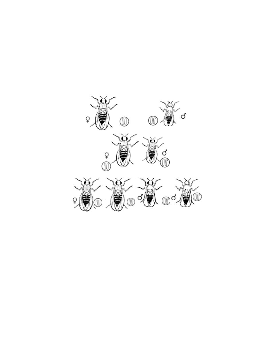
192
eyed male individuals of F
1
are intercrossed the F
2
generation possessed 50 %
red eyed and 50 % white eyed females. Similarly the male population of F
2
included 50 % red eyed and 50 % white eyed flies.
Sex linked inheritance in Humans
Most of the sex linked characters in humans are X - linked. There
are 150 confimed X- linked traits known. Most of them are recessives.
Colour blindness :
The human vision is basically due to cells called rods and cones found
on the retina of the eye. The cone cells are sensitive to red, green and violet
light. The formation of colour sensitive cones is controlled by a dominant X-
linked gene.
The recessive form of this gene is incapable of producing colour sen-
sitive cones. Hence homozygous recessive females (X
C
X
C
) and Hemizygous
Parents
F1
F2
Fig.4.4.1. Drosophila - Red eyed female x White eyed male
Red eyed female
White eyed male
Red eyed female
Red eyed male
White eyed male
Red eyed female
Red eyed female
Red eyed male

193
recessive males (X
C
Y) are unable to differentiate between red and green colour.
The frequency of colour blind women is less than colour blind men.
Colour - blind man x normal visioned woman
When a colour-blind man marries a normal woman in their F
1
progeny
all children would be normal. However the female will be a carrier for the
recessive gene. If that female gets married to a normal male in the F
2
genera-
tion normal and colour-blind nature will occur in 3 : 1 ratio.
4.5 Pleiotropy
It is an established fact that a specific gene controls a specific
phenotypic trait. This finding is not always true. Studies on ‘gene expression’
have revealed that a gene often influences more than one phenotypic trait.
This phenomenon of multiple effects of a single gene is called pleiotropism.
In such a genic influence more conspicuous expression of a phenotypic trait
by a gene is called its major effect. If the gene causes other less conspicuous
phenotypic changes, it is known as secondary effect. Such genes responsible
for multiple effects are called pleiotropic genes.
Vestigeal wings in Drosophila are caused by a recessive gene
in homozygous condition. A keen observation has shown that this gene affects
other traits as well. They are (i) the small halters or balancers behind the wing
(ii) structure of reproductive organs (iii) egg production (iv) life duration and
(v) bristles on the body.
Hardy-Weinberg Law
During the 20th century, scientists showed great interest in applying
the theories of Mendel to human traits. They were able to identify a dominant
trait namely brachydactyly that agreed with the Mendelian theories and showed
Mendelian ratio in the human populations. Brachydactyly is characterised by
abnormal shortening of the phalanges while the rest of the portion of the arms
remain normal.
An objection to this example was raised. If the trait brachydactyly is a
dominant character, three-fourth of the human populations should possess the
abnormally shortened phalanges. But in reality brachydactyly is seen rarely in
human population. This occurrence appeared to contradict Mendelian
theories. However, until 1908 it remained unexplained among the
scientists.
194
Two scientists G.E. Hardy and Wilhelm Weinberg (worked
independently) put forth the theory Hardy-
W
einberg Law. This theory explains
why some characters though dominant (like brachydactyly) appear rarely in a
population.
Fig.4.5.1. Human traits
brachydactyly
polydactyly
hitchhiker’s thumb
This law states that under specified conditions the genotypic
frequency in a population remains constant. It simply means, that the
common traits remain common and the rare traits remain rare. It
further explains that the Mendelian terms dominant and recessive refer to
the phenotypic appearance of the heterozygotes and not to the abundance of
any trait in the population. The relative abundance of a genotypye in a
population is referred to as its genotypic frequency.
The genotypic frequency of any genotype in a population could be
expressed mathematically between 0 to 1 (or 100%) Let us consider that a
particular trait has two alleles ‘A’, ‘a’. ‘A’ remains dominant to ‘a’. Let us
assume that the number of individuals in a new population (with AA and aa) is
100 or 1.Then the number of individuals (or frequency) with AA is ‘p’ and the
number of individuals with ‘aa’ (or frequency of aa) is ‘q’. With that we can
Fig.4.5.2. Hardy-Weinberg equili brium
parents
g a m e t e s
offspring
g a m e t e s
offspring
(homozygotes)

195
draw the equation p+q =1 (or 100). As both the characters are considered as
homozygotes the individuals with AA will produce all gametes with ‘A’. Like
wise the ‘aa’ individuals will produce all gametes with ‘a’.
If all the gametes are viable, combine at random, all have the chance
of survival and no new gametes are introduced in to the population, we can
calculate the proportion of next generation with genotype AA using the usual
Punnet Square method.
gametes frequency
pA
qa
pA
p
2
AA
pq Aa
qa
pqAa
q
2
aa
The new generation will have p
2
AA + 2pq Aa+q
2
aa =1.
Problems: This could be proved mathematically p + q = 1 on both sides.
P + q = 1
(P + q) x (p + q) = 1 x 1 = 1
(P + q)
2
= P
2
+ 2pq + q2 =1
A geometrical representation can also be given. Here each side of the
square represents one unit. It is divided into segments representing ‘p’and ‘q’
(or the proportions of A and a gametes). The points are chosen randomly. This
represents the random meeting of the gametes. The area of each rectangle
represents the frequency of the resulting progeny (or) the proportion of the
next generation having the genotype specified in each cell. The total area
represents 1x1 (or) 1 square unit, representing 1.00 (or) 100% of the
population.
Numerical verification:
A new population is founded by 40% AA and 60% of aa eidividuals.
p
q
q2
p
q
p2
pq
pq
p + q = 2/5 + 3/5 = 1
(p + q)2 = 4/25 + 12/25 + 9/25 = 1

196
Substitute these values for the symbols p and q and find out the percentage of
heterozygotes found in the population when it reaches the equilibrium.
Reminders:
Convert the percentage values into decimal fractions before
multiplying them.
p = 40% , q= 60%
p + q = 1 or 100
i.e 40
+
60
= 1
(or)
2
+
3
= 1
100 100 5 5
P + q = 1 = p
2
+ 2pq + q
2
=
+
+
+
=
+
+
=
+
+
p
2
= 16%
2pq = 48 %
q
2
= 9%
Where the percentage of population having Hetero zygotes is 48.
It is clear that this law is applicable only to populations with following
features
a) Infinitely large populations
b) No mutation
c) No selection
d) Random mating
e) Isolated from any other population of the same species.
This law could be mathematically proved not only to a new population
but also to the populations already existing with equilibrium. Such populations
that has reached equilibrium remain unchanged from generation after
generation. This means that they remain constant and have no chance for
evolution.
1.If a population of fruit flies has a gene frequency of 20% for the recessive

197
allele causing vestigial wings, what proportion shows this trait phenotypic ally?
What proportion of the population is heterozygous?
2.Assume that brown eyes are dominant to blue eyes. On a certain island
about 9% of the population is blue eyed. What proportion of the population is
heterozygous.
Self Evaluation
Part-A
1. The skin colour agouti in rabbits has the genotype
(a) CC
(b) Cc
a
(c) Cc
h
(d) all the above.
2. The alternative forms of a gene are
(a) allelomorphs
(b) multiple alleles
(c) heterozygous
(d) homozygous.
3. The universal recipient is
(a) ‘O’ Group
(b) ‘AB’ Group
(c) ‘A’ Group
(d) ‘B’ Group.
4. ABO blood group in man is an example for
a) Pleiotropism
b) multiple allelism
c) x - linked inheritance
d) y - linked inheritance
5. The inheritance of skin colour was worked out by.
(a) Yule
(b) Mendel
(c) Galton
(d) C.B.Davenport.
6. Rh. factor in blood was discovered by
a) Galton
b) Davenport
c) Landsteiner and Wiener
d) Clarence Mc clung

198
7. The type of sex determination in moths and butterflies is
a) xx - xo type
b) xx - xy type
c) zo - zz type
d) zw - zz type
8. The Y chromosome in Drosophila is
(a) longer
(b) straight
(c) curved
(d) rod like
9. The poly ‘X’ females are
(a) super females
(b) hermaphrodites
(c) bisexuals
(d) intersex
10. An example for ZW - females is
(a) Gypsymoth
(b) Man
(c) Honeybee
(d) Drosophila
11. Most of the sex linked characters in humans are
(a) Y-linked
(b) XY linked
(c) X linked
(d) all the above.
12. X/A ratio in super females is
a) 1.5
b) 1.0
c) 0.6
d) 0.5
13. Holandric genes occur exclusively on
a) x - chromosomes
b) y - chromosomes
c) autosomes
d) both x and y chromosomes
14. The genotypes better adapted for malaria is
(a) Hb
A
Hb
A
(b) Hb
A
Hb
S
(c) Hb
S
Hb
S
(d) all the above
15. A
y
allele in mice is
(a) pleiotropic
(b) co-dominant
(c) lethal
(d) yellow colour

199
Part - II
Give very short answer.
1. What are multiple alleles ?
2. Provide the genotypes for himalayan albino rabbits
3. Mention the possible genotypes of the offsprings if the parental blood groups
are B and B.
4. What is the cause for the death of a child in erythroblastosis fetalis?
5. What was the opinion of the biometricians as geneticists?
6. Who are mulattoes?
7. What is Hermaphroditism?
8. What is arrhenotokus parthenogenesis?
9. What are holandric genes.
10. What are modifying genes.
11. What are secondary sexual characters?
12. What are autosomes and Sex-Chromosomes?
13. Write down the symptoms of Turner ’s syndrome.
14. Explain multiple factor hypothesis.
15. Differentiate a normal RBC from a diseased RBC.
Part - III
Answer briefly
1. What will be the nature of the F
2
progeny, if a coloured rabbit is crossed with
an albino?
2. Mention the possible genotype for ABO blood group.
3. Discuss how ‘O’ blood group is considered as an universal donor.

200
4. What is erythroblastosis fetalis?
5. Write notes on quantitative inheritance.
6. Provide an account on turner’s syndrome and klinefelter’s syndrome.
7. Describe the process of sex determination in Bonellia
Part - IV
Answer in detail
1. Give an account of ABO blood groups in human beings.
2. Explain genic balance mechanism of sex determination.
3. What is sex - linked inheritance ? Give an account of x - linked inheritance in
Drosophila.
4. Explain Hardy - Weinberg law.

201
5. Developmental Biology
The process of sexual reproduction ensures the formation of a diploid
zygote which could constitute the next generation. A zygote is a single celled
structure. By an ontogenetic process the zygote undergoes various develop-
mental phases resulting in multicellular embryonic organisation. These phases
include cleavage, gastrulation, neurulation, organogenesis and the pe-
riod of growth and histological differentiation. Inspite of the fact that or-
ganisms vary in their structure, form and mode of life, the processes of
embryogenesis, development and differention are remarakably similar in all
metazoans. Till later stages of development a fundamental uniform pattern in
development can be observed. The ontogenetic stages also reflect the
historical development of species or phylogenetic development.
Realising the mode of formation of a young individual of the next gen-
eration has always interested human mind. There is a recorded history of
human natural curiosity in sexual reproduction from very early period. The
‘Susruta samhita’, a monumental Indian medical book, written during
second or third century A.D., describes the development of a human child in
the mother’s womb.
The earliest recorded work had been done by Aristotle (384-322 BC).
His classical work De Generatione Animalium is concerned with the gen-
eration of animals. It describes the reproduction and development of many
kinds of animals. In his another work “De Historia Animalaium”, Aristotle
provides an account of the development of the hen’s egg. He compared
reproductive methods of different animals and provided a classification based
on that. By observing the development of hen’s egg he
concluded
that the
development always proceeds from simple formless beginning to the
complex organization of the adult. For this
speculative
idea he provided the
name epigenesis. Through his remarkable observation and speculations
Aritotle established ‘embryology’ as an independent field in science. Thus
to-day he is regarded as the founder of the science of embryology.
After the period of early Greek thinkers this discipline once again got
the attention of the scientists from the beginning of the 17
th
century. Through
the contributions made by various workers like Von Baer ,
E. Haeckel, O. Hertwig, E.B Wilson, Spemann, C.M Child, Maclean
and others rapid advancements were being made in the understanding of de-
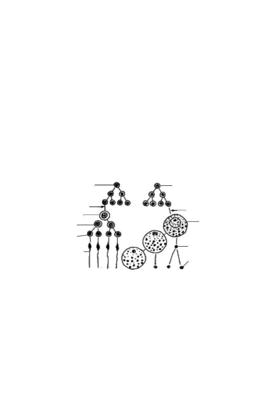
202
velopmental processes in animals. Modern embryology has utilised all tools
made available from other branches of science and diversified into branches
such as ‘Experimental embryology’, ‘Chemical embryology’, ‘Compara-
tive embryology’ and Descriptive embryology. Such studies have paved
the way for meeting the challanges of to-day’s world through works on clon-
ing techniques, tissue culture, stem cell researches, ‘in vitro’ fertilisation,
organ transplantations, regeneration, tissue grafting and other medical and
non-medical fields.
Gametogenesis :
The process of embryonic development in sexually reproducing
multicellular organisms is made possible through processes of
gametogenesis and fertilization. Gametogenesis is the formation of sex
cells or reproductive cells or gametes. It happens in primary sex organs
called gonads. The male and female gonads, namely the testis and ovary
contain primordial germ cells. These cells are responsible for the produc-
tion of gametes.
Spermatogenesis
:
In the testis of vertebrates the specialised tissue for the process of
spermatogenesis are located in the seminiferous tubules. The primordial
germ cells of these tubules produce cells which ultimately become sperm
3 polar bodies
Fig.5.1.1. A-Spermatogenesis - B-Oogenesis
spermatogonium
growth phase
primary
spermatocyte
secondary
spermatocyte
A
B
mitosis
meiotic division
spermatid
spermatozoan
oocyte
1 ripe egg
ootid
growth phase
oogonium
first polar body
second polar body

203
mother cells or spermatogonia. Through a growth phase the speramtogonia
get converted into primary spermatocytes. These are diploid cells. They
undergo meiotic cell division. Initially the I Meiosis results in the formation of
secondary spermatocytes. Through II Meiosis they form spermatids. The
spermatids are haploid in nature. By a process of spermiogenesis or
spermioteliosis they get differentiated into specialized cells called
spermatozoa.
Oogenesis
:
A similar process happens inside the female gonad, namely the ovary
for the production of Ova. This process that happens in the primordial germ
cell of the ovary passes through stages of primary oogonia, primary oocyte
and secondary oocyte. These stages are conducted by meiotic cell divisions.
Thus the final product, namely the ovum is a haploid female reproductive cell.
Fertilization
:
Embryogenesis could occur only after the fertilization of the ovum or
egg. Fertilization provides the diploid nature to the cell. Thus all the somatic
cells of the embryo will remain diploid. Further, the process of fertilization
triggers or initiates the initial stages of embryogenesis. During the process of
fertilization the sperm and ovum of the same species approach and come in
contact with each other. The entry of sperm initiates further changes in the
egg. The haploid nuclei of the sperm and ovum fuse, resulting in the formation
of a diploid zygote nucleus. This process of nuclear fusion is known as syn-
gamy or amphimixis.
5.1. Types of eggs
For the embryo to develop inside a fertilised egg nutrition is needed.
The amount of food needed varies for different organisms. It normally
depends on the duration of development. Food is provided in the form of yolk.
It may be ‘fatty yolk’ or protein yolk’. It is provided by the ovary during
differentiation of the egg. Due to accumulation of yolk a maturing egg rapidly
increases in size. In amphibian eggs yolk occurs in the form of large granules,
called yolk platelets. Chemically, the yolk platelets contain two main pro-
teinaceous substances namely phosvitin and lipovitellin.
The amount of yolk is an important determining factor for further pat-
terns in embryological stages. The amount of yolk influences cleavage and
gastrulation methods.
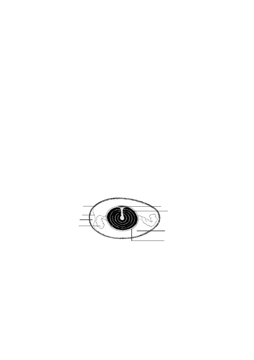
204
The eggs can be classified based on amount and distribution of yolk.
Amount of yolk and egg types :
In certain animals the developmental stages are not very elaborate.
The final ‘young one’ born may be very simple in structure and organization.
Such conditions remain in animals like Hydra, Sea urchin, Amphioxus and
Placental mammals. In the eggs of such organisms due to brevity of the
growth period the amount of yolk is much reduced. Such eggs are said to be
Microlecithal or oligolecithal.
In certain other animals the eggs need to release young ones in a more
self supportive condition. Hence for such eggs the amount of yolk is consider-
able in quantity. Such eggs with moderate amount of yolk are called meso-
lecithal eggs. Such eggs are produced by annelid worms, molluscs and am-
phibians.
In some animals the growth and differentiation of the embryo is much
more elaborate. The growth period is sufficiently long. Hence for supporting
the embryo in development the eggs contain large quantity of yolk. Such eggs
are termed as Megalecithal or Macrolecithal eggs. The eggs of reptiles
and birds are considered as macrolecithal. Further these eggs are covered by
a calcareous shell. It is a protective structure for laying the eggs on lands.
Such eggs are called cleidoic eggs or land eggs.
Distribution of yolk.
The pattern of cleavage and the consequent gastrulation processes
are affected by the distribution of yolk within the egg. According to the pat-
tern of dispersal of yolk the following egg types had been identified.
Fig.5.1.2. Hen’s egg
shell
yolk
chalaza
shell membrane
air space
disc of cytoplasm
albumen
vitelline membrane
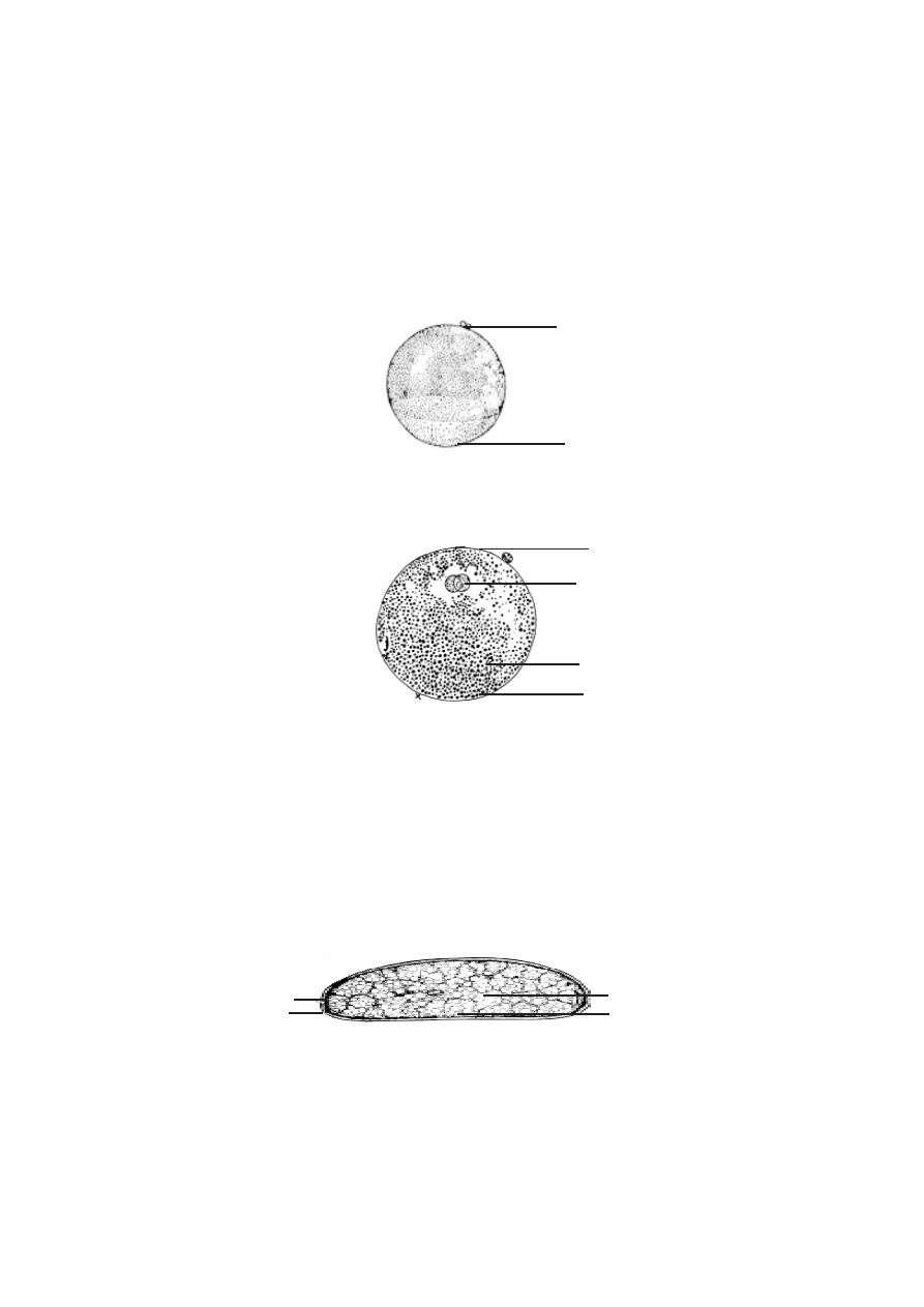
205
Fig.5.1.3. Isolecithal egg
animal pole
vegetal pole
1. Homolecithal or isolecithal eggs.
Eggs of this type have the yolk disbursed in the entire cytoplasm. The
distribution is somewhat uniform in animal, vegetal poles and the equatorial
region. In such eggs the cleavage will be deeper and may bisect the eggs
connecting the two poles. All microlecithal eggs have this nature.
2. Telolecithal eggs.
All eggs have polarity. In polarity, the eggs have an innate nature for
to be differentiated into upper animal pole and lower vegetal pole. The polar
nature is mainly due to the denser material in the cytoplasm, namely yolk. The
yolk in the egg will normally get concentrated in the vegetal pole. The
cytoplasm with the nucleus will occupy the upper animal pole. The extent of
vegetal pole is determined by the amount of yolk. Thus the eggs having a
polarised distribution of yolk in the cytoplasm are referred to as Telolecithal
eggs. Mesolecithal and macrolecithal eggs remain as telolecithal eggs.
3. Centrolecithal eggs.
Fig.5.1.4. Telolecithal egg
yolk
nucleus
animal pole
vegetal pole
Fig.5.1.5. Centrolecithal egg
yolk
cytoplasm
nucleus
shell
•

206
An egg need not be spherical always. In invertebrate animals oval
shaped eggs are seen. The pattern of cleavage and further gastrulation also
deviate from that of the vertebrates. In insects the eggs are oval in shape and
the yolk remains in the centre of the egg. However, the eggs of echinoderms
are similar to that of the vertebrates.
5.2 Cleavage and types - Frog’s egg.
The process of cleavage reamains one of the earliest mechanical ac-
tivity in the conversion of a single celled egg into a multicellular embryo. It is
initiated by the sperm during fertilization. However in parthenogenetic eggs
cleavage can commence without the influence of fertilization.
The process of cleavage or cellulation happens through repeated mi-
totic divisions. These divisions result in cells called blastomeres. In later
stages of development the blastomeres occupy different regions and differen-
tiate into several types of body cells.
The first cleavage of frog’s egg was observed by Swammerdam in
1738. The entire process of cleavage in frog’s egg was studied by Prevost
and Dumas in 1824. With the development of microscopes cleavages and
further stages were observed in the eggs of sea urchin, star fishes, amphioxus
and hen’s eggs.
From all these studies it has become clear that all divisions in cleavage
are mitotic. The mitotic process is very rapid. In the eggs of sea urchin divi-
sion of the blastomeres can be observed every 30 minutes. As the cleavage
progresses the resultant daughter cells, namely the blastomeres get reduced
in size. During cleavage there is no growth in the blastomeres. The total size
and volume of the embryo remains the same. The cleavages result in a com-
pact mass of blastomeres called morula. It gets transformed into blastula.
While the wall of the blastula is called the blastoderm, the central cavity is
called the blastocoel.
The planes of cleavage
An egg can be divided from different planes during cleavage. De-
pending on the position of the cleavage furrow the planes of cleavage are
named.
1. Meridional cleavage: The plane of cleavage lies on the animal vegetal
axis. It bisects both the poles of the egg. Thus the egg is divided into two equal
halves.

207
2. Vertical cleavage: The cleavage furrows may lie on either side of the
meridional plane. The furrows pass from animal to vegital pole. The cleaved
cells may be unequal in size.
3. Equatorial cleavage: This cleavage plane bisects the egg at right angles
to the main axis. It lies on the equatorial plane. It divides the egg into two
halves.
4. Latitudinal cleavage: It is similar to the equatorial plane, but it lies on
either side of the equator. It is also called as transverse or horizontal
cleavage.
Influence of yolk on cleavage
Yolk is needed for embryonic development. However the fertilized
egg has to undergo all stages of development and result in a suitable ‘young
form’ initiating next generation. Somehow with all the influences of yolk the
developmental procedures are so adapted and modified that a well formed
embryo will result. The initial influence of yolk is felt during the process of
cleavage.
The amount of the yolk and its distribution affect the process of cleav-
age. Accordingly several cleavage patterns have been recognised.
1. Total or holoblastic cleavage - In this type the cleavage furrow bisects
the entire egg. Such a cleavage may be either equal or unequal.
(a) Equal holoblastic cleavage - In microlecithal and isolecithal eggs, cleav-
age leads to the formation of blastomeres of equal size. Eg: Amphioxus and
placental mammals.
(b) Unequal holoblastic cleavage - In mesolecithal and telolocithal eggs,
cleavage leads to the formation of blastomeres of unequal size. Among the
blastomeres there are many small sized micromeres and a few large sized
macromeres.
2. Meroblastic cleavage - In this type the cleavage furrows are restricted to
the active cytoplasm found either in the animal pole (macrolecithal egg) or
superficially surrounding the egg (centrolecithal egg). Meroblastic cleavage
may be of two types.
(a) Discoidal cleavage - Since the macrolecithal eggs contain plenty of yolk,
the cytoplasm is restricted to the narrow region in the animal pole. Hence
cleavage furrows can be formed only in the disc-like animal pole region. Such
a cleavage is called discoidal meroblastic cleavage. Eg: birds and reptiles.
208
(b) Superficial cleavage - In centrolecithal eggs, the cleavage is restricted to
the peripheral cytoplasm of the egg. Eg: insects.
Laws of cleavage
Apparently there are several cleavage patterns. However, all cleav-
ages follow a common procedure. The cleavages are governed by certain
basic principles or laws.
1. Sach’s laws - These laws were proposed by Sach in 1877.
i) Cells tend to divide into equal daughter cells
ii) Each new division plane tends to intersect the preceding plane at right
angles.
2. Balfour’s law (Balfour 1885) - “The speed or rate of cleavage in any
region of egg is inversely proportional to the amount of yolk it contains”.
Cleavage of fertilized egg in Frog.
In frog’s egg the cleavage is holoblastic and unequal. The cleavage
occurs as follows.
1. The first cleavage plane is meridional. Initially, a furrow appears at the
animal pole. It gradually extends towards the vegetal pole of the egg. It cuts
the egg through its median animal-vegetal polar axis and results in two equal-
sized blastomeres.
2. The second cleavage furrow is again meridional. It bisects the first cleav-
age furrow at right angles. It is a holoblastic cleavage affecting both the
blastomeres of the first cleavage. It results in the formation of four blastomeres.
Fig.5.2.1. Cleavage in frog’s egg
first cleavage
second cleavage
third cleavage
meridional
meridional
latitudinal
blastula-entire
blastula-L.S
blastoderm
blastocoele
209
3. In the next stage a latitudinal furrow is formed above the horizontal fur-
row nearer to the animal pole. Such a furrow is due to the influence of yolk
concentration in the vegetal pole. The latitudinal furrow uniformly affects all
the blastomeres. It results in the formation of eight blastomeres. Four of them
remaining in the vegetal pole are large. They are named as macromeres.
Another four blastomeres remain in the vegetal pole. They are named as
micromeres. The micromeres are smaller in size than the macromeres.
4. The fourth set of cleavage planes are meridional and holoblastic. They are
unequal. They divide yolkless micromeres more rapidly than yolk-rich mac-
romeres. These cleavages result in the production of 16 blastomeres.
5. As a result of further cleavages, a ball of several small blastomeres result.
A closer observation reveals that, while the blastomeres above the equator
are small and remain as micromeres, the blastomeres of the vegetal pole re-
main progressively larger. The larger blastomeres are called the macromeres.
At the final stages of cleavage the embryo acquires a characteristic,
mild, oblong shape. In this stage it is called the morula. The morula initially
contains a shallow cavity called the blastocoele. Gradually the blastocoelic
space increases into a large cavity occuping the middle of the blastula. How-
ever the blastocoele mostly remains in the animal pole region in the middle of
the micromeres.
The blastomeres gradually adhere to each other, and arrange them-
selves into a true epithelium called the blastoderm. The blastoderm remains
two cell thick in the animal pole. The embryo having a fluid-filled blastocoele
and blastoderm is called the blastula.
It has been reported that around 12
th
cleavage the blastula possesses
about 4096 cells. The blastula moves to the next stage, namely gastrulation at
a stage in which it has about 20,000 cells.
The ultimate blastula is a ball of blastomeres which have to form dif-
ferent embryonic body layers and organs of the body. The fate of each and
every blastomere has been observed and marked. A map showing various
Fig.5.2.2. L.S of Frog blastula
animal pole
vegetal pole
micromere
macromere
blastocoele
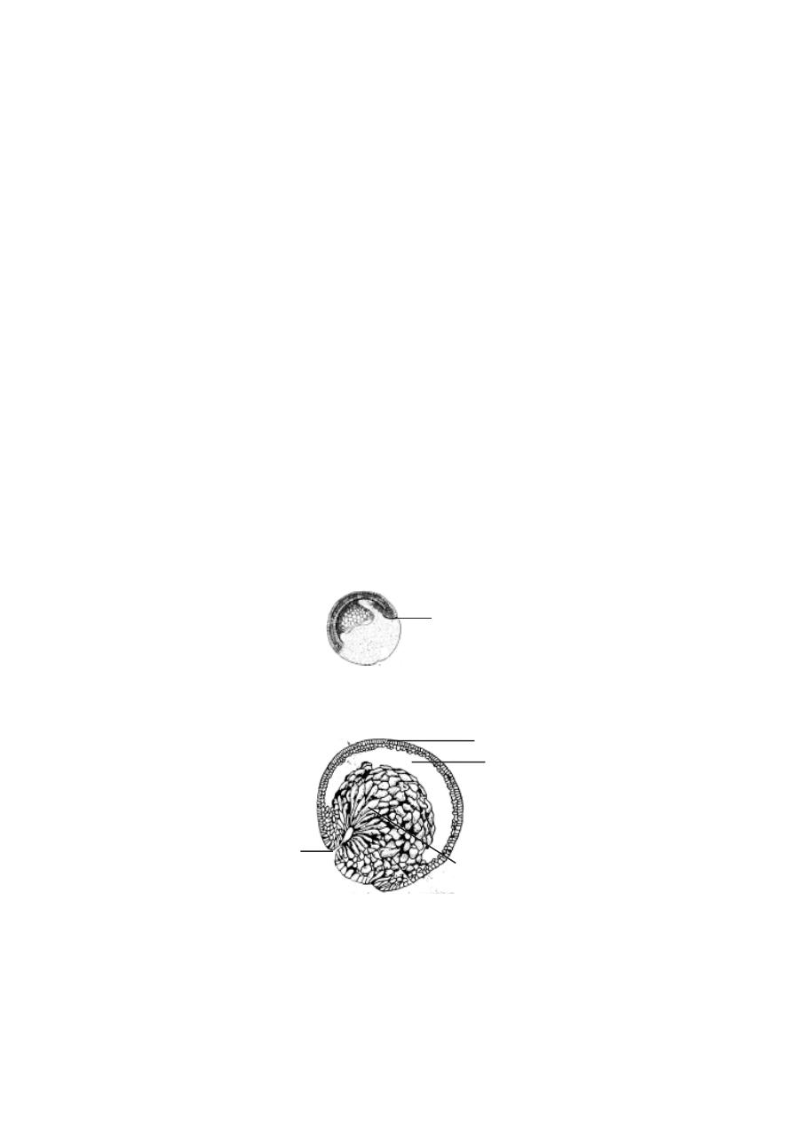
210
organ forming, areas on the blastula is called the ‘fate map’. This map shows
prospective ectoderm, mesoderm and endodermal areas. It also shows the
‘zone of involution’ and ‘zone of invagination’ for the next stage of gas-
trulation.
5.3. Gastrulation in frog embryo
The process of gastrulation is a continuous activity succeeding, cleav-
age. During this process the blastodermal cells begin to move. They wander
and occupy their prospective organ forming zones. During this movement at
one region on the blastula, the cells wander inside and occupy the blastocoelic
cavity.
At a specific region below the equator the blastoderm cells assume an
elongated bottle like shape. They move toward the interior of the blastula. As
the cells move further inside, an invagination happens. A deepening of the
invagination results in a cavity called the archenteron or gastrocoel.The
opening of the archenteron on the surface of the blastula is called the
blastopore.
The blastopore gradually assumes a crescentic shape. Finally it be-
comes circular. The region dorsal to the blastoporal opening is called the ‘dor-
sal lip’. The lower edge may be called the ‘ventral lip’.
The surface cells representing several prospective zones of the em-
bryo begin to wander inside through the blastopore. These inwandering of
cells is termed as involution.
Fig.5.3.2. Invagination of Endodermal cells
dorsal lip of blastopore
invagination of macromeres
blastoderm
blastocoele
Fig.5.3.1. Formation of dorsal lip
dorsal lip
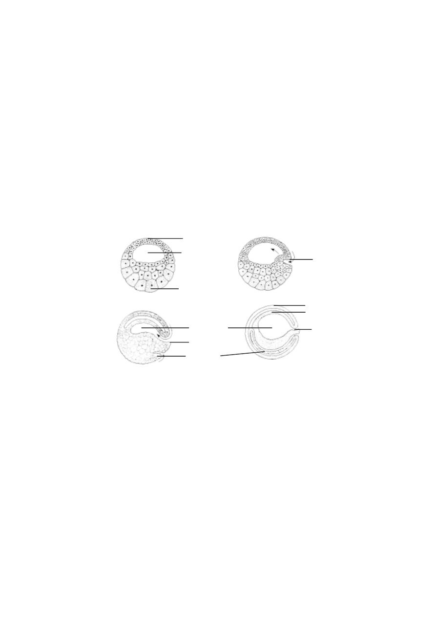
211
Initially, the first pharyngeal endodermal cells undergo invagination over
the dorsal blastoporal lip. These cells move to the interior. They are followed
by other cells. The inwandering cells gradually occupy the region of the blas-
tocoele. Thus the blastocoelic cavity gets reduced. A new cavity among the
involuted cells results. It is called the gastrocoel. The gastrocoel later be-
comes the archenteron. The interior region of the archenteron gradually trans-
forms into the pharyngeal region. This region remains as the foregut. The
mesodermal and endodermal cells gradually occupy their positions.
The inward movement of the exterior cells through the blastoporal
region is called involution. The involution results in the positioning of chor-
damesodermal cells and pharyngeal endodermal cells.
The mesodermal cells occupy the region between inner endodermal
and outer ectodermal cells. While the exterior chorda-mesodermal cell invo-
lute inside, their place is taken up by the ectoderm. The expansion of the
ectoderm is due to epiboly. Epiboly causes overlapping or ‘the roofing over’
of the gastrula by the ectoderm.
The blastopore is gradually covered by certain endoderm cells. The
closing cells of the blastopore constitute the yolk-plug. Gradually the
yolk-plug withdraws to the interior and the blastopore gets reduced into a
narrow slit.
Fig.5.3.3. Gastrulation of Frog
micromere
macromere
yolk plug
ectoderm
blastocoele
gastrocoele
dorsal lip
blastopore
endoderm
mesoderm
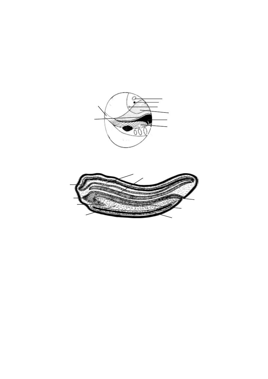
212
The process of gastrulation converts the blastula into a spherical, bi-
laterally symmetrical, triploblastic gastrula. Gradually the gastrula undergoes
the process of tubulation or neurulation to become a neurula.
Neurulation
The process of neurulation is the formation of a neural tube. However
during this process mesoderm and endoderm also undergo differentiation.
During neurulation the embryo lengthens along the anteroposterior axis.
The dorsal side of the gastrula is lined by ectodermal cells. The presumptive
area of the nervous system gets differentiated from the rest of ectoderm. It
remains as medullary plate or nerual plate. The neural plate later thickens
and it gets raised above the general level as ridges called neural folds. In the
middle of the neural fold a neural groove appears. The neural groove deep-
ens inside. The neural folds above the groove. The neural groove gets con-
verted into a neural tube. This tube gets detached from the surface. The
neural tube remains as the prospective nervous system. The embryo at this
stage is called the neurula.
Fig.5.3.4. Fate map of frog blastula
sucker
neural plate ectoderm
tail mesoderm
notochord
somatic mesoderm
lateral plate mesoderm
ear
lens
Fig.5.3.5. Neurula of frog
digestive tract
brain
neural canal
notochord
pharynx
yolk
coelom
anus
liver
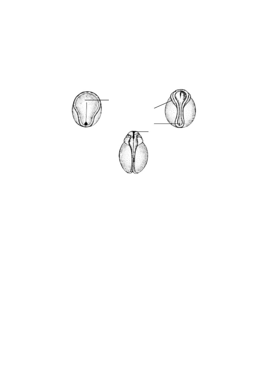
213
During neurulation, the tubulation of chorda-mesoderm and tubulation
of endoderm also happen.
The post-neurular development of frog involves the formation of all
body organs.
5.4. Organogenesis of Frog
The primary organ rudiments from ectoderm, mesoderm and endo-
derm get well established during the processes of gastrulation and neurula-
tion. In the next stage the primary organ rudiments subdivide into secondary
organ rudiments. These rudiments get differentiated into various organs and
organ systems.
The development of ectodermal organs
The neurula of frog has three kinds of ectodermal tissues namely, epi-
dermal ectoderm, neural ectoderm and neural crest cells.
Epidermal ectoderm
The epidermal derivatives are the skin, olfactory sense organs, ear,
lateral line sense organs, median fins, external gills and lining of mouth and
anus.
A
B
C
Fig.5.3.6. Neurulation of frog embryo
neural plate
blasto pore
neuropore
neural fold

214
Neural ectoderm
This layer of cells form the central nervous system and peripheral
nervous systems.
Development of eye
The eye is a photoreceptor. It is an ectodermal derivative. Its
development begins even at the gastrulation stage. However the first sign of
eye formation appears with the development of two optic vesicles from the
lateral walls of the embryonic diencephalon.
Formation of optic cup
The eyes develop as two lateral outgrowths of the prosencephalon
called optic vesicles. The cavity of the optic vesicle is called optocoel. The
connection of the optic vesicle with the brain becomes a narrow stalk like
structure called optic stalk. The optic stalk becomes connected with the
ventral side of the optic vesicle rather than at its centre. The optic vesicles
extend outward and reach the ectoderm. The wall of the optic vesicle next to
the ectoderm is gradually flattened and later invaginates to form a double
walled cup called optic cup.
The optic cup consists of two layers. The inner layer (derived by in-
vagination) gives rise to the nervous region, the retina. The outer layer will
be a thin, black pigmented layer (tapetum - nigrum) for the absorption of
light. Initially the opening of cup is very large. Soon its rim bend inward and
converges, so that the opening is reduced. This opening is called the pupil.
The rim of the optic cup surrounding the pupil becomes the iris. Later on,
large amount of pigment is deposited in the outer epithelial layer of iris. A
groove extends along the ventral side of the optic cup. It is called the
choroid fissure. It extends to the middle of optic stalk. It serves for the
entry of blood vessels and mesenchyme cells into the posterior chamber of
eye.
The retina develops a membrane on its inner most surface called the
internal limiting membrane. The outermost cells of the neurosensory retina
differentiate into rods and cones. The inner cells of the retina differentiate
into neuroblasts or nerve cells.

215
Development of lens :
When the lateral surface of the growing optic vesicle comes in con-
tact with the ectoderm it gives off stimulus of some kind, which causes the
ectodermal cells to elongate, forming a disc shaped thickening. This is called
the lens placode or lens rudiment. It curves into a cup and finally
separates from the ectoderm. The free edges of the cup fuse to form a globular
hollow lens vesicle. The lens vesicle comes to lie in the cavity of the optic
cup.
The cells of the inner side of the lens vesicle elongate, become
columnar and are finally transformed into long fibres. Their nuclei degenerate
and cytoplasm becomes hard and transparent making it refractile. These cells
are called lens fibres. The outer layer of the lens remains unchanged and
becomes the lens epithelium. The junction between the lens fibres and the
lens epithelium represents the growing point of lens. Here the epithelial cells
are continuously transformed into lens fibres.
When the lens is formed, the free margin of optic cup touches the
edges of the lens and grows in front forming iris. Thus lens hangs in the
opening of optic cup.
Soon after the development of lens the overlying ectoderm closes
over and differentiates to become the cornea. It is continuous with the skin.
The transformation of the skin into cornea is caused by an induction arising
from the optic cup and lens. The ectodermal cells covering cornea form an
extremely thin, transparent membrane. This is known as conjunctiva of the
eye ball. In adult this becomes continuous with the inner lining of upper and
lower eyelids.
The space between lens vesicle and the overlying presumptive
anterior epithelium of cornea represents the anterior chamber. It contains
cellular material called anterior vitreous body.
The choroid and sclerotic coat of eye develop from the mesenchyme
cells accumulating around the eye ball. The interior layer of mesenschyme
cells give rise to a net work of blood vessels surrounding the pigmented
retinal layer and is called choroid coat. The outer layer of mesenchyme form
fibrous capsule, the sclerotic coat or sclera around the eye. The sclera
provides protection to eye and the eye muscles.
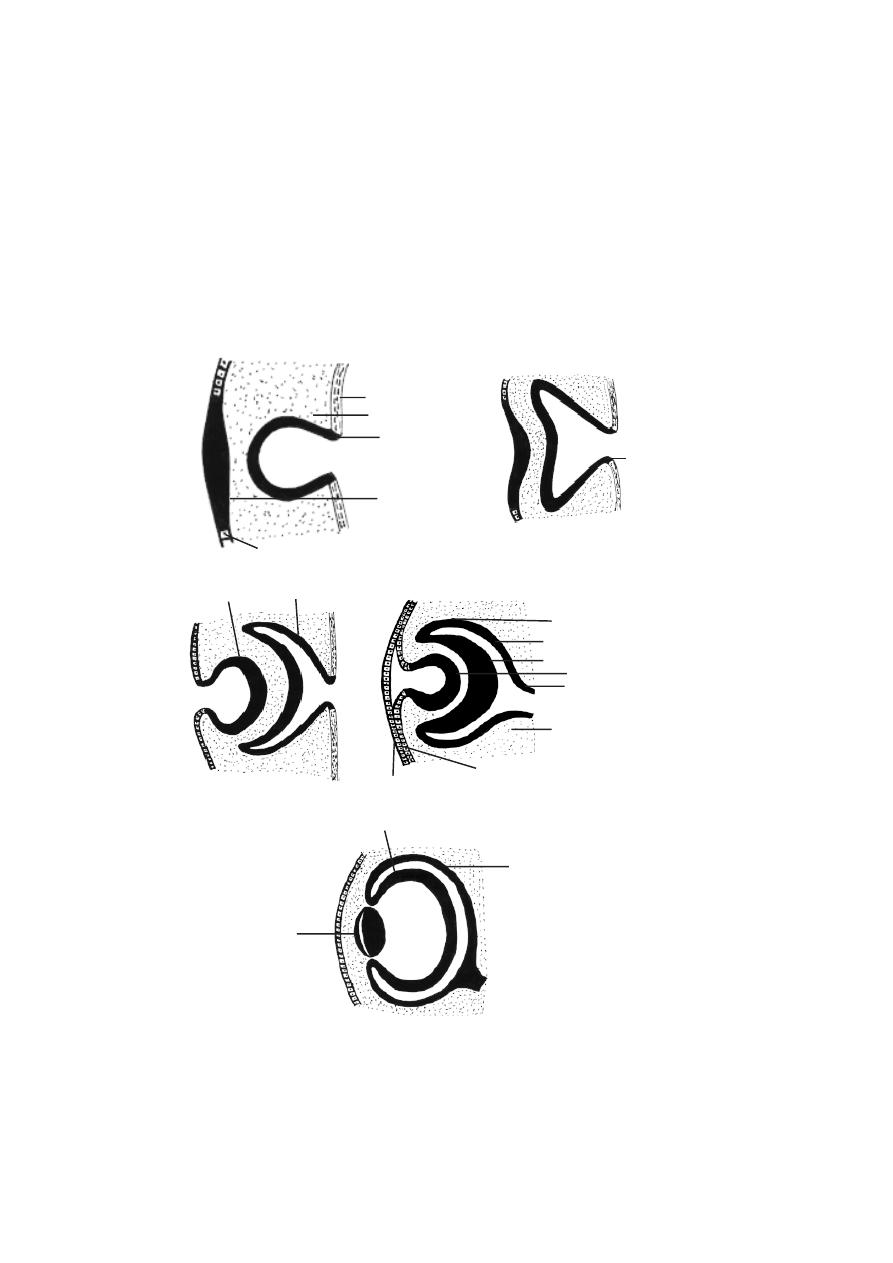
216
The ectoderm from above and below the original lens placode region
grows out as two folds. These folds grow over conjunctiva and come to
touch each other forming a complete layer of ectoderm. At a later stage
these folds separate along the line of fusion to form the regular upper and
lower eye lids.
optic stalk
optic vesicle
mesenchyme cells
brain
ectoderm
lens placode
optic cup
lens vesicle
pigmented layer of retina
sensory layer of retina
lens
inner ectoderm
outer ectoderm
optic cup
mesenchyme cells
optic stalk
lens vesicle
sensory layer of retina
pigmented layer of retina
Fig.5.4.1. Frog - Eye development

217
The development of mesodermal organs
The mesodermal derivatives are the limbs, endoskeleton, heart, blood
vessels, kidney, coelom and reproductive organs.
Development of heart in Frog
The heart is the first organ to start functioning because it has to
circulate blood which brings nourishment from the endodermal cells to the
other parts of the embryo.
The initial development of heart is similar in all vertebrates. The first
rudiment of heart develops after neurulation in the region beneath the
pharynx, from the mesenchyme and mesoderm.
The heart of frog develops at the ventral side of pharynx. It is formed
by mesenchyme cells. The mesenchyme cells accumulate at the mid-ventral
line of pharynx and form a longitudinal strand of tissue called the rudiment
of endocardium. Soon the endocardial cells get arranged in the form of a
thin walled tube, the lumen of which later develops into the cavity of the
heart.
While the endocardial tube is being formed, the mesoderm of both
sides unite above and under the endocardial tube and a cavity called
pericardial coelom is formed around the latter. The visceral layer of
mesoderm envelops the endocardial tube and gives rise to its muscular walls
or myocardium and also its close fitting covering called the visceral
pericardium.
Differentiation of Heart
The heart thus formed is a straight tube, situated longitudinally in the
middle line below the pharynx and surrounded by the pericardium on all the
sides. Its wall is formed of three layers, outer epicardium, middle
myocardium and inner endocardium. The myocardium grows in thickness.
The tubular heart elongates rapidly in antero-posterior direction. The
small size of pericardial cavity and the anterior and posterior
attachments do not permit its increase in antero-posterior direction. The
increase in size causes the originally straight tube to curve and fold forming-
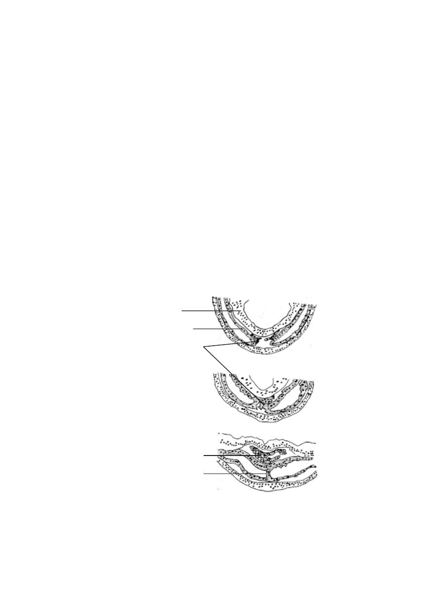
218
S- shaped coiled tube.The anterior part is folded ventrally and posteriorly
and the posterior part becomes folded dorsally and anteriorly.
At the same time different regions of the tube grow differently in
thickness and constrictions appear demarcating different presumptive
chambers of the adult heart. The posterior most limit of the heart, where the
vitelline veins converge, is the sinus venosus. This is followed by a thin
walled atrium which opens infront into a thick walled ventricle.
Initially the atrium is single chambered. Later on, a partition grows
downward from the dorsal wall of the atrium dividing it into larger, right and
smaller left atrial chambers. The partition forms inter-atrial septum. As the
septum is developed on the leftside of the opening of the sinus venosus, the
sinus venosus communicates only with the right auricle. The pulmonary veins
which are also formed at this stage of development now pour the blood into
the left auricle. The valves of the heart arise as folds of endocardium.
Formation of endocardial tube
endoderm
parietal layer of lateral
plate mesoderm
rudiment of endocardium
endocardial tube
ventral mesocardium
A
B
C
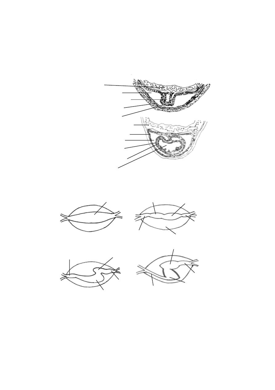
219
endoderm
myocardium
pericardium
endocardial tube
pericardial cavity
endoderm
pericardium
pericardial cavity
cavity of heart
myocardium
endocardium
Fig.5.4.2. Development of the heart in forg’s embryo
D
E
Fig.5.4.3. Transformation of tubular heart of frog’s embryo into adult heart
tubular heart
pericardial cavity
truncus
ventricle
atrium
sinus venosus
atrium
truncus
sinus venosus
ventricle
atrium
truncus arteriosus
sinus venosus
ventricle

220
The development of endodermal organs
The predominant endodermal organs are the organs of the alimentary
canal, lungs, pancreas and urinary bladder.
Self Evaluation
Part - I
Choose the right answer
1. The founder of the science of embryology was
(a) Aristotle
(b) E.Haeckel
(c) O.Hertwig
(d) E.B.Wilson
2. The process of spermiogenesis involves
a) production of sperms
b) differentiation of spermatids into spermatozoa
c) formation of testis
d) differentiation of nucleus
3. Centrolecithal eggs are produced by
a) frog
b) human beings
c) reptiles
d) insects
4. In amphian eggs yolk occurs in the form of
(a) yolk platelets
(b) blood platelets
(c) phosvitin
(d) lipovitelline
5. Macrolecithal eggs are found in
(a) amphibians
(b) birds
(c) fishes
(d) echinoderms
6. Superficial cleavage is seen in
(a) amphibians
(b) insects
(c) reptiles
(d) birds
7. Discoidal cleavage is seen in the eggs of
a) birds
b) amphibians
c) insects
d) amphioxus
8. Sach’s law is related to
a) gametogenesis
b) cleavage

221
c) gastrulation
d) organogenesis
9. The cavity formed in a gastrula is called as
a) gastrocoel
b) blastocoel
c) blastopore
d) cavity
10. The embryo having a fluid -filled cavity and a layer of cell is called
(a) blastocoel
(b) blastula
(c) morula
(d) blastoderm
11. The gastrorcoel later becomes
(a) archenteron
(b) blastula
(c) blastocoel
(d) ectoderm
12. The heart is derived from
(a) ectoderm
(b) mesoderm
(c) endoderm
(d) all the above
13. The cavity of the optic vesicle is
a) optic cup
b) optocoel
c) optic stalk
d) optic cavity
14. The valves of the heart arise as folds of
a) myocardium
b) epicardium
c) endocardium
d) pericardium
Part - II
Give very short answer
1. Provide a list of various stages in the embryology of animals
2. Why do we consider Aristotle as the founder of the science of embryology?
3. What are the several diversified fields formed from modern embryology?
4. What are microlecithal eggs?
5. Define gametogenesis and spermatogenesis.
6. What is syngamy?
7. What is a morula?
8. Name the proteinaceous substances of yolk platelets.
9. What are mesolecithal eggs? Give an example.
10. Define meridional cleavage.

222
11. Mention Balfour’s law regarding cleavage
12. What is a ‘fate map’ ?
13. Name the two types of meroblastic cleavage.
14. What is involution ?
15. What is a gastrula?
16. What is epiboly?
17. Define tubulation?
18. What is epidermal ectoderm?
19. Name the organs developed from the mesoderm?
Part - III
Answer briefly
1. Give an account of the megalecithal egg.
2. What is a centrolecithal egg?
3. Provide a general account on cleavage.
4. Give an account of the planes of cleavage.
5. Explain the process of equal and unequal holoblastic cleavage.
6. What is sach’s law?
7. Draw a neat labelled sketch of gastrula.
8. Draw a neat labelled sketch of hen’s egg.
9. Give an account of the development of ovum.
10. What are the ectodermal and endodermal derivatives in the organogenesis of
frog embryo?
Part -IV
Answer in detail
1. Provide a detailed account on the types of eggs.
2. Give an account on the cleavage of fertilized egg.
3. Give a detailed account of the cleavage in frog with neat diagrams.
4. Describe how the process of gastrulation occurs in the egg of an amphibian
5. Describe the structural organisation in the neurula of frog.
6. Describe the development of heart in frog.
7. Explain the process of development of eye in a frog embryo.

223
6. ECONOMIC ZOOLOGY
Since time immemorial, human beings have used animals for food and
other purposes. While some animals are very useful to mankind certain others
cause loss to the economy of man. Though every organisms has its own im-
portance in nature, some of them such as a few mammals, birds, fishes, prawns
and insects have become valuable. Some pests are competitors of human be-
ings for natural resources and food. Thus a study of economically important
animals will always be useful.
6. 1. Beneficial animals
The animals contributing to our economy and welfare are known as
beneficial animals. Many animals provide us with nutritious food like meat
and milk, clothing materials like silk and wool and the luxurious items like
pearls and corals. The silk worms, the honey bees, the lac insects, fowls,
fishes, prawns and crabs belong to this category.
6. 1. 1 Corals - reef builders
Most of us know about corals and are not familiar with their biology.
Coral rocks are actually the skeletal remains, primarily of calcium carbonate
secreted by living coral polyps. The coral organisms belong to the phylum :
Coelenterata.
All reef building corals live as large colonies. Reef corals are typically
shy, nocturnal feeders. During the day, polyps are withdrawn into skeletal
cups and the corals appear more or less lifeless. But at night time the whole
reef magically comes to life and coral polyps stretch out their tentacles, prob-
ing the waters for food. The reef looks like a field of flowers.
Reef forming corals grow only in shallow, tropical seas where the
temperature of the water never falls below 20
o
c. Only in warmer waters, the
coral polyps can extract calcium from the sea water and deposit it as calcium
carbonate in their skeletons. Corals thrive only in crystal clear water.
Small plants like Zooxanthella living in the coral tissue contribute to
the yellow, brown and green colours of some reef forming corals. The brighter
red and orange colours are created by pigment cells in the body wall.
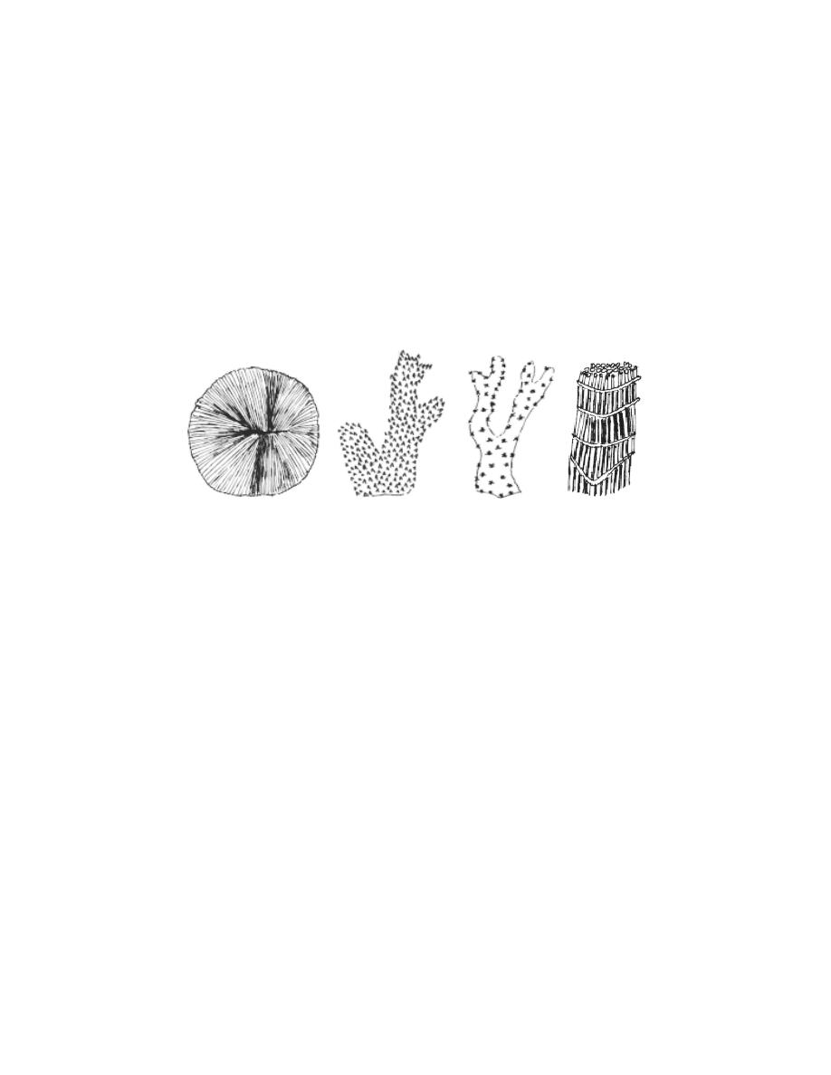
224
Formation of coral reefs
Corals are best known for the massive rocky reefs they build in trop-
ics. The corals spread by producing vast number of minute ciliated larvae
called Planulae, through sexual reproduction. These larvae initially lead a
free swimming life. Later they settle on rocks and start new colonies of pol-
yps by repeated fission or budding. A coral reef is thus a result of the activity
of millions of coral polyps over several thousand years.
Coral reefs are of three types namely, Fringing reefs, Barrier reefs
and Atolls. In India coral reefs occur in the Lakshadweep, Andaman and
Nicobar Islands and in the south east-coast.
Fringing reefs form shallow shelves in shallow waters at or near the
shore of the mainland or around offshore Islands. At the southern end of
Indian peninsula, this type is seen as a chain of well developed reefs starting
from Rameshwaram Island and extending beyond Tuticorin. They are also
present in the Gulf of Kutch on the west coast of India.
Barrier Reefs are situated away from the coast and form off-shore
break waters parallel to the coasts or isolated islands. The famous Great
Barrier reef of North Eastern Australia which is 2012 kilometres long, is the
largest of its kind.
Atolls consist of a ring shaped reef, encircling a shallow lagoon which is
connected to the outside by an opening. Hawaii and Carribian Islands are
famous for Atolls.
Fig.6.1.1. Corals
Fungia
Acropora
Oculina
Tubipora
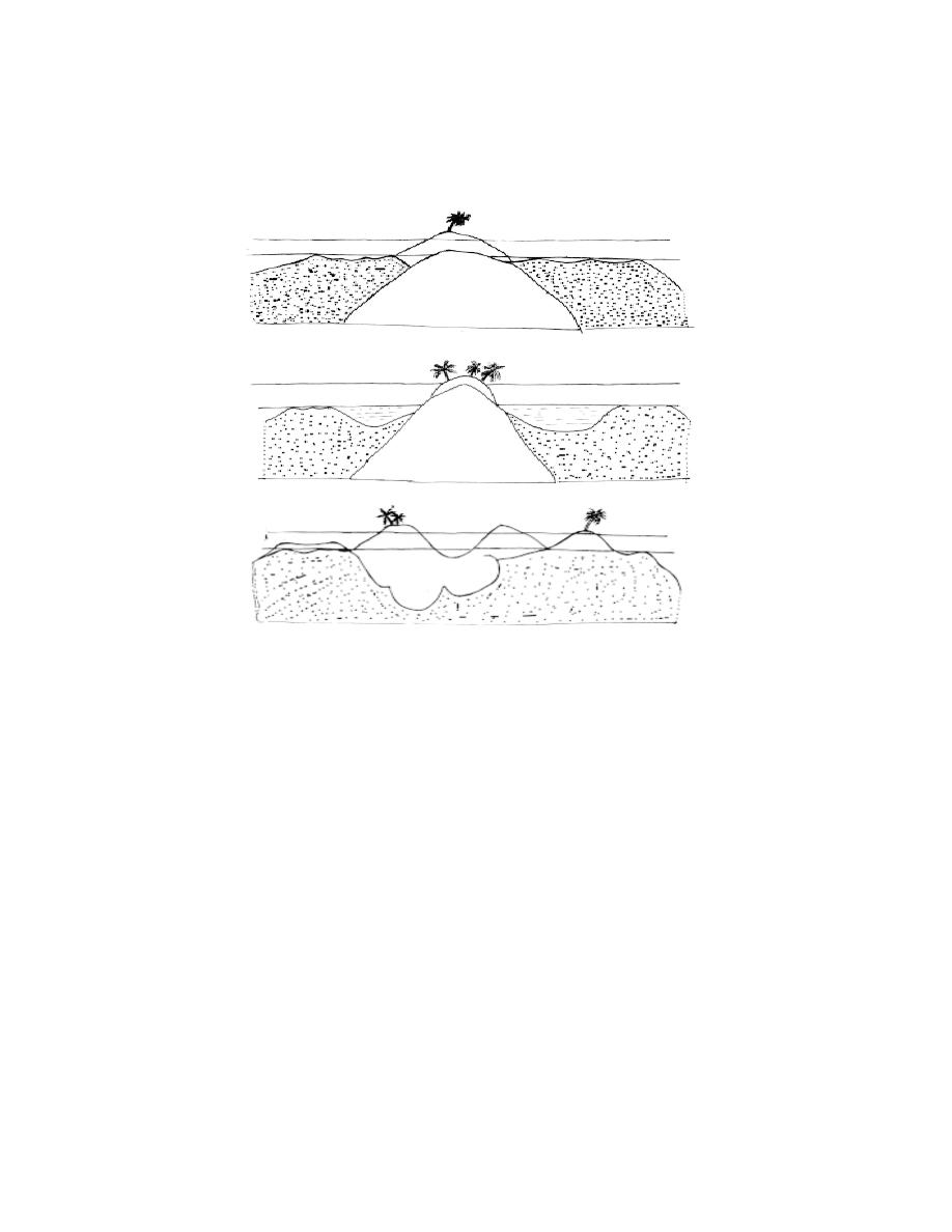
225
Economic importance
Some corals are highly priced for their decorative value. Precious corals
like Corallium nobile (= C. rubrum ) are used in jewellery and
ornaments. Corals are also important in building the coral reefs and islands,
some of which are used as habitation by human and other animals. The organ
pipe coral (Tubipora) is used in indigenous system of medicine in South In-
dia.
Coral skeletons especially of species like porites are used in as
building construction and for metalling their roads. Corals serve as raw
materials for the preparation of lime mortar and cement because of their
calcium carbonate content. Some older coral lime stones are rich in magne-
sium, hence they are of great value in making cement. Coral skeletons act as
natural barriers against sea erosion and cyclonic storms. Old reefs have long
served as sea bird sanctuaries and have collected huge deposits of bird drop-
pings (guano).
Fig.6.1.2. Coral reef - Types
Coral reef
Coral reef
Volcanic island
High tide
Low tide
Cross section of fringing reef
Coral reef
Lagoon
Lagoon
Coral reef
Low tide
High tide
C. S. of Barrier reef
Coral reef
Coral reef
Low tide
High tide
C. S. of an Atoll reef
Lagoon

226
Coral reefs provide a unique habitat for large and diverse variety of
organisms. The richness of the invertebrate fauna and the complex structure
of the coral reefs have provided opportunities for the evolution of a number of
fishes associated with coral reefs.
In several countries Fringing reef, Barrier reef and Atolls are helping
in the tourism industry.
6. 1. 2 Earthworm - Vermiculture
The growth of industries, urban cities and the ever increasing human
population have led to an increased accumulation of waste materials. Waste
materials introduced into the environment are of two types namely
non-degradable and degradable wastes. The disposal of these solid wastes
remains serious challenge in most of the countries.
Waste biomass from agriculture, domestic, urban and industrial sources
remains the main cause of organic pollution in many countries, including India.
Degradable or decomposable materials constitute a major percentage of the
refuse (more than 60%).
New technologies are now available for recycling some of these solid
wastes. These wastes can be utilized in vermitechnology for the production of
earthworm casts and earthworm tissue protein.
India produces about 2500 million tonnes of organic wastes annually.
If properly managed about 400 million tonnes of plant nutrients can be
produced from this huge organic wastes.
Vermiculture :-
In recent years vermiculture has received much attention in many
countries. The spiralling cost of fish meal and the low supply of soyabean
meal are responsible for the utilization of earthworms as an alternative low
cost protein meal in aquaculture and poultry industry. Further the worms also
play an important role in waste disposal. They decompose natural organic
wastes into rich compost fertilizer. A single earthworm can produce 1000 to
1500 offsprings in a year. 2000 mature breeders can produce more than 1
million worms in a year.
Selection of earthworm species and their culture
Lampito mauritii and Perionyx excavator are cultured in India and
Thailand. Helodrilus foetidus and Lumbricus rubellus are distributed world

227
wide. Amynthas hawayana, Eisenia foetida and Eudrilus engeniae are
also commonly used for waste management.
The methods of culturing earthworms can vary from place to place. A
cultured worm must be able to adapt to substrates, grow fast and breed or
multiply readily under controlled conditions.
A compost pile bed of 2.4m long, 1.2m wide and 0.6m deep appears
to be very satisfactory for a population of more than 50,000 earthworms. The
boxes can be made from wood (51cm long, 36cm wide and 15 cm deep) to
accomodate 5000 to 6000 worms. Soil, organic matter, manure, leaves, rice
straw, dried water hyacinth, saw dust and any fermented substrate can be
used as a culture medium to raise worms in boxes or containers.
The worms in the worm beds can grow and reproduce faster if they
are given sufficient food. Any decayed organic matter appears to be good
food for worms but the feed should not be contaminated by detergents or
insecticides.
Since the earthworms are rich in protein (65%), fat(14 %),
carbohydrate (14 %) and ash (3%) they are used as a feed component for
fish, prawn and shrimp. They are also used as livestock feed in poultry
industry.
Vermicompost
Many species of earthworms are easily adaptable to agricultural wastes
like after harvest strubble, sugarcane thrash, coirwaste, dung of cow, horse,
sheep and poultry droppings. The breakdown of these materials or the
degrades of organic matter by worm activity is called ‘Vermicompost’ It is a
better source of organic manure.
6. 1. 3 Beneficial Insects
Insects affect man’s welfare in many ways. Many insects are benefi-
cial to man. Beneficial insects are divided into two main groups according
to the nature of benefit derived from them. They are Productive and Help-
ful insects.
I. Productive Insects
These insects produce certain substances which are useful to humans.
The important useful insect products are Honey, Silk and Lac.

228
A. Honey bee
Honey bees are social insects. They live as colonies. They are active
throughout the year. They feed on the pollen and nectar of flowers. In India
there are four different varieties of honey bees. Of these, only three species
are useful in collecting honey.
1. Apis dorsata (Rock bee). This is the largest of the Indian honey bees. It
produces plenty of honey. It builds large open single combs which may often
be four feet long on tall forest trees. It is not possible to domesticate them for
the bee keeping industry.
2. Apis florea : This is the smallest of the three species and is known as the
little bee. It builds single combs, which are very small. They are found hanging
from bushes and corners of roof.
3. Apis indica : This is the common Indian honey bee. Its size is intermediate
between A. dorsata and A. florea. This bee builds several parallel combs,
generally in hollows of trees, on the walls, inside wells, caves and similar
protected spots. This is the only Indian honey bee capable of domestication.
The honeybees collect nectar from various flowers. The nectar is
swallowed by the bees. In its stomach due to the action of enzymes certain
changes happen to the nectar. Later in the bee hive it is regurgitated and
stored in chambers as honey.
Apiculture or Bee keeping is the technique of rearing honey bees
for honey and wax from their comb or beehives.
Uses of Honey and Bees wax
Honey has a high nutritive value. Its colour and smell varies in
accordance with the nectar collected from different flowers.
It is estimated that 200g of honey provides as much nourishment as
11.5 litres of milk or 1.6kg cream or 330g meat. One gram of honey provides
approxmately 33k.cal of energy. Honey has laxative, antiseptic and sedative
charcteristics. It is used in Ayurvedic and Unani systems of medicines. It is
helpful in building up the haemoglobin of the blood. It prevents cough, cold,
and fever. It cures ulcers on tongue and alimentary canal. It is also used in the
preparation of bread, cakes and biscuits.
Beeswax is also a natural secretion of the worker bee from the glands
located in the abdomen. It is used in the manufacture of cosmetics, face creams,
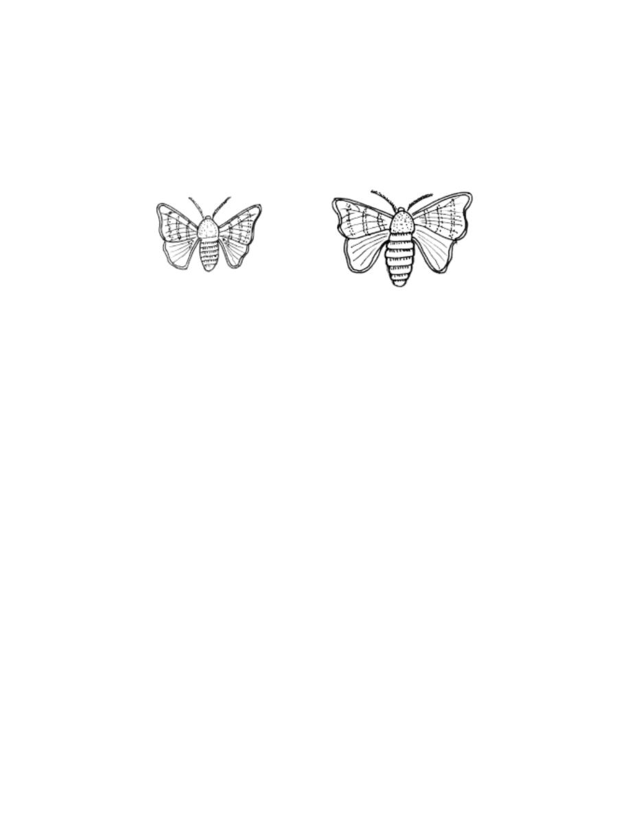
229
paints, ointments, insulators, plastic works, polishes, carbon paper and many
other lubricants. It is also used in microtomy for block preparation of tissues.
B. Silk worms
Silk is another valuable product from the insect world. The silk is ob-
tained as fine threads from the cocoons of various species of silkworms. Seri-
culture is the scientific management of production and marketing of natural
silk from silkworms.
Types of Silk worms
1. Mulberry silkworm - Bombyx mori : This is a completely domesticated
insect. Since the natural food of this worm is mulberry leaves, it is called
mulberry silk worm. The silk produced by this moth is white in colour. It is
called the mulberry silk.
2. Tasar Silk worm : Antheraea paphia
This caterpillar feeds on ber, oak, sal and fig plants. The cocoon pro-
duced by this worm is smooth and hard. It is of hen’s egg size. The cocoon
yields reelable, brown coloured Tasar silk.
3. Muga Silk worm: Antheraea assamensis: The native place of this spe-
cies is Assam where it has now become a good source of cottage industry.
The silk produced by this moth is known as Muga silk.
4. Eri Silk worm : Attacus ricinii
The caterpillar of this worm feeds on castor leaves. The cocoons of
this worm have very loose texture and the silk produced is called as Arandi
silk locally. The threads are not glossy but much durable.
The sericulture plays a significant role in the rural economy of our
country. The Chinese have the credit of discovering silk. It was kept as a
Fig. 6.1.3. Silkmoth
Adult male
Adult female
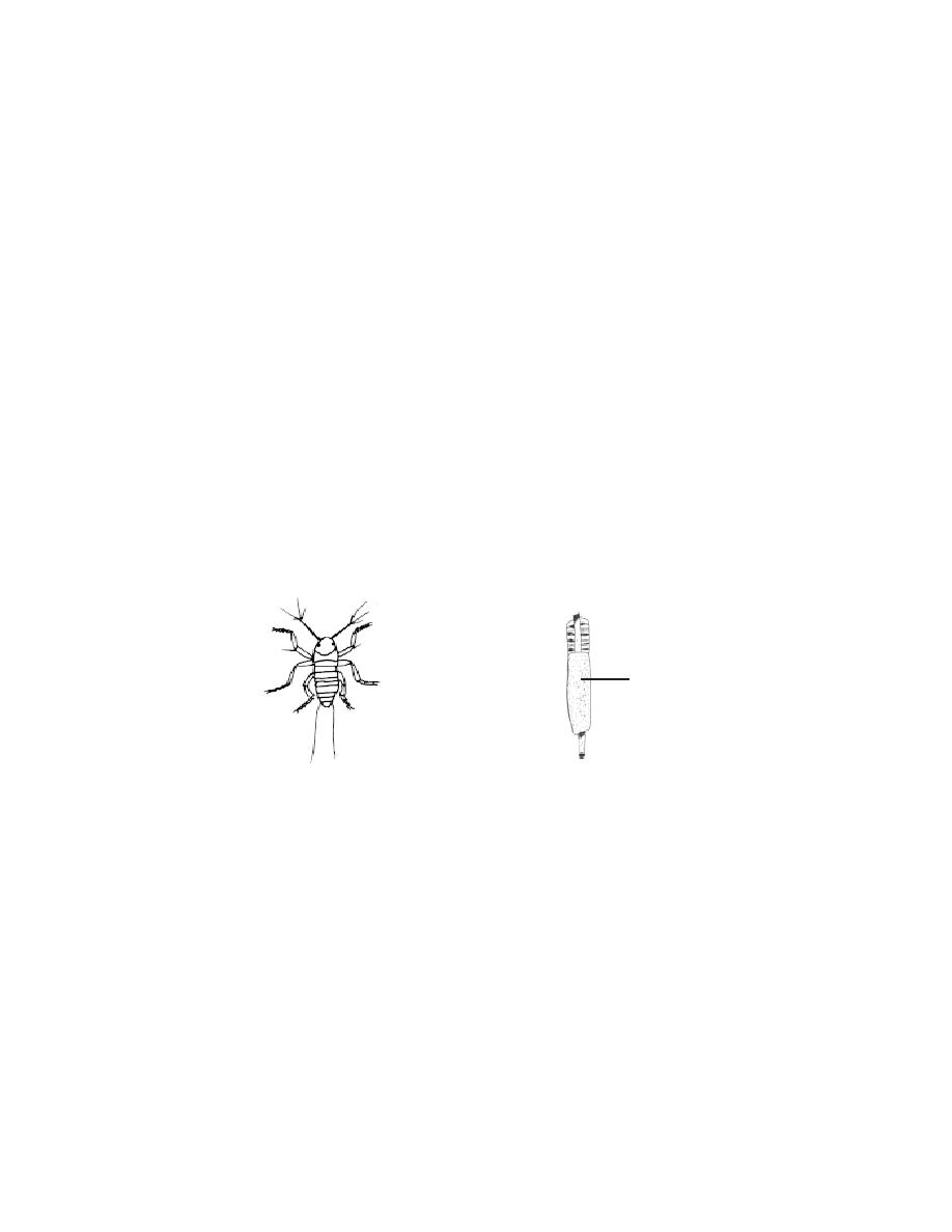
230
secret for several centuries. India now occupies a unique position in the world
for its rich sericigenous fauna to produce pure mulberry silk as well as the
three other varieties of non-mulberry silks.
Uses of silk
The raw silk is used in the manufacture of woven materials, knitted
fabrics and garments. It is also used in parachutes, parachute cords, fishing
lines, as sieves in flour mills, insulation coil for telephones and wireless
receivers, and tyres of racing cars.
C. Lac insects
Another useful product we get from insects is lac. Lac is the resinous
protective secretion produced by a kind of scale insect called Laccifer lacca.
They secrete a brown resinous substance called the lac. The minute red
coloured larva of this insect, settles on succulent shoots of the host plants.
While growing they secrete a resinous material which covers them. The twigs
are harvested and the encrustations scraped, dried and processed to yield the
lac of commerce.
The important trees needed for lac encrustation are the kusum, the
Ber, Palas, Babul and sal. These trees are common in the western ghats.
Uses of lac
Lac is one of the most versatile natural resinous material. It has a
unique combination of properties which render it useful in the plastics,
electrical, adhesive, leather, wood finishing and other industries. In the
electrical industry it used in the form of insulating varnishes and moulded insu-
lators. It possesses very good adhesion to mica. It is an ingredient of var-
nishes, polishes, finishes wood used for protective and decorative purposes. It
Fig. 6.1.4. Lac insect
Lac incrustation
Nymph
lac encrustation
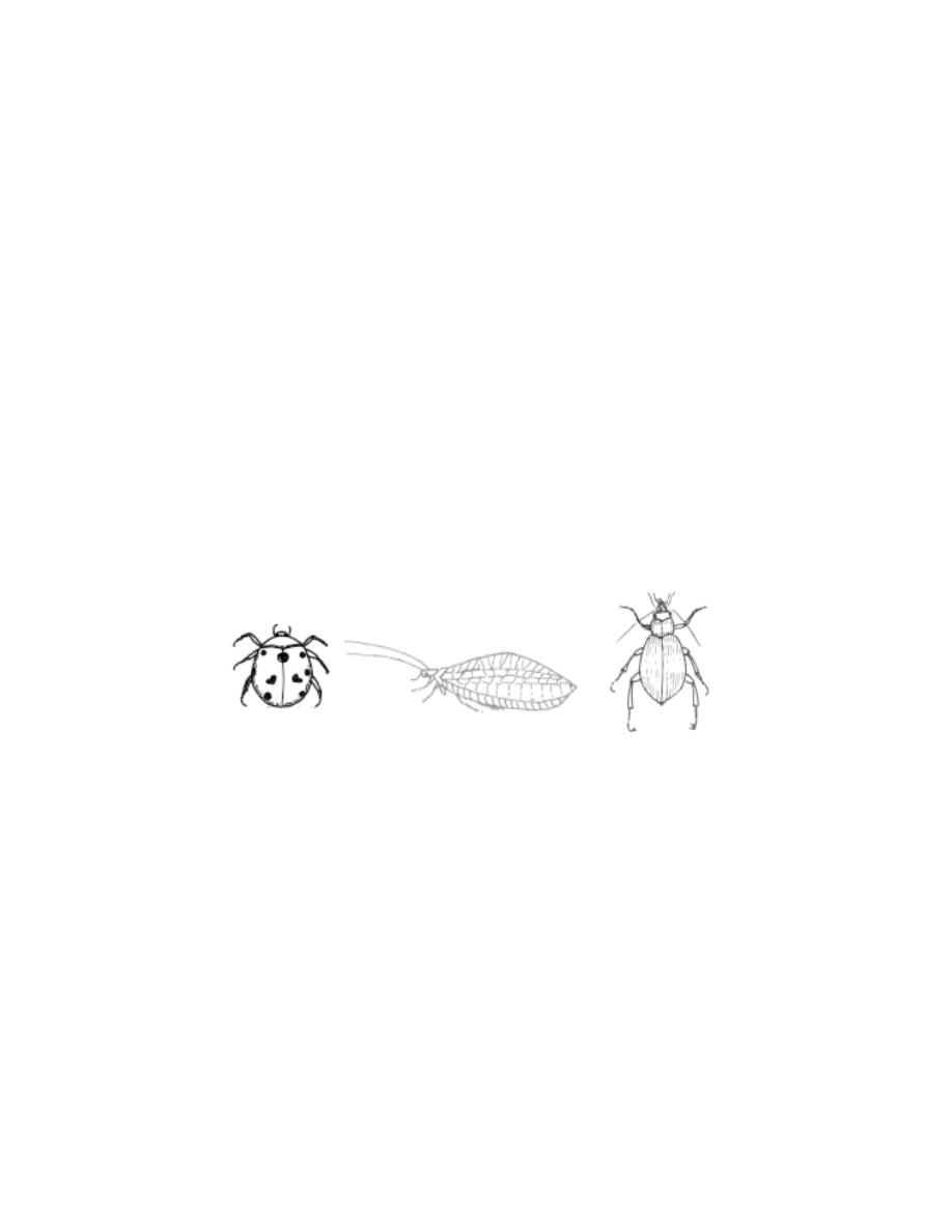
231
is a principal ingredient of sealing wax. It is also used in the
manufacture of glazed paper, printing and water proof inks, nail polishes, den-
tal plates, ammunition, bangles, wax crayons and optical frames.
II. Helpful Insects
This category of insects includes all forms which by their life
activities help man in controlling the plant and animal pests. The most
important of these helpful insects are the insect feeding or entomopha-
gous forms including predators and parasites. These are important in main-
taining a balance in insect populations.
Insect - Predators
These are generally larger than their prey. They can pounce upon and
devour the prey easily. Among the insect predators, lady bird beetles are
more useful to the farmers and gardeners. These small beetles are voracious
and are highly prolific and long-lived. Both larvae and adults feed on a wide
range of insects. Other common and well known predators are Aphis lions
and ground beetles.
Insect parasites
Insect parasites are smaller than their hosts. They live continuously
for atleast a part of their lifecycle on or within the body of the host. The
tachinid flies parasitize caterpillars, beetles and other groups of insects. The
braconids are a large group of small wasp-like insects that parasitize a wide
range of insects including plant lice and caterpillars. Most group of insects are
plagued by ichneumon parasites.
Employing insect predators and parasites for controlling the popula-
tion of insect pests is known as biological control.
Fig. 6.1.5. Insect - predators
Ladybird beetle
Ground beetle
Aphis lion
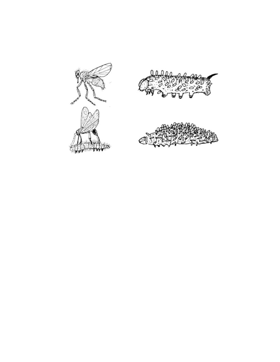
232
Insects as pollinators
Insects play an important role in the pollination of plants. Bees, wasps,
ants, butterflies, beetles and thrips render valuable service in pollination. The
services of honey bees are needed in the production of cultivated crops, such
as apples, pears, plums and vegetables. Some plants like the yucca and smyrna
fig are completely dependent upon insects for pollination.
Other useful insects
A number of insects feed on plants and they may aid in keeping plant
weeds under control. The most successful use of an insect in the control of a
plant was the introduction of the caterpillar of a moth (cactoblastis cactorum)
to control the prickly pear (Opuntia spp) in Australia.
6. 1. 4 Prawns, Lobsters and Crabs
A. Prawns
The prawns are one of the most economically important fishery
organism of India. It helps to earn a sizeable amount of foreign exchange. The
prawns are the most esteemed food among the marine food organisms. There-
fore, they are in great demand both in the local and international
markets.
Fig. 6.1.6. Insect parasites
Tascinid fly
Pupae of Tacinid fly on a caterpillar
Ichneumon wasp laying eggs
in a caterpillar
Pupae of a braconid fly on the
larva of a moth

233
Export of ‘prawn pulp’ to Burma and Malaya from earlier times and
‘frozen and canned’ prawns to USA and Japan in recent years has made
Indian prawns a major foreign-exchange earner. Apart from being a delicacy,
prawns are a rich source of protein and vitamins (A and D). They contain
considerable quantities of glycogen and free aminoacids in their muscles
imparting their flesh a sweet taste. As they contain very little fat, they have
become a favourite protein food for the weight conscious persons.
Indian prawns of commercial importance
Penaeus indicus, P. monodon, P. japonicus, Metapenaeus dobsoni,
M. monoceros M. affinis, M. brevicornis, Parapenaeopsis stylifera,
P. sculptilis, Macrobrachium rosenbergii , M. malcomsonii, Palaeomon
tenuipes and P. styliferus
Freshwater prawns inhabit rivers and lakes across the entire country.
They migrate to brackish water for breeding. Eg. Macrobrachium,
Palaeomon. Marine prawns occur in shallow coastal waters. They form large
shoals close to malabar coast during the monsoon season. Penaeus,
Parapenaeopsis and Meta penaeus are the important genera of the Indian
coast. The practice of rearing prawns as a ‘secondary crop’ between
November and April in the paddy fields along the coastal areas in India should
be a step towards increased production of fresh water prawns.
Fresh prawns are packed in ice and sent to inland markets for
consumption. Large specimens are frozen directly between layers of ice.
Smallar varieties are boiled, shelled and then packed between ice. Prawns are
also cured. This includes sundrying, salting and pickling.
Fig. 6.1.7. Prawns
Marine prawn
Fresh water prawn
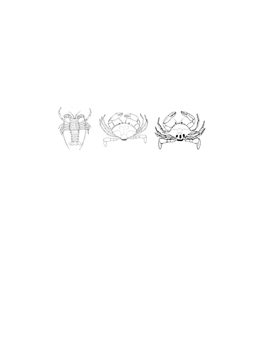
234
B. Lobsters
Lobsters belong to four main groups. They are clawed or true
lobsters, spiny or rock lobsters, sand or slipper lobsters and coral lob-
sters. The lobsters of our country are called spiny lobsters. The economically
important species of spiny lobsters are Palinurus polyphagus P. homarus,
P. ornatus and P. versicolors. The lobsters are called ‘fš Ïuhš’(Kal Eral)
in Tamil.
The lobster fishery of India gained importance only recently. Its food
value (proteins 15-24 %) was realised following the demand for lobsters in
western countries. India is one of the prominent countries in spiny lobster
marketing. The main lobster landing centres in India are Mumbai, Veraval,
Kolachal, Tuticorin, Chennai, Mandapam and Kozhikode. North west coast
contributes 80-90 percent of the landings with P. polyphagus being the
dominant species. On the west coast P. polyphagus and P. homarus are very
common and on the east coast P. polyphagus and P. ornatus are common.In
the Gulf of mannar the lobster fishery is confined to the areas where coral
reefs are present. The lobsters are caught throughout the year but the peak
fishing season is December to January.
The lobsters are esteemed as good food particularly in foreign
countries. Therefore most of the lobster catches are exported to USA, Canada,
UK, France, Spain, Belgium, Gulf countries, Nepal and Singapore.
The central Marine fisheries Research Institute (CMFRI) has carried
out studies on fishery, recruitment, biology, physiology, breeding, larval
rearing and culture of economically important spiny lobsters in India.
Fig. 6.1.8. Lobsters and crabs
Lobster
Scylla serrata
Portunus sanguinolentus
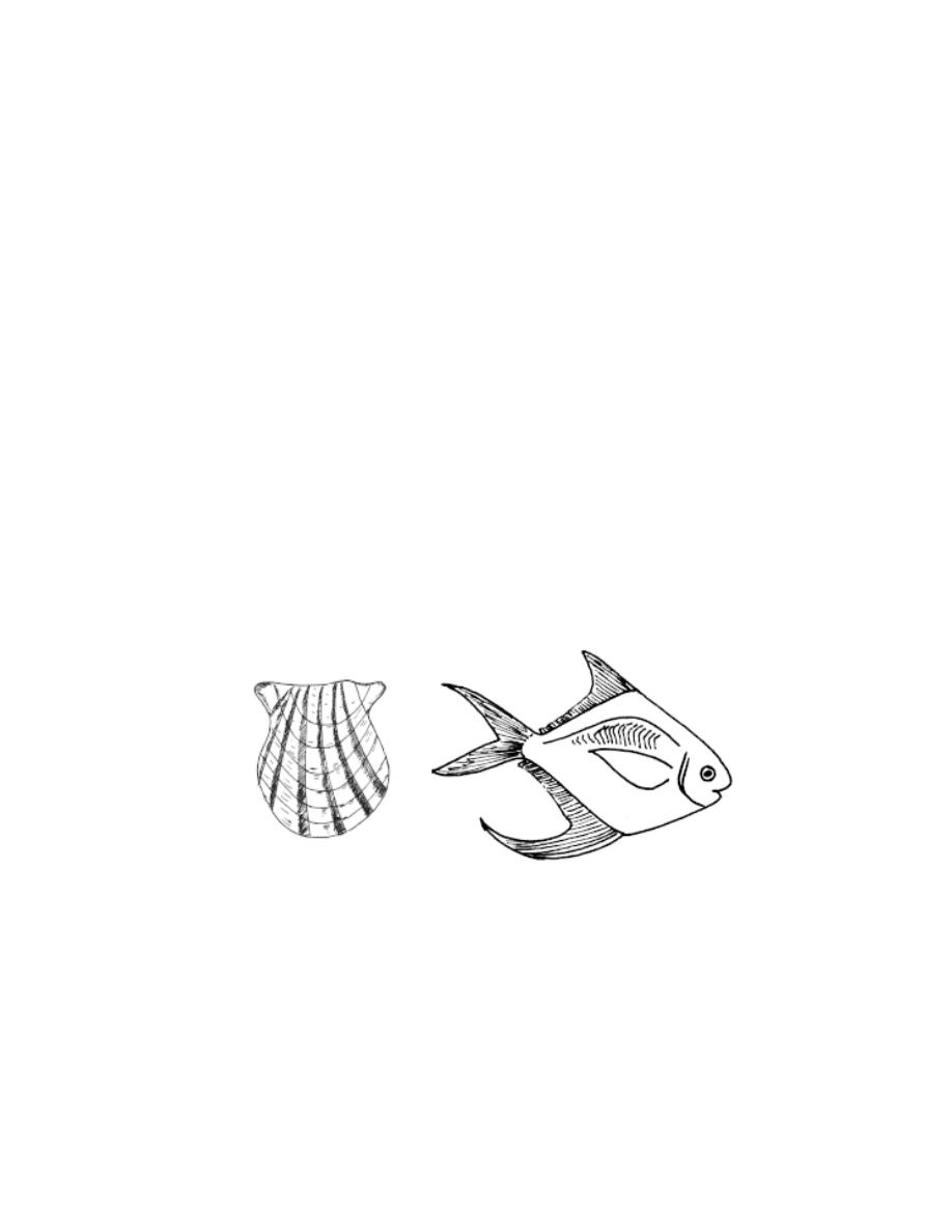
235
C. The Crabs
The crabs are decapod crustaceans. They are characterised by the
greatly enlarged cephalothorax. The abdomen in crabs shows sexual
dimorphism. In males the abdomen is narrow. In female crab the abdomen is
broad. and it carries eggs during breeding season.
There are nearly six hundred crab species occurring in the Indian waters
but only very few of them are being used for food purposes. The
important species of crabs supporting the fishery are Matuta lunaris, Scylla
serrata, Portunus sanguinolentus, and Charybdis cruciata.
6. 1. 5 Pearl Oyster
Pearls are one of the rarest and highly esteemed gems. Pearls are
produced by the pearl oysters of the genus Pinctada under class Bivalvia.
From the point of view of pearl production in Indian waters, the most
important species is P.fucata. It has a wide distribution in the Persian Gulf,
Red sea, Gulf of Kutch, Gulf of Mannar and the Palk Bay. The oysters occur
on ridges of rock or dead coral in the Gulf of Mannar along both coasts of
India and Sri Lanka. These formations are known as ‘pearl banks’ or ‘paars’.
They extend from Cape comorin to Rameshwaram Island. Heavy production
had been recorded in Tuticorin.
From each pearl oyster bed thousands of oysters are taken out. Every
oyster contains a pearl. Most of the pearls obtained are too small. Some of
them are large, perfectly round with fine lustre fetching high prices. Hence it
is worthwhile farming oysters and induce pearl formation. In recent years
India has succeeded in developing farming techniques for the production of
cultured pearls of good shape and lustre.
Fig. 6.1.9. Pearl Oyster
Fig. 6.1.10. Pomfret

236
The CMFRI in India gives necessary training in pearl culture tech-
niques. In this process shell beads are introduced into the soft tissues of the
oyster along with a strip of the mantle so that the latter may secrete the pearly
substance around the bead. The treated oysters are well taken care of in
cages suspended from floating rafts in shallow waters of the sea. Thus, cul-
tured pearls are produced in the same way as the natural pearls. The pearl is
a concretion of calcium carbonate in an organic matrix. It is like the nacreous
layer secreted by the mantle on the inner surface of the shell valve.
Shells having a brilliant silvery sheen are known in commerce as the
“mother of pearl”. They are collected for the manufacture of buttons and
other fancy articles.
6. 1. 6 Fishes - Nutritive value
The marine fisheries of India are of importance in increasing the
country’s food resources and fetching a considerable amount of foreign ex-
change through the export of frozen and processed marine products. Besides,
the major capture fisheries, a breakthrough has been effected in recent years
to initiate the culture of selected species of finfishes and crustaceans. The
establishment of Central Marine Fisheries Research Institute (CMFRI), Cen-
tral Institute of Inland and Brackish water Aquaculture (CIBA), National In-
stitute of Oceanography (NIO), Central Institue of Fresh water
Aquaculture(CIFA), National Institute of Ocean Technology (NIOT) and
Marine Products Export Development Authority (MPEDA) has led to the gen-
eration of considerable information on various aspects of Marine biology,
Marine, Fresh water and Brackish water Fisheries and Oceanography.
Consumption of fish for food has appreciably increased in recent years in all
countries. The declaration of Exclusive Economic Zone (EEZ) has provided a
great opportunity and challenge to coastal nations. In India there is good scope
for development of marine resources to derive economic, social and
nutritional benefits.
The nutritive and medicinal value of fish have been recognized from
time immemorial. Fish flesh is an excellent source of protein in human diet.
The principal biochemical contents of fish flesh are protein, fat and water.
Protein constitutes about 20 percent.
The nutrional value of fish flesh is comparable and even higher than
that of the flesh of birds and mammals. Fish flesh remains a good source for
all essential aminoacids in needed concentrations.

237
The Indian pomfret (Stromateus argentius) is ranked high on the
basis of its aminogram. As a child food, fish is easily tolerated by infants.
Lean fish is recommended to convalescing patients.
The nutritive value of preserved and processed fish and fishery
product is generally lower than that of raw fish.
Medical and Economic importance of fish
Apart from direct consumption, contents in the body of fish are
processed into a number of valuable products. Chief among these are :
Fish liver oil
Fish liver contains vitamin A and D in considerable quantities. Eg.
shark liver oil and cod liver oil.
Fish liver oil can cure or prevent occurence of deficiency diseases
such as rickets, xerophthalmia, impaired vision and eye defects,
abnormalities in skin, mucous membrane and vertebrae. Fish liver oil will en-
sure healthy growth of bones and teeth.
Fish body oil
Fish body oil is obtained from the entire body of fish. Fish body oil is
generally extracted from oil sardines or from less edible varieties of fishes. It
is also extracted from wastes discarded from fishery industries. Some of the
important uses of oil are : 1) manufacture of cheap soaps, paints and var-
nishes 2) tanning of leather 3) steel and chemical industries 4) manufacture
of lubricants and candles.
Fish Meal :
Fish meal is the cooked, ground and dried preparation of the fish body.
It makes an excellent poultry and animal feed. It enhances egg and milk pro-
duction.
Fish flour :
Fish flour is considered an ideal protein source to supplement diet of
both adults and infants. It may also be mixed with wheat and maize flour. It is
also used to enrich bakery products such as cakes, breads, biscuits, soup and
sweets.

238
Fish manure and guano :
All rejected and thrown out products from fishery industry are made
use of as fish manure. Fish guano is obtained as waste from fish oil
industries. It is a good manure.
Fish Glue :
It is a kind of good glue obtained from fin trimmings, bones and skins
of fishes. Fish glue is considered as a good adhesive.
Isinglass :
Isinglass is a high grade collagen produced from the air bladders of
certain groups of fishes. It is used in the clarification of wines, beer and
vinegar. It is also used in the preparation of plasters and special cements.
Fish skin :
Tanned skin of sharks and rays are used in the manufacture of shoes,
handbags and wallets.
Omega Fatty acid
The unique feature which differentiates fish food from other animal
protein sources is the presence of omega-3 fatty acids such as linolenic acid,
decosahexaenoic acid (DHA) and eicosapentaenoic acid (EPA). DHA
promotes learning ability in children and improved memory in elders. DHA is
essential for the foetal growth and development.
Omega fatty acid is also good for heart. It helps to control diabetis by
improving insulin action. It is also reported to be good for arthritis.
6. 1. 7 Guano (Bird excreta)
Guano is the accumulated excrement or droppings of fish eating sea
birds such as gannets, cormorants and pelicans. It has been used as a
fertilizer. These sea birds populate some islands off the west coast of Peru,
lower California and Africa. Their population strength may go upto 5, 600, 000
per square mile. They may consume 1000 tonnes of fish daily. Exports of
guano from the Peruvian deposits began in 1810 and reached 50, 000 tons in
1856. The Government protects the seafowls and processes the guano which
contains about 11 to 16% nitrogen, 8 to 12 % phosphoric acid and 2 to 3%
potash. The fertilizing properties of the phosphoric acid and nitrogen
contained in fish were not recognised until the guano became a stimulus to

239
intensive agriculture. The real guano is found in vast stratified accretions on
rainless islands off the coasts of Peru.
6. 1. 8 Aquarium
Next to birds fishes are perhaps the most colourful of animals. They
are graceful in their shape, bodyform and movement. The Chinese first con-
ceived the idea of growing fishes in and around their homes. Thus, varieties of
gold fishes which are popular with aquarium lovers were bred by them from
ordinary carplike fishes. Gold fishes are varieties of the genus Carassius, a
fresh water group of fishes related to the carps.
An Aquarium is a container made of glass, or with glass walls. It
permits watching the fishes swimming easily and for prolonged periods of
time. It is a hobby which appeals to young and old and has opened up a
flourishing business in all big cities.
Setting up an aquarium tank
Tank selection : The choice of an aquarium tank, depends largely on where
it is to be housed, the extent of one’s enthusiasm and the money one wants to
spend. While selecting a tank it is very important to note that it is of good
construction. Shallow and wide tanks are preferable as they have more
surface area to facililate oxygenation through atmospheric contact. Further
such tanks can accomodate more number of fishes.
Before setting up the tank, a suitable place has to be selected. In
order to get a good view of both fishes and plants in an aquarium tank, the
light should come obliquely. Hence, instead of the window sill, a place nearer
to its side may be selected. To avail maximum sunlight an eastern exposure is
preferable. The support or table for positioning the tank must be strong and in
level. It is also essential to have a cover with a provision for installing lights
and feeding.
If the tanks are purchased they should be checked for leakage. Their
sides should be cleaned with 1 % potassium permanganate solution, besides
repeated washing with tap water. The next step in the setting up of an aquarium
is the collection of sand that is necessary to make the bed at the bottom of the
tank. The sand for the tank bottom can be collected from the sea shore or a
river. This sand has to be washed to remove the dirt. It is advisable to put the
sand in a shallow pan or aluminium vessel and heating it. The cleaned sand
may be exposed in the bright sun. These processes ensure the killing of all

240
bacteria or germs. The cleaned sand should be spread evenly in the tank
bottom. Over the top layer of sand a thin layer of stone chips or very small
pebbles may be placed.
Quality of water for Aquarium tank
Rain water free from contaminants or tap water is ideal for aquarium
tanks. A tap water of domestic supply is likely to have chlorine which is
harmful to fish. This water may be stagnated for about one or two days before
use. If the water is hard it must be properly treated. The tank should be filled
with the desired water without disturbing the bottom sand.
Planting : The aquarium tank after being filled with water can be planted with
selected varieties of aquatic plants. The plants provide shade, shelter and sanc-
tuary for fish. Besides decorating the aquarium tank, they also help in spawn-
ing and as food for certain fishes. The plants provide oxygen through photo-
synthesis. Over crowding of plants has to be avoided to ensure an
adequate supply of oxygen to fishes.
Several types of plants are available. Among them the important ones
are the tall rooted plants such as Vallisneria and Myriophyllum. Before
planting, the plants must be washed well otherwise disease causing parasites
may attack the fish at a later stage. Plants may be disinfected by rinsing in 0.1
percent solution of alum (Potassium aluminium sulphate) followed by a good
washing in water. The roots are trimmed and are kept between wet newspa-
per sheets to prevent them from drying before they are set. The plants are
carefully installed using a forked wooden piece. Large plants like vallisneria
may be planted at the back while the bushy plants can be placed in the corners
or at sides. The floating plants may be necessary in tanks where gouramies
are bred. The fragments of these plants are used by these fishes in the
construction of bubble nests for breeding.
Lighting : Light besides beautifying the aquarium helps in the photosynthesis
of aquarium plants. Fishes also require light to trace their food. Further the
light is known to influence the growth of fish. Strong sunlight destroys bacte-
ria and keeps the tank healthy. For a moderate tank two bulbs of 60 watts
each may be lit for eight hours a day. It is better to use fluorescent lighting for
promotion of plant growth and for even distribution of light.
241
Common ornamental fishes
Live bearers
Guppy
-
Lebistes reticulatus
Platy
-
Xiphophorus maculatus
Sword tail
-
Xiphophorus helleri
Black molly
-
Mollienesia sphenops
Egg Layers
Siamese fighter - Betta splendens
Giant gourami - Colisa fasciata
Kissing gourami - Helostoma temmineki
Angel fish - Pterophyllum scalare
Gold fish - Carassius carassius
Fig. 6.1.11. Ornamental fishes
Angel fish
Siamese fighter
Guppy
Gold fish
Sword tail

242
Introduction of fish in an aquarium tank.
The number of fishes suitable for stocking in an aquarium tank
depends mainly on the surface area of the tank, its dissolved oxygen content
and size of the fish. It is reported that 1 cm long fish may need about
75 cm
2
of the surface area. Based on the above, a tank of 75 X 30 cm size for
example may hold three fishes each of 10 cm length.
The fishes are generally introduced two to three days after planting
when the water would be clear and well oxygenated by plants. Before
introduction into the aquarium tank, the fish may be treated with 2 percent
potassium permanganate solution to avoid parasitic attack.
Feeding
Algae which often grow around stones and in water serve as a good
food source to swordtails, kissing gourami and mollies. Live protein rich foods
such as tubifex worms, Chironomid larvae and mosquito larvae are
considered excellent. Artificial fish feed meant only for aquarium fishes can
also be used.
Fishes may be fed once or twice a day according to their preference
and satiation. Excess food and detritus may be removed 30 minutes after
feeding, by siphoning out using a rubber tubing. If the level of the water gets
reduced in course of time it should be restored by the addition of some rain
water or chlorine free tap water.
Aesthetic value :
A large number of fishes are cultured in aquaria for their beauty and
graceful movements. The movements of colour fishes in an aquarium would
certainly please the ailing and convalescing people. The aquarium may
gladden one’s heart.
Vivarium ( Zoo )
The growing awareness for nature and wild life conservation has made
zoos a popular institution. Estimates indicate that 10% of the world’s human
population visit zoos every year. There are about 350 animal collections in
India, which are visited by more than 50 million people annually. Most zoos
until the last two decades were set up mainly for entertainment and recre-
ation, with the result the scientific management of wild animals in captivity
has evolved slowly.

243
The need for making conservation as one of the main objectives of
management of zoos was realised by Government of India soon after
independence. Indian Board of wild life made important recommendation in
this regard. Today wildlife habitats are under severe pressure and a large
number of species of wild fauna have become endangered. The zoos apart
from sustaining their own populations have to augment the depleting popula-
tions of endangered species in the wild. As zoos are visited by a large number
of visitors, they are potent tools for educating people about the important of
wild life management as it remain a life supporting processe of nature.
Objectives
The main objective of the zoos shall be the conservation of the rich
biodiversity of the country, particularly the wild fauna. This objective can be
achieved through the following methods.
1. Supporting the conservation of endangered species, which have no chance
of survival in wild.
2. To inspire amongst zoo visitors empathy for wild animals, an understanding
and awareness about the need for conservation of natural resources and for
maintaining the ecological balance.
3. Providing opportunities for scientific studies useful for conservation.
Safari :- Safaris are specialized zoos where the captive animals are housed in
very large naturalistic enclosures and the visitors are allowed to enter the
enclosure to view the animals in a mechanised vehicle or a predetermined
route and watch the animals from close quarters.
List of important Zoos
1. Indira Gandhi Zoological park, Visakha patnam, A. P.
2. Nehru Zoological park, Hydrabad, A. P.
3. Assam state Zoo-Botanical Garden, Guwahati, Assam.
4. Sanjay Gandhi Biological park, Patna, Bihar.
5. National Zoological park, Delhi.
6. Kamla Nehru Zoological Garden, Ahmedabad, Gujarat.
7. Sri Chamarajendra Zoological Garden, Mysore.
8. Nandankanan Biological park, Orissa
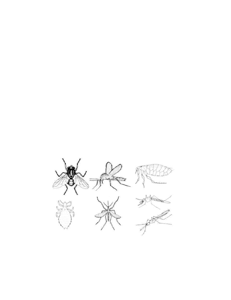
244
9. Mahendra Chaudhury Zoological park, Punjab.
10. Arignar Anna Zoological park, Vandalur, Tamil Nadu.
11. Kanpur Zoological park, Kanour, U. P.
12. Zoological Garden, Calcutta.
6. 2 Harmful Animals
The harmful animals are those that cause injury to plants and domes-
tic animals. Human beings are affected directly or through bites or stings or
by transmission of various kinds of pathogens. The nature of harmful insects
ranges from simple nuisance value of cockroaches to spreading of epidemic
diseases, such as malaria, filariasis by mosquitos. For the convenience of our
study the harmful animals are grouped under the following categories namely
disease causing organisms, poisonous animals, fouling organisms and pests.
6. 2. 1 Disease causing organisms - Vectors
Some insects are injurious to man as vectors of human diseases.
Through the ages millions of people have died of dieases transmitted by
insects. There are a number of insect-borne diseases, and they may be
transmitted in different ways.
Fig. 6.2.1. Insect - vectors
House fly
Sand fly
Rat flea
Human louse
Mosquito
Culex
Anopheles
Sitting position of
mosquito species

245
1. Housefly - Musca domestica
House flies are cosmopolitan in distribution. They are closely associ-
ated with humans and thrive best where people are careless in the disposal of
wastes. Adult flies are non-parastic.
They feed on all kinds of decaying and decomposing matter. It is an important
mechanical vector in the transmission of diseases like typhoid (Salmonella
typhosa - a bactericum), dysentery (Entamoeba histolytica) and cholera
(Vibrio sps.) The housefly cause diseases through food contaminations.
Control : Housefly control is normlly done in 3 different ways, namely
sanitary, mechanical and chemical methods. Populations of houseflies can be
controlled by proper disposal of manure, garbage, sewage, food waste,
human excreta and other organic materials. Mechanical practices such as
screening, using of traps or sticky paper or baits can be valuable in excluding
houseflies. Insecticides may be used against larvae. Spraying with 2%
malathion 1% chlordane or lindane, 0.5% tremephos are effective.
2. Sand flies - Phlebotomus papatasi
These flies are 4 mm long. Only the female possess piercing-sucking
mouth parts and are haematophagous. The males are non parasitic, feeding on
moisture. They are small slender insects with hairy bodies. Through biting this
fly transmits the disease called kala-azar. The causative oraganisms is Leish-
mania, a parasitic protozoan. During the day time the flies remain hiding. At
night they come out to feed. The sand fly attacks during night times. The
insect sucks the parasite from an infected person, along with blood. In side the
body of the fly, the parasite undergoes changes. When an infected fly bites
man, the parasites pass into the blood and fresh infection is effected. The
parasites mostly concentrate in the capillaries of spleen, liver and bone mar-
row. The disease is characterized by the symptoms like anaemia and emacia-
tion.
Control :
Spraying of 5 % DDT / BHC easily kills the flies. The pyrethrum
ointment used on exposed part of the body works as a repellent.
3. Rat fleas - Xenopsylla cheopis
The insect parasite, Xenopsylla cheopis is commonly known as the
Asiatic rat flea. Both male and female fleas take in the bacillus pasteurella

246
pestis from infected rats during feeding. This rat - flea is responsible for the
transmission of plague from man to man, or from rat to man. When this
bacterium is introduced into the skin, the lymph glands become inflammed.
This is known as bubonic plague. Frequently, the bacilli become established
in the victims blood. The condition is then referred to as septicemic plague.
If the victim’s lungs become involved, it is referred to as pneumonic Plague
When the rat flea sucks the blood of man or a rat infected with plague,
the bacilli enter into its stomach and grow there into large numbers. The flea
thus heavily laden with the bacilli, may bite a healthy man and introduce the
bacilli into the wound and cause infection. The bacilli are deposited by the flea
on the skin along with the faeces. The bite of the flea causes scratchings and
the bacilli are introduced into the blood when the skin is scratched.
Control :
Destruction of rats and other rodents is an effective method. Dusting
of 1 to 2 % chloradane, or 2 % Y - BHC is very much effective in the
elimination of fleas on the body of pet animals. Application of 5% DDT is
recommended for spraying at the time of the spread of plague in all the areas.
4. The human louse - Pediculus humanus:
Louse is a blood sucking ectoparasite of man. It is cosmopolitan in
distribution .
The human louse is a major vector for three important human dis-
eases, relapsing fever, typhus and trench fever.
Disease
-
Parasite
Relapsing fever
-
Borrelia sp
Typhus
-
Rickettsia sp
Trench fever
-
Rickettsia sp
Control : Wearing clean clothes, and having regular bath avoids infestation.
5. Mosquitoes : Anopheles, Culex, Aedes sp.
Mosquitoes are cosmopolitan in distribution. They are nocturnal in habit
and are found in abundance in damp, marshy lands near stagnant water. Only
female mosquitoes are adapted to suck the blood of human beings and func-
tion as carrier of viral, protozoan and nematode diseases.
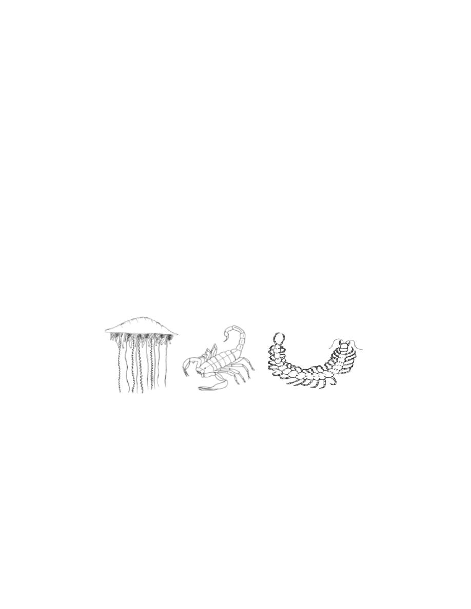
247
Culex mosquitoes serve as the vectors for filariasis or elephantia-
sis. This disease is caused by the nematode parasite, Wuchereria bancrofti.
It is commonly known as filarial worm. It is found in the lymphatic vessels and
lymph glands of man. The female worms give birth to living embryos known
as microfilariae. The microfilariae normally circulate at night(10 to 2 am) in
the peripheral blood. At that time they are ingested by the mosquito along with
blood, the mosquito is not just a mechanical carrier of the parasite.
Developmental changes take place in the body of the parasite. When the
infected mosquito next bites another person, the larvae penetrate the
superficial skin to find their way into the lymphatic vessels, and attain sexual
maturity. In severe infection the adults cause blocking of lymphatic system
which results in the enlargement of legs, arms, scrotum, and mammary glands.
It is known as elephantiasis.
The Anopheles mosquito transmits plasmodium, a causative protozoan
for malaria (Refer : Plasmodium)
Another type of mosquito, Aedes transmits yellow fever through a
virus.
6. 2. 2 Poisonous Organisms
Free living organisms have developed some device to protect
themselves against predators. These protective devices ranges from the simple
stinging cells of Physalia to the massive poison glands of the snakes. The list
of poisonous organisms is exhaustive. A few of the important poisonous
organisms are mentioned here.
Fig. 6.2.2. Poisonous animals
Physalia
Scorpion
Centipede
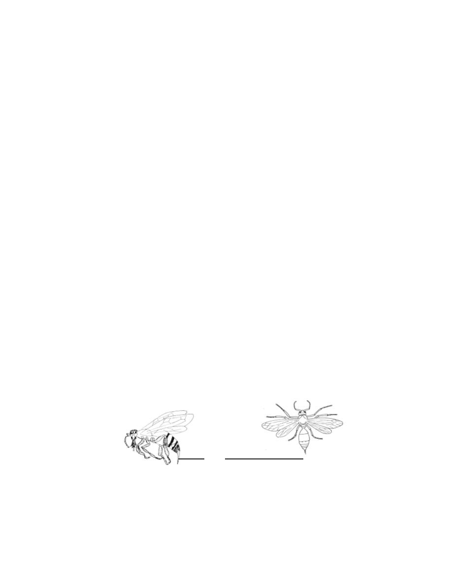
248
Physalia
These are marine coelenterates. They are notorious for the painful
sting they can inflict on unsuspecting swimmers who accidently brush against
them. They attack using stinging cells on their trailing tentacles. Their
powerful stings cause painful local inflammation and can even be fatal.
Scorpion.
In scorpion the sting is attached to the posterior part of the last
segment. It consists of bulbous base and a sharp curved barb that injects the
venom. The venom is produced by a pair of oval glands. The scorpion raises
the posterior abdomen over the body making it curved forward. A stabbing
motion is used in stinging.
The venom of most scorpions is sufficiently toxic to kill a vertibrate.
The venom of the scorpion Androctonus is equivalent in toxicity to cobra
venom.
The neurotoxic venom of scorpions is very painful and may cause
paralysis of the respiratory muscles or cardiac failure in fatal cases. Anti
venoms are available for these species.
Centipede :
Centipedes are distributed throughout the world. They live in soil and
humus and beneath stones. The largest centipede is the tropical American
Scolopendra gigantea which may reach 26 cm in length. They have a large
pair of poison claws sometimes called maxillipeds. Each claw bears a terminal
pointed fang. The venom although painful is not sufficiently toxic to be lethal
to man even to small children. However S. gigantea has been known to cause
human death.
Honey bees and Wasps :
Fig. 6.2.3. Poisonous insects
Worker honeybee
Wasp
sting
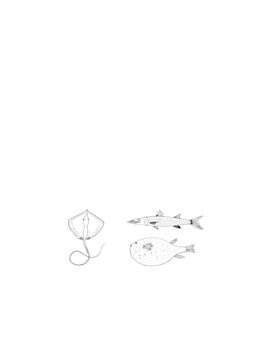
249
In worker honey bee (undeveloped females) the poisonous sting is
situated at the hind end of the body. It is a pointed structure provided with
minute hooks or barbs at its free end. On stinging the tip of sting gets
detached. Hence a bee can sting only once.
Unlike the bee the wasp is able to withdraw its sting from the wound.
Hence it can sting again. In wasp the sting is a modified ovipositor and once it
has penetrated the skin of the victim poison is injected as in a hypodermic
syringe. The wasp’s poison is a histamine.
The sting by honey bees and wasps lead to pain and inflammation.
Poisonous fishes
More than 700 species of fishes have poison glands. Venom in fishes
is of two kinds. One kind of venom is produced by specialized glands which
may occur in various parts of the body. In the second, the flesh itself may
secrete some toxic substance and the fish becomes poisonous and inedible.
There are several poisonous cartilaginous fishes. The poison glands
are usually associated with a spine or sting as in the case of sting ray. In the
sting ray(Trygon), the poison glands lie along a lateral groove on each side
of the spine on its tail. The spine causes pain and numbness in the flesh of
victim.
The large Barracuda of Cuba and other tropical islands have
poisonous flesh, which when eaten cause pain in joints and extremities,
nausea, vomiting and general trembling.
Fig. 6.2.4. Poisonous fishes
Sting ray
Barracuda
Puffer fish
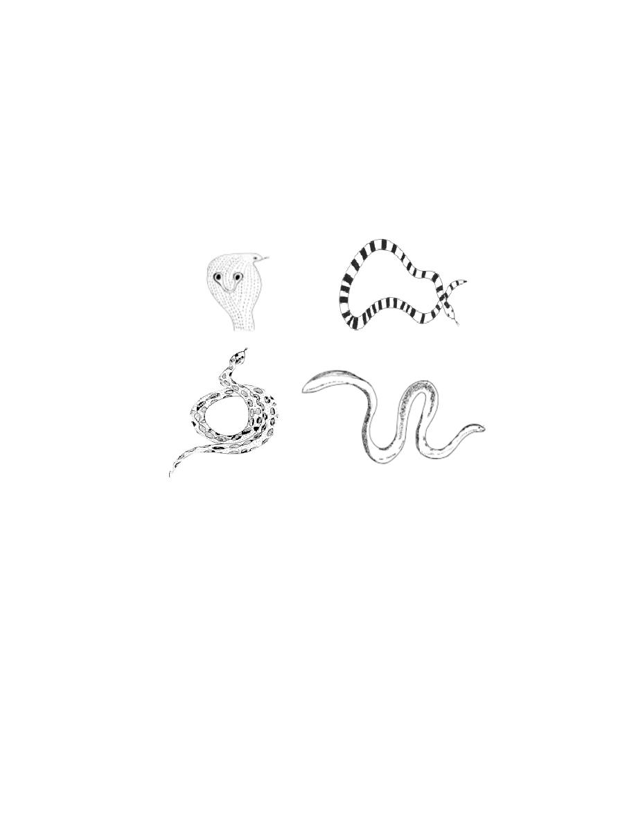
250
The Puffer fish, (Tetrodon) is considered to be world’s most
dangerous fish. Its ovaries, intestine, kidneys, skin and eyes contain a
neurotoxin called Tetradoxin. This toxin has no antidote. It is several times
deadlier than cyanide. In a dilute form, tetradoxin is used as a pain killer for
victims of neuralgia, arthritis and rheumatism
Poisonous snakes
Indian poisonous snakes are the cobras, the kraits, the vipers and
the sea snakes. These can be distinguisted from the non poisonous by the
tail, the arrangement and size of scales, plates and shields found over the
body.
Cobra : It is well known all over India. When provoked it raises its
head and expand the skin of the neck region in the form of a characteristic
hood. The hood may bear a spectacle mark. Such cobras are called two
ringed or spectacled forms. In others there is a oval spot surrounded by an
ellipse. These are known as the one ringed or monocled variety. They are
found in Bengal. In still others there is no mark on the hood. Only two species
of cobra are found in India. They are Naja naja (Indian cobra) and
Ophiophagus hannah (king cobra).
Fig. 6.2.5. Poisonous snakes
Binocellate
Banded krait
Russell’s viper
Sea snake
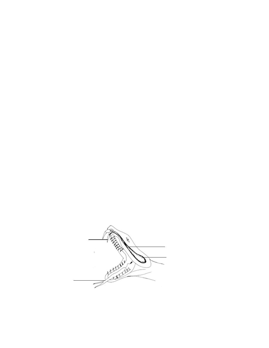
251
Krait : These are common poisonous snakes of India. There are two com-
mon Indian kraits. They are the common krait (Bungarus coeruleus) and the
banded krait (B. fasciatus).
Vipers : There are two classes of vipers. Some have a distinct pit on the sides
of the head between the nostril and the eye in the region called ‘lore’. These
are called pit vipers. The other one is the pitless viper.Vipers are vivipa-
rous in nature.
The vipers have movable upper jaw, so that the fangs when not in use can be
folded backwards. It gets erected with the opening of the mouth while inflict-
ing injury. It produces a loud hissing sound by expelling air through nostrils.
Pitless Viper - Vipera russellir (Russell’s viper) Echis carinata (The little
Indian viper).
Pit viper - Trimeresurus sp
Sea Snakes : Sea snakes can always be distinguished from other snakes by
their laterally compressed tails. This is an adaptation to their life in the sea. All
sea snakes are highly poisonous.
Eg. Hydrophis sp Enhydrina sp
Poison Apparatus of a Snake
The poisonous snake possesses a poison apparatus comprising of a
pair of poison glands, a pair of poison ducts and a pair of fangs. The poison
glands are situated on either side of the upper jaw below and behind eyes.
They are specialized salivary glands. A duct carries the venom secreted from
each gland to the fang. A fang is meant for injecting the venom into the body
of the prey. Fangs are specialized teeth of the upper jaw which are tubular or
grooved.
Fig. 6.2.6. Snake jaw showing fangs and poison glands
poison gland
poison duct
fang
tongue
252
Biting mechanism in Cobra
Cobra is not an aggressive snake. When disturbed, it attempts to
escape. When the snake attacks, the mouth opens by lowering the lower jaw.
This makes the fangs to be erect to penetrate the muscles of the victim. When
the mouth is closed the poison glands are pressed. The venom thus reaches
the fangs and is injected into the body of the victim. This whole process takes
place in no time.
Snake Venom
There are two types of snake venoms. One type acts mainly on the
nervous system (neurotoxic). It affects the optic nerves (causing blindness)
or the phrenic nerve of the diaphragm (causing paralysis of respiration). The
other type is haemolytic. It breaks down the red blood corpuscles and blood
vessels and produces extensive extravasation of blood into the tissue spaces.
6. 2. 3 Fouling Organisms
Several aquatic organisms cause damages to submerged surfaces.
Since this infestation has an economical importance, several studies are being
made. Marine sedantary organisms may affect piles, floats, wooden dry docks
and boats. These organisms are called foulers. Most of these organisms are
distributed all over the world through the agency of ships.
They are of economic importance, since fouling of ships results in
increased resistance to movement through water, reducing the efficiency, low-
ering of speed, increasing fuel consumption and leading to wear and tear of
engine. The efficiency of underwater sound equipments fitted on to commer-
cial and naval vessels is also seriously affeceted as result of the accumulation
of fouling organisms.
Fig. 6.2.7. Fangs of snakes and marks left by the bite of poisonous and
non-poisonous snakes
poisonous snake non-poisonous snakes
Canalised teeth
of russell’s viper
Gutter teeth of
cobra
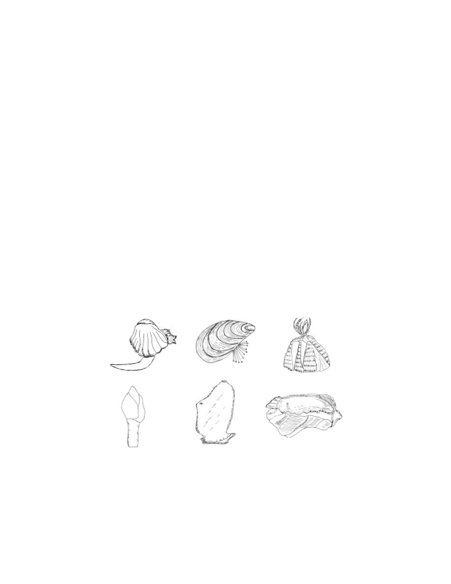
253
Pipes and conduits used to convey salt water in ships, industrial plants,
oil refineries, nuclear power plants and aquaria become clogged and the flow
is interupted by the growth of these organisms inside the pipe. Thus the
problem of fouling organims is serious.
Factors influencing the settlement of fouling organisms
Several factors are known to influence the settlement of larvae of
fouling organisms. The most important of which is recognised to be the
formation of the primary film. It is composed chiefly of diatoms and algal
spores with a relatively small portion of bacteria. The thin filaments of algae
may afford foothold on the substratum for the larvae to settle. The algae and
diatoms serve as food for these larvae.
Members of the fouling community
The most important members of the fouling community include algae
and representatives from almost all the invertebrate groups and the tunicata.
Among the molluscs the mussels are the most important foulers. They settle
heavily, especially in the pipe systems carrying seawater and obstruct the flow
of water. Among Arthopoda, the barnacles are the dominant representatives
of the fouling community. There are over 100 species of barnacles as foulers.
Prevention of fouling.
Fig. 6.2.8. Common foulers
Cardium
Mussel
Balanus
Lepas
Tunicate
Oyster
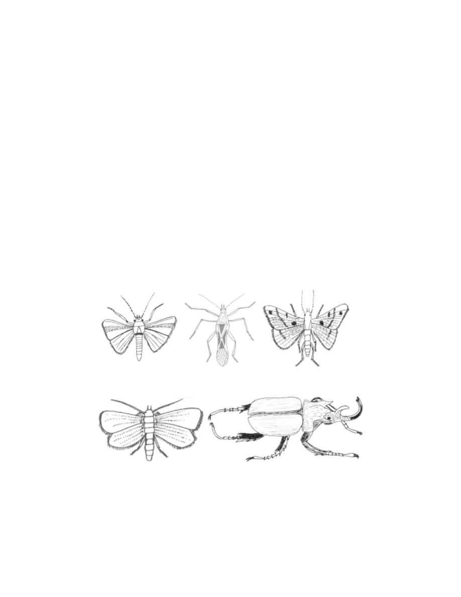
254
1. Using copper sheathing on wooden hulls of boats.
2. Applying suitable antifouling paint.
3. Chlorination in condenser system.
6. 2. 4 Pests
Any animal which becomes a source of trouble or loss to human is
called a pest. Among insects such pests are numerous and are of different
kinds. An insect is usually called as a pest when it causes appreciable damage
and loss to the crops or other belongings. The pests may be classified as
major or occassional. The insects damaging standing crops cereals, fruits and
other plant products of commercial importance are designated as crop pests.
Those insects destroying stored grains are called the store pest. Insects
causing damage to household articles are called the household pests.
Pests of Crops
Fig. 6.2.9. Common pests of crops
Spotted bollworm
Rice bug
Sugar cane top shoot borer
Brinjal shoot and fruit borer
Rhinoceros beetle

255
I. Pest of Cotton
a. Pink boll worm - Pectinophora gossypiella
This pest causes damage to the buds, flowers and seeds.
b. Red cotton bug - Dysdercus koenigii
This bug sucks the cell sap of green bolls and leaves.
c. Spotted boll worm - Earias vitella, E. insulana
These two species are the most important pests of cotton in India
causing heavy losses to the crop every year. E- vitella is abundant in high
rainfall areas, where as E- insulana abounds in areas receiving scanty rains.
The caterpillars of Earias. bore into the stem portion of young seedlings and
apical shoots and later eat into buds, flowers and bolls. The attacked shoots
wither, droop and ultimately die.
Control : The attacked shoots and bolls should be collected and destroyed.
Avoid growing lady’s finger during the offseason in the vicinity of the cotton
fields. Insecticidal control of the pest consists of spraying is done every
15 - 20 days of monocrotophos, endosulfan or malathion.
II. Pests of Paddy
a. Rice stem borer - Tryporyza incertulas
This pest bores into the stems of the young mature stages of paddy.
b. Rice bug - Leptocorisa acuta
It is the most important pest of paddy in India. It mainly feeds on
paddy but is also found feeding on millets, maize, sugar cane and some grasses.
The adults and nymphs feed on the milky juice of the forming grains which as
a consequence become chaff.
Control : In nurseries and fields , the leaves of paddy plants containing eggs
should be clipped and burnt. As the bugs feed and breed on various types of
grasses, removal of such grasses from the fields will help in reducing the
population. Collection of bugs with a hand net and their destruction is a useful
mechanical method. Among the insecticides BHC and malathion as dusts
and carbaryl and methyl parathion as sprays just before flowering of the plants
are effective.

256
III. Pest of Sugarcane
a. Indian sugarcane leaf hopper - Pyrilla perpusilla.
This pest sucks the cell sap of leaves.
b. Sugarcane root borer - Emmalocera depressella.
This pest causes damage to the stem below the soil surface.
c. Sugarcane shoot borer - Chilo infuscatellus.
This pest causes damage to the shoot.
d. Sugarcane top shoot borer - Scirpophaga nivella
It is one of the most destructive pests of sugarcane.It is found all over
India. The damage by the borer actually starts from the mid-rib of the top leaves
into which it bores and makes tiny holes. When the growing point is damaged, side
shoots are formed in the young plants and bunchy tops in the olders ones. The
quality of the juice is also affected.
Control : Effective control of this pest is possible only by integrating mechanical
and chemical control methods. Mechanical methods include the collection and de-
struction of egg masses and affected top shoots and sowing of resistant varieties.
The chemical methods involve application of 4 % carbaryl or endo sulfan granules
in the leaf whorls or spraying of 0.05 % monocrotophos or 0.1 % endrin.
IV. Pests of Vegetables.
a. The red pumpkin beetle - Raphidopalpa foveicollis.
This pest causes damage to the leaves, flowers and buds of younger plants.
b. Cabbage butterfly - Pieris brassicae .
This pest causes damage to the leaves.
c. The Hadda beetle - Epilachna dodecastigma.
This pest causes damage to the leaves of the brinjal, potato and tomatoes.
d. Brinjal shoot and fruit borer - Leucinodes orbonalis.
It is the most important and destructive pest of brinjal and has a country-
wide distribution. The pest starts damaging the brinjal plant a few weeks after its
transplantation. When the shoot is attacked by the caterpillar it droops and withers
, finally drying up. When the petioles of the leaves are bored into by the larva the
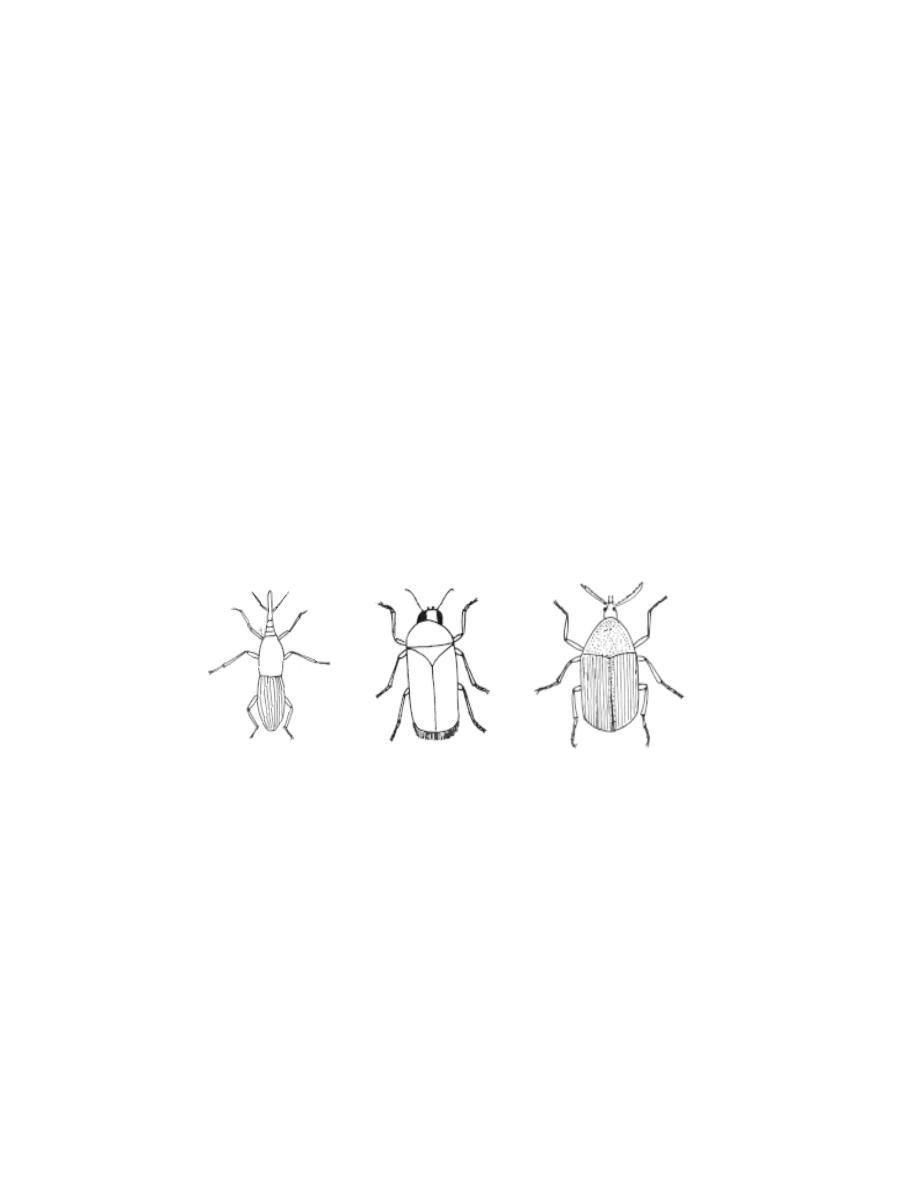
257
leaves wither and drop. The attacked fruits show holes plugged with excreta on
them. Upto 70 % loss of crop is caused by this pest.
Control : Prompt collection and destruction of the plant parts harbouring larvae
help in reducing the infestation. Insecticides such as carbaryl, endosulfan, Lindane
and diazinon, when applied at regular intervals give relief from heavy infestation.
V. Pest of Coconut palm
Rhinoceros beetle - Oryctes rhinoceros. It is distributed throughout South East
Asia, Southern China, Philipines and south Pacific Islands. The adult causes infes-
tation by feeding the young leaf fronds. They make burrows and throw out a
fibrous mass. The infestation is marked by a number of holes on the fronds, when
they open out . The attack results in the destruction of growing plant as a result of
which the tree dies.
Control : The beetles should be destroyed by inserting specially designed hooked
rod. In earlier developmental stages beetles should be destroyed by tackling the
breeding places like manure pits near gardens by spraying 0.01% aldrin.
VI. Pests of Stored grains
a. Rice weevil - Sitophilus Oryzae
This is a very serious major pest of stored grains in farm storage. It is
worldwide in distribution. Generally, infestation starts in grains only during storage
which may lead to heat spots in the grain. The grains are hollowed and the weight
is reduced.
Control : The weevil is unable to breed at a grain moisture content of 9 % or less.
Hence dry storage of grains can aviod infestation by the pest. The larvae and
adults are killed by exposing them for 48 hrs to the vapours of ethylene dichloride
Fig. 6.2.10. Pests of stored grains
Rice weevil
Khapra beetle
Pulse beetle

258
- carbon tetra chloride mixture under gas - proof covers. Fumigation of infested
grains with methyl bromide is also effective and kills all stages of pest including
eggs.
b. Khapra beetle - Trogoderma granarium
Khapra beetle is a very serious pest of wheat and other stored grains all
over India. Only the larval stage is destructive, adult beetle being harmless. The
grubs attack the germ portion of the grain. In severe infestation the cereals are
reduced to mere frass.
Control : Stocks of grains should be stored in thoroughly clean and insect-free
stores which are regularly aerated. Before storage of grains, the godown should
be properly disinfected with benzene hexachloride smoke or fumigants.
c. Pulse beetle - Callosobruchus Chinensis
This is a very important pest of various pulse crops in India. It affects both
in fields and in stores. The pest attacks leguminous pods in the field from where
they are carried to godowns. The larvae bore into the pulses and grains. They
feed and grow inside. The damaged grains are unfit for human consumption.
Control : Control can be achived by growing suceptible crops atleast a kilometre
away from storage godowns which are the main source of infestation. Fumiga-
tion with methyl bromide in the godown is very effective but proper care must be
taken because of the high toxicity of this compound.
VII. Pests of household goods
Fig. 6.2.11. Pests of household goods
Silver fish
Termite

259
a. Termites (white ants) - Odontotermes obesus
There are more than 2000 species of termites. The food of termites con-
sists primarily of wood (cellulose). This habit of termites is the cause for very
serious losses, particularly in tropical countries. They destroy wood work, furnitures,
buildings, fences and other wooden structures that come into contact with the soil.
The losses caused by termites to Indian agriculture and other commercial crops
are extensive. About 40 species of termites are injurious to economic plants such
as wheat, barley, maize, gram, sugarcane, groundnut, several vegetables, fruit trees
and coconut in India.
Control : The seriousness of their attack demands immediate control. For termite
control, insecticides should be applied to the soil. Emulsions of 1 % chlordane, 0.5
% aldrin and 0.5 % heptachlor are suitable for soil treatment. 5 % pentachlorophe-
nol is good for wood preservation. The application of a mixture of BHC and aldrin
to soil in the foundations of a building is effective.
b. Silverfish - Lepisma saccharina
It is cosmopolitan in distribution. It is commonly found living in moist warm
places and among old books. It is a whitish wingless insect of about 13 mm in
length It mostly attacks old books and magazines. It infests starched clothes, rayon
fabrics, book labels or bindings where glue has been used.
Control : The books should not be kept in damp places. Books should be exposed
to sunlight frequently. Dusting of 5% malathion has proved to be an effective
control measure for heavily infested cases.
6.3. Regeneration studies in Planaria
Tissue culture, nuclear transplanting, stem cell research, tissue
response to hormones are all recent interesting experimental fields in
biology. An application of such practices requires a basic knowledge of regular
processes as such as cell divisions, cell organization, cell transformation, cell
initiations, cell’s responsiveness, immune systems, cell differentiation, organ
repairing and wound healing. These inquisitive studies have paved the way
for applicability of biological knowledge towards human welfare.
‘Regeneration’ has remained as one such study, that could provide a
wealth of knowledge regarding the behaviour of somatic cells in growth and
differentiation. The animals possess the ability to repair extensive damages
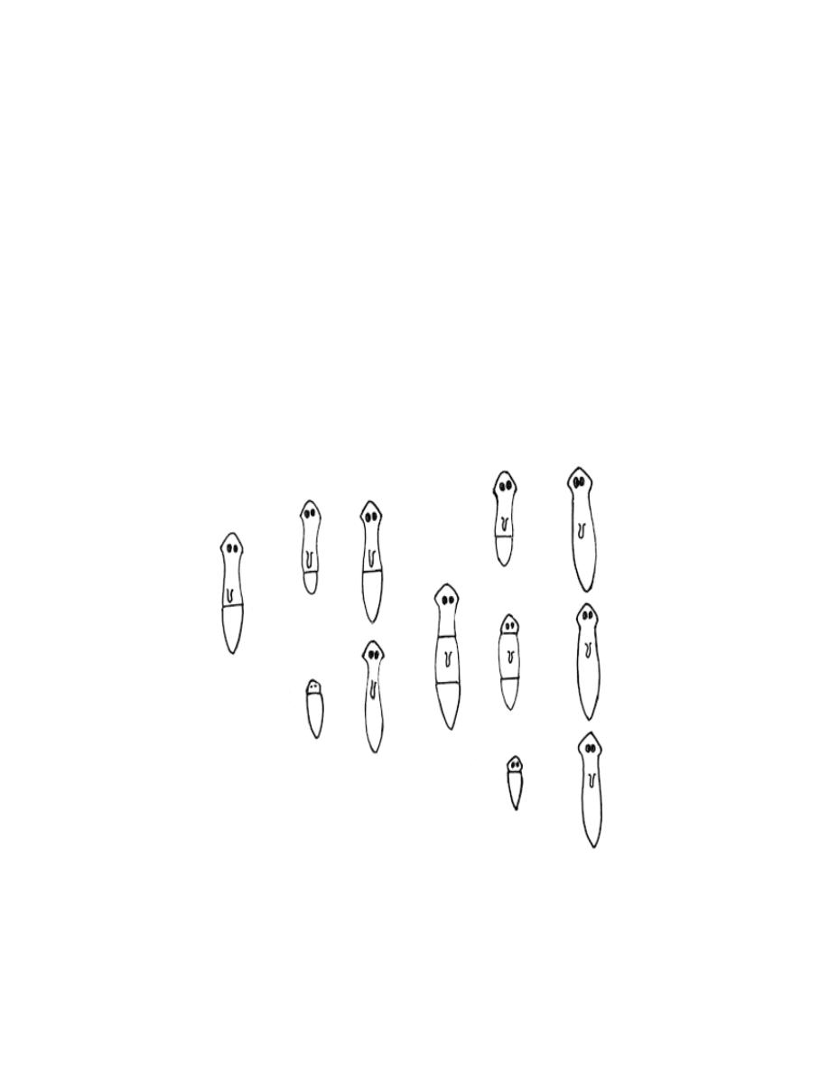
260
incurred by the body either accidentally or in natural conditions. The ability
of a living organism to regrow a portion of its body that has been injured or
lost is known as regeneration. The capacity to regenerate lost parts differs
in various groups of animals.
Organisms like sponges and coelenterates have exceedingly high
ability for regeneration. As the organization of animals becomes advanced
the capacity for regeneration gets restricted. Crabs can replace severed legs.
Mammals can regenerate blood cells. Other specialized cells are replaced
with scar tissue that does not regain the function of the lost or damaged
tissue.
Among multicellular and triploblastic animals the Planarians are
remarkable for their high regenerative ability. They may be cut across or
lengthwise, and each part of the body will regenerate the missing half.
→
→
→
→
→
→
→
→
→
→

261
Mechanism of regeneration
The process of regeneration happens through epimorphosis and
morphallaxis. When a cut is made, a regenerative blastema is formed, at
the cut surface. This process is termed as epimorphosis. Later the remaining
part gets reorganized on a small scale resulting in a smaller regenerated
organ. This reorganization is called as morphallaxis. The processes of
regeneration are controlled by many factors. A knowledge of all factors and
the mechanisms of regeneration even to-day remain as potential fields for
further research, study and application.
Self - Evaluation
Part I
Choose the correct answer
1. Reef forming corals normally grow in
(a) cold waters
(b) deep seas
(c) shallow, tropical seas
(d) polluted and muddy waters
2. The Great Barrier reef occurs in
(a) Gulf of kutch
(b) Carribean islands
(c) Andaman islands
(d) the coast of Australia
Fig.6.3.1. Regeneration experiments in planaria

262
3. The larva of corals is a
(a) trochophore
(b) planula
(c) medusa
(d) polyp
4. A ring shaped reef with a lagoon connected to the sea is a / an
(a) atoll
(b) barrier
(c) fringing
(d) gulf
5. The coral used in jewellery making is
(a) Corallium nobile
(b) Tubipora
(c) Organ pipe
(d) Guano
6. The earthworm commonly employed in Indian vermiculture is
(a) Lampito mauritii
(b) Apis indica
(c) Penaeus indicus
(d) Pinctada fucata
7. The degrades of organic matter by worm activity is
(a) lac
(b) isinglass
(c) vermicompost
(d) guano
8. The number of offspring, a single earthworm could produce a year
(a) 1000 to 1500
(b) 10,000 to 20, 000
(c) 5000 to 10,000
(d) 80 to 100
9. The honey bee used commonly in bee-keeping industry is
(a) Apis dorsata
(b) Apis florea
(c) Apis indica
(d) all the above
10. The scientific management and production of silk is
(a) Sericulture
(b) horticulture
(c) Apiculture
(d) aquaculture
11. The secretion from the abdominal glands of the worker bee is
(a) honey
(b) nectar
(c) beeswax
(d) all the above
12. The silk produced by Bombyx mori is
(a) tasar silk
(b) muga silk
(c) arandi silk
(d) mulberry silk
13. Assam is famous for

263
(a) Muga silk
(b) Eri silk
(c) Tasar silk
(d) Mulberry silk
14. Lac insects are cultred in
(a) Kusum
(b) Ber
(c) Babul
(d) All the above
15. The predatory insects are said to be
(a) entomophagous
(b) larvivorous
(c) parasitic
(d) pests
16. An example for insect predator is
(a) Eudrillus
(b) Laccifer
(c) Lady bird beetle
(d) Tachynid fly
17. The organization involved in pearl oyster culture is
(a) CIBA
(b) CMFRI
(c) NIO
(d) MPEDA
18. The most common freshwater prawn used in aquaculture is
(a) Macrobrachium sp
(b) Metapenaeus sp
(c) Penaeus sp
(d) Panulirus sp
19. The name Kal Eral in Tamil refers to
(a) Prawns
(b) crabs
(c) a fish
(d) lobsters
20. The common Indian Lobster is
(a) spiny Lobster
(b) coral Lobster
(c) slipper Lobster
(d) sand Lobster
21. Fish meal is used as in
(a) animal feed
(b) manufature lubricants
(c) preparation of cakes
(d) all the above
22. The fish Stromateus argenteus is popularly called as
(a) guppy
(b) pomfret
(c) gold fish
(d) angel fish
23. Gold fishes are the varieties of the genus
(a) Xiphophorus
(b) Betta

264
(c) Carassius
(d) Lebistes
24. Which part of the body in fish provides isinglass
(a) skin
(b) liver
(c) air bladder
(d) bone
25. Which country was economically benefitted by marketing bird excreta
(a) USA
(b) Peru
(c) Australia
(d) West Indies
26. Guano are important in the usage as
(a) fish meal
(b) fertilizers
(c) adhesives
(d) lubricants
27. The idea of aquarium maintenance was first conceived by
(a) Chinese
(b) Japanese
(c) Red Indians
(d) Africans
28. Washing the aquarium tanks with 1% potassium permanganate solution is
necessary for
(a) raising the nutritive value
(b) avoiding small
(c) making the glass clear
(d) disinfecting
29. Nandankanan Biological park is situated in
(a) Delhi
(b) Orissa
(c) Bihar
(d) Assam
30. Example (s) for mechanical vectors
(a) sand fly
(b) rat - flea
(c) house fly
(d) all the above
31. The filarial worms attain sexual maturity in the
(a) human blood vessels
(b) human lymphatic vessels
(c) culex mosquito
(d) all the above
32. The pneumonic plague affects
(a) liver
(b) brain
(c) lymph glands
(d) lungs
33. Which is the best time to have blood test for filariasis
(a) morning
(b) evening
(c) noon
(d) mid-night

265
34. The defensive structure found in physalia is
(a) neurotoxins
(b) fangs
(c) stinging cells
(d) poison glands with claws
35. The intestine and kidneys of the puffer fish has a dead ly poison called
(a) tetraoxin
(b) cyanide
(c) haemotoxin
(d) cystamine
36. The characteristic feature of tail in sea snake is
(a) round
(b) pointed
(c) compressed
(d) cylindrical
37. Ophiophagus hannah refers to
(a) Indian cobra
(b) King cobra
(c) Russell’s viper
(d) Sea snake
38. Oysters, tunicates, Lepas and cardium are
(a) Entamophags
(b) foulers
(c) parasites
(d) vectors
39. The caterpillar of the butterfly causes
(a) cabbage fertilization
(b) cross pollination
(c) damage to leaves
(d) damages to roots
40. Fumigation is a method of
(a) disinfection
(b) harvesting
(c) food processing
(d) watering
41. The silver fishes commonly live among
(a) rocks
(b) algae
(c) other fishes
(d) old books
Part II
Give very short answer
1. Give any two importants uses of corals.
2. What is a fringing reef ?
3. Mention the use of vermicompost.
4. What are productive insects? Give an example.
5. Differentiate nectar and honey.

266
6. What are the uses of beewax.
7. What is meant by biological control?
8. How are insects useful to flowering plants in reproduction ?
9. Name any two edible crabs.
10. Provide the names of one live bearer and one egg layer from among the
ornamental fishes.
11. Name any two fish liver oils.
12. Name any two rooted plants used in aquarium.
13. What are the controlling measures taken to control pests in coconut palm.
14. How do the haemolytic toxins cause death in an organisms.
15. Provide the names of zoos in the states of Tamilnadu and Andhra Pradesh(one
each).
16. Name the two types of venom released by poisonous snakes.
17. Provide atleast one major role of CMFRI.
18. Name any two cultivable animals.
19. What is Corallium nobile ?
20. What is primary film?
Part III
Answer briefly
1. Give the names of two common pests of cotton. By what way they affect
the crop? Give any two mechanical methods of controlling them.
2. Write notes on pearl culture.
3. Light is very important for the survival of the aquarium fishes - explain.
How this could be achieved?
4. What is Biological control ? Explain with an example.
5. Provide an account on Indian prawns of commercial importance.
6. Give an account of cultured pearls.
7. Write notes on omega fatty acids.
8. What is guano ? comment on its economic importance.
9. Write notes on Xenopsylla cheopis
10. Give an account on fouling organism.
11. Draw and label the poison apparatus in a snake.
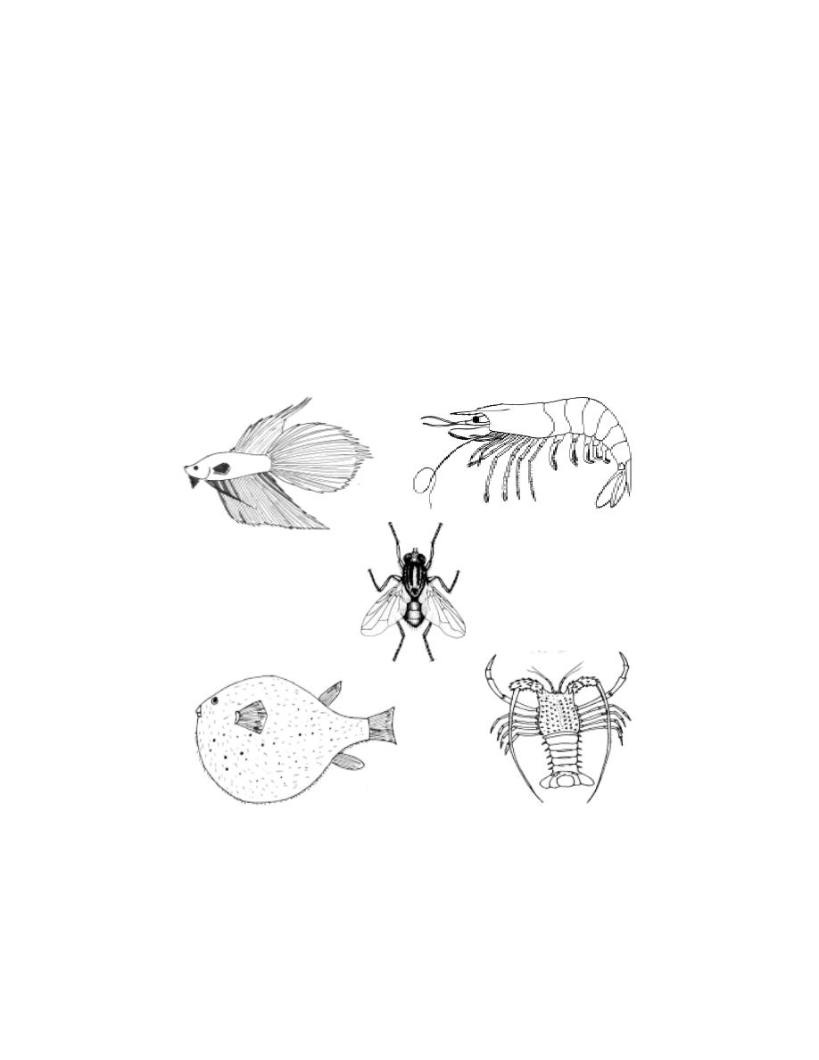
267
Part IV
Answer in detail.
1. What is Vermiculture? Name two cultivable animals in Vermiculture. Explain
the process Vermiculture.
2. Give an account of insects and insect products of commercial importance.
3. What are the nutritive values of fishes.
4. Give a detailed account on setting up an aquarium and maintaining the same.
5. Write an essay on insect pests of stored products and household goods.
Identify and name

268
7. ORIGIN OF LIFE
7.1. Theories
Origin of life
Our planet earth is inhabited by an enormous variety of living organ-
isms. The plants and animals show remarkable biodiversity. The diversities
mostly appear as adaptations for diverse habitats and living methods adopted.
This vastness has always made people wonder about the origin of life and
diversity. To a certain extent Charles Darwin’s natural selection concept pro-
vides an explanation towards an understanding of evolution of various kinds of
life forms. However the theory of origin of life is still a matter of speculation.
Several intelligent explanations had been provided to account for the origin of
life on earth.
1. Theory of special creation
According to the proponents of this theory all living forms with their
richness in diversity were created abruptly by a super natural power. The
support or acceptance of this theory is mostly due to faith rather than experi-
mental or scientific evidences. However, supporters of this theory have re-
cently created a new discipline called “creation science”.
2. Cosmozoic theory
This theory states that life came to earth from some other planet or
star. The resistant spores that lead to life on earth are named as ‘cosmozoa’.
They reached earth accidentally. Under favourable conditions they evolved
and produced all forms of life on earth. An acceptance of this theory needs
evidence for the existence of extra-terrestrial life.
3. Theory of spontaneous generation or abiogenesis
The theory that life originated from non-living material is now called
abiogenesis. An earlier version of this theory was spontaneous genera-
tion or the origin of life without apparent cause. The ancient Greek philoso-
phers like Thales, Empedocles and Aristotle supported this concept.
Thales (624-548 BC) suggested that oceanic water was the mother
from which all living forms originated. According to Empedocles (540-433
BC) life originated by itself from non living matter and imperfect forms were

269
replaced by perfect forms. Aristotle (384-322 BC) proposed that living forms
are animated forms of non-living matter. The activation was due to vital forces
or guiding intelligence. He further stated that the vital forces operate con-
stantly and improve the living world.
In the 17
th
century the idea of abiogenesis was opposed by an Italian
physician Francisco Redi (1621-1697). For the first time he proposed through
experiments, that life could arise only from pre-existing living things. He tested
his hypothesis by sealing meat inside four closed flask, while leaving another
four meat filled flasks open. Soon the meat in the open flasks was full of
maggots. Flies were entering and leaving the flask. Even after many days no
worm appeared in the closed and sealed flask. This experiment confirmed his
idea that new life can come only from early life.
4. Big Bang theory
This theory is concernced with the origin of earth and other planets.
According to this theory the universe originated in a ‘big bang’. All matter
was created in this big bang including the matter of which stars and planets
are composed of. According to this theory as a young star increases in den-
sity, it gets heated up due to increasing pressure. When a certain critical tem-
perature is reached thermonuclear reactions will begin and cause a ‘big bang’.
It is believed that our sun was formed in this way.
5. A. I. Oparin’s theory
Alexander I. Oparin, a Russian chemist published a paper in 1924. He
advanced a new theory regarding origin of life. This theory is called the “theory
of primary abiogenesis”. According to Oparin conditions on Earth today are
no longer suitable to the production of life from non living matter. The primor-
dial earth, about 2600 million years ago provided suitable atmosphere for the
origin of life. The earth during that period had many simple chemical com-
pounds. More and more complex organic compounds were formed under the
influence of electric charges and Ultra violet rays. The origin of complex
organic molecules made the origin of life easier.
6. J. B. S. Haldane’s hypothesis
Haldane was a British biochemist. He was the first to propose that life
originated on earth, when the atmosphere was devoid of O
2
gas. Today’s earth
is surrounded by oxygen and its derivative ozone. The ozone layer protects
earth by preventing the entry of harmful cosmic radiations. In 1929, he
suggested that an atmosphere lacking O
2
would have no ozone layer. Hence
ultra violet rays were freely entering earth’s atmosphere. In a reducing atmo-
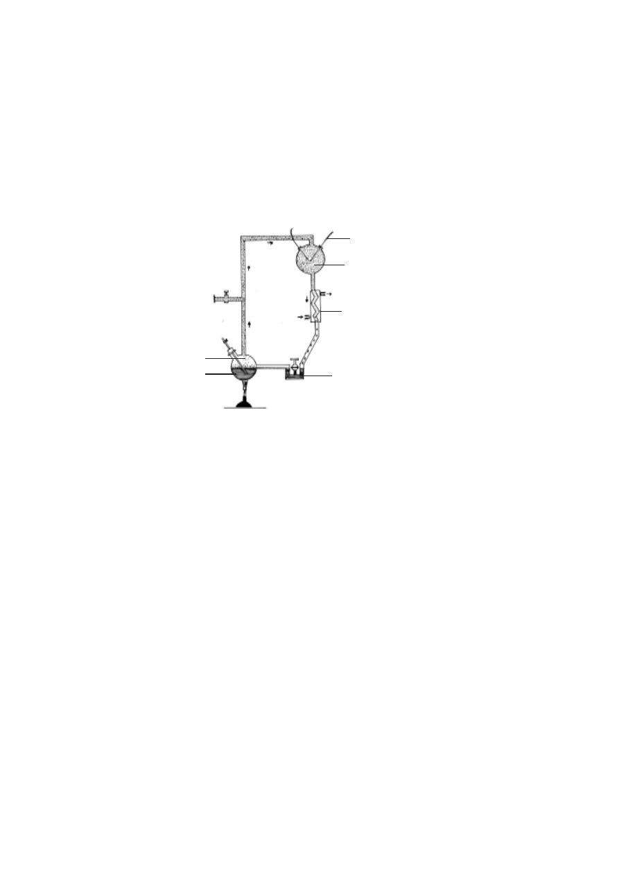
270
sphere this could have caused photochemical reactions resulting in the gen-
eration of organic molecules. These molecules gradually accumulated in the
oceans as a dilute ‘soup’.
7. Urey - Miller hypothesis
U.S scientists Harold Urey and Stanley Miller in 1950’s proposed and
attempted to prove that amino acids can be synthesised outside living
systems. They conducted experiments in which a gas mixture containing
hydrogen, ammonia, methane and water vapour was subjected to electric spark.
This trial yielded aldehydes, amino acids and carboxylic acids. They visualised
the existence of a similar situation and happening of events resulting in a large
scale accumulation of diverse biomolecules in the primordial earth. These
chemical incidents could have paved the way for the origin of a cellular
organisation.
8. Coacervation theory
Coacervates were considered to represent the protocell model. The
tendency of biological polymers to form polymer-rich droplets called coacer-
vates has been proposed by Oparin. He reported that coacervates are readily
formed from aqueous suspension of proteins, polysaccharides and nucleic ac-
ids. Biochemical processes like enzyme action and electron transport can happen
within a coacervate. Such systems were considered the forerunner of living
cells.
Fig.7.1.1 Spark-charge apparatus of Stanley Miller
condensate having amino acids
nitrogen, ammonia, methane
tungsten electrodes
condenser
water vapour
boiling water

271
7.2. Geological time scale
The oldest known rocks are about 3.8 billion (3800 million) years old.
It corresponds to 38, 000, 000 centuries. According to Geologists and Geo-
physicists our planet earth is 4.7 billion (4,700 million) years old.
In this ancient time scale evidenes showed that the first life originated
2.5 billion (2,500 million) years ago. Hence from the formative stage for nearly
2,200 billion years there was no life on earth. Such a
period in earth’s history is known as the Azoic era (2,200 to 5,000 billion
years ago). Evidences show that during this era the earth was a hot sphere.
Gradually the upper surface of the earth cooled down. It resulted in solidifica-
tion and formation of rocks and rocky terrain. Further water molecules were
formed resulting in accumulation of water and water reservoirs as water
masses and land surface were established. These transformations provided a
suitable condition for the origin of the first life.
Once the life originated and established, evolutionary changes took
place. Changes happened in the structure, organisation and living methods of
organisms, depending on natural surroundings and changes in natural surround-
ings. Thus fauna and flora started flourishing on earth. The water masses
were fully exploited. Later land surfaces were invaded by plants and animals.
The Geological time succeeding Azoic Era, was dramatic and rich in
life. This period is divided into three eras. These were significant periods in
earth’s history. Of these, the oldest era was the Paleozoic era. It ranged
from 600 to 210 million years ago. Thus its duration was nearly 390 million
years. This era saw the origin and adaptive radiation of sponges, starfishes,
snails, insects, crabs, and terrestialised amphibians and reptiles.
The Paleozoic era was followed by middle period named as Meso-
zoic era. This era ranged from 65 to 210 million years ago. Its duration was
145 million years. During this era, among animals the reptiles came to promi-
nence. Hence this era is known as the golden age of reptiles. Further this
era saw the origin and development of birds and reptiles.
The period ranging from 65 million years till date is named as the
Coenozoic era. This era is characterised by rapid evolutionary changes in
mammals. This era is known as the Age of mammals.
Geological time scale
A geological calendar has been formulated by assessing the age of
rocks and rock sediments. Based on age, and events, the ancient period from

272
earth’s history is formulated into eras-periods-epochs. Each division in the
geological calender is clearly identified and demarcated. Incidents pertaining
to earth surface, plant and animals life are neatly recorded. The influence of
geological and climatic changes on the life and the evolution of the living or-
ganism had been well analysed.
Era
Duration
Significane
Palaeozoic
600 - 210 m. years ago
“Cradle of anicent life”.
Mesozoic
210 - 65 m. years ago
“Golden age of reptiles”.
Cenozoic
65 - 1 m. years ago
“Age of mammals”.
Era
Period
Epoch
Duration
Quarternary
Pliestocene
2 - 1
CENOZOIC
Tertiary
Pliocene
7 - 2
Miocene
26 - 7
Oligocene
38 - 26
Eocene
54 - 38
Palaeocene
65 - 54
Cretaceous
130 - 65
MESOZOIC
Jurassic
160 - 130
Triassic
210 - 160
Permian
235 - 210
Pennsylvanian
255 - 235
Mississipian
275 - 255
PALAEOZOIC
Devonian
315 - 275
Silurian
350 - 315
Ordovician
440 - 350
Cambrian
600 - 440
Precambrian
440 and before
(million years ago)

273
Periods
Events
Quarternary - Pleistocene 2 - 1 m.years ago
Human evolution
TERTIARY - Pliocene
7 - 2 m. years ago
Rodents were successful
Mammals increased
TERTIARY - Miocene
26 - 7 m. years ago
Priaries were formed
Horses evolved.
Carnivorous mammals
were dominant
TERTIARY - Oligocene 38 - 26 m. years ago
Monkeys and apes
originated
TERTIARY - Eocene
54 - 38 m. years ago
Horses originated
TERTIARY - Palaeocene 65 - 54 m. years ago
First flowering plants
Mammals originated
Dinosaurs disappeared.
CRETACEOUS
130 - 65 m.years ago
Dinosours became extinct
JURASSIC
160 - 130 m. years ago
Birds originated
Modern bony fishes.
TRIASSIC
210 - 160 m. years ago
Dinosaurs evolved.
Mammals originated.
PERMIAN
235 - 210 m. years ago
Origin of reptiles.
PENNSYLVANIAN
255 - 235 m. years ago
Land living insects.
MISSISSIPPIAN
275 - 255 m. years ago
Origin of Amphibia.
Land living insects,
Forests.
DEVONIAN
315 - 275 m. years ago
Age of fishes. Fernsand
cycas.
SILURIAN
350 - 315 m. years ago
Jawed fishes originated
ORDOVICIAN
440 - 350 m. years ago
First vertebrates,
Jawless fishes,Bryophytes.
CAMBRIAN
600 - 440 m. years ago
Thallophytes,
Arthopods,Molluscs,
Echinoderms.
PRECAMBRIAN
before 440 m. years ago
Protozoans,
Poriferans and Annelids
lived.
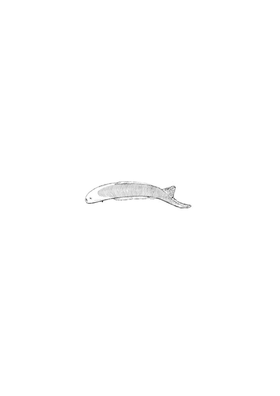
274
I. Paleozoic era :- This era produced revolutionary changes in the
biosphere. Further this era saw the origin and the radiation of several groups
of animals and plants that remained as the forefathers for the modern groups.
Thus this era is known as the Cradle of ancient life.
1. Cambrian period :- (600 to 440 million years ago)
The period preceeding cambrian is known as Pre-cambrian period.
During precambiran time simple algae, protozoans, poriferans, annelids, were
well established. Thus the cambrian started with the plants and animals that
were suceessful during the precambrian period. During cambrian among plants
thallophytes were well establised. They diversified into various groups.
(Chlorophyceae, Rhodophyceae etc.,). Among animals the aquatic arthropods
and echinoderms came to prominence. The fossils of such organism were
obtained from several places.
2. Ordovician period :- (440 to 350 million years ago)
This period was marked by formation of coral rocks and molluscs and
echinoderms. Among plants the semi terrestrial bryophytes were getting es-
tablished. Interestingly this period saw the origin of first vertebrates. These
were the now extinct agnatha. (Jawless, armoured fishes). The origin of early
vertebrates was the major event that happened in the evolution of animals.
Among arthopods, the trilobites were more prominent during this period.
3. Silurian period :- (350 to 315 million years ago)
The oldest land plant originated in this period. These plants possesed
conducting tissues. They colonised the land. Among invertebrates except for
insects all others flourised. The corals diversified. Several coral islands were
formed. Jawed fishes originated. The fishes developed scales and paried fins,
for the first time jaws originated in fishes. Origin of paired fins and jaws is
considered as major events in chordate evolution.
Fig.7.2.1 Jamoytins - an earliest jawless fish
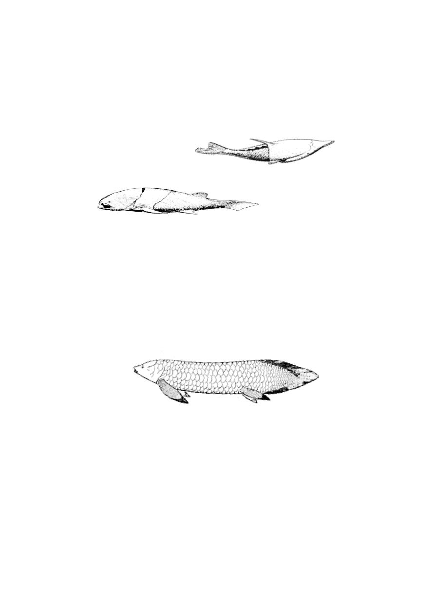
275
4. Devonian Period :- (315 to 275 million years ago)
It is a significant period in the paleozoic era. During this period land
living plants were more successful. The forests were filled with varieties of
ferns and cycas. (non-flowering plants). Among aquatic animals fishes be-
came dominant. They diversified by adapting themselves to live in various
aquatic ecosystems. The forefathers of almost all modern fishes lived during
this period. Due to these reasons this period is called as the Age of fishes.
5. Mississippian Period :- (275 to 255 million years ago)
Several changes happened to the land structure. There were massive
upraising of land in several places. This resulted in the formation of several
mountain ranges. Huge water bodies were broken into smaller lakes. These
major changes on earth’s surface were known as revolutions(eg. Caledonian
revolution). Such changes were the cause for the origin of lungs in fishes.
Lungs evolved for the purpose of living temporaily on land. It helped such
fishes to find new water bodies. Such practises encouraged the origin of the
Fig.7.2.3 The Australian lung fish - Neoceratodus
Fig.7.2.2 Ostracoderms - the early armoured, jawless
fishes and Placoderms - the early jawed fishes.
Coccostens (Placoderm)
Pteraspis (Ostracoderm)

276
amphibians. The origin of land living amphibians were further increased by
the proliferation of several land living insects.
6. Pennsylvanian :- (255 to 235 million years ago)
The land living forms became more successful during this period. There
were huge forests of ferns and cycas. Due to geotectonic changes several
forests got burried under the soil. Today’s coal and petroleum are obtained
from such resources only. Hence the Pennsylvanian and the earlier Mississip-
pian were collectively krown as Carboniferous (carbon bearing) period.
7. Permian Period :- (235 to 210 million years ago)
It was the last period in the Paleozoic era. This period was marked by
extinctions of several older groups of animals and plants. Nearly 60% of the
organism that survived at that time became extinct. Some of the
amphibians dramatically laid land eggs (cleidoic eggs). Specifically the group
of organisms that laid such eggs are identified as Seymouria. These are
considered as inter-connecting links between amphibians and reptiles.
II. Mesozoic Era
:-
This middle period in the history of life was marked by the
prominence of land living forms. Among animals the reptiles became more
dominant. They increased in size and in number. Hence this era is named as
the Golden age of reptiles.
1. Triassic Period :- (210 to 160 million years ago)
For the first time fossils of turtles, crocodiles, and dinosaurs have been
obtained from this period. Fossil evidences show that aquatic and
flying reptiles thrived during this time. The mammals orginated from reptiles
during this period.
2. Jurassic Period :- (160 to 130 million years ago)
There was a marked adaptive radiation among dinosaurs. They diver-
sified into carnivorus and herbivorus forms. The first birds originated from the
reptiles. The earlist bird thus originated is known as the Archeopteryx. The
origin of birds was a major physiological change among animals. From a more
common poikilothermic condition through feathers the birds became homeo-
thermic.
The modern bony fishes were diversified into several groups.
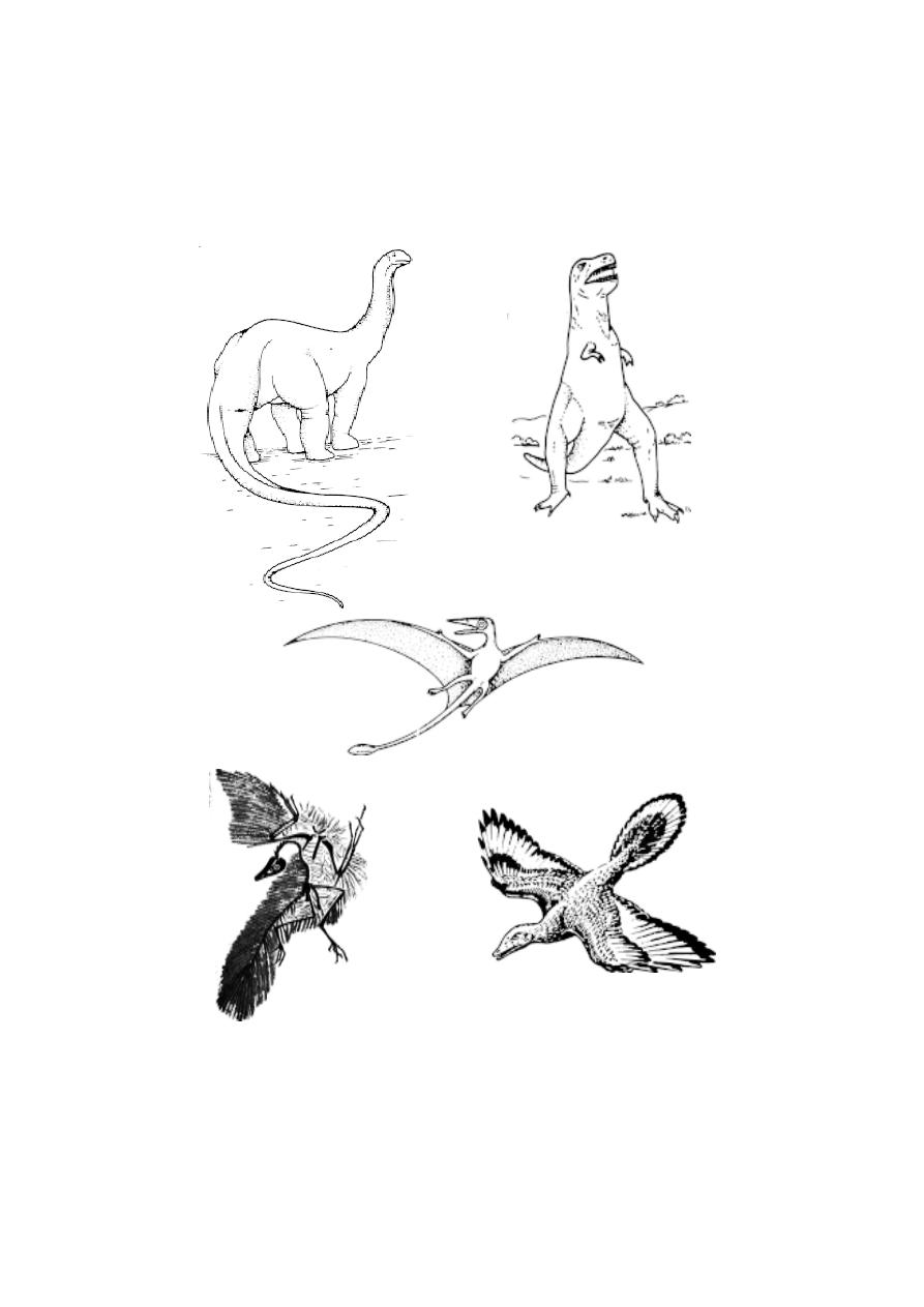
277
Fig.7.2.4 Mesozoic Birds and Reptiles
Brontosaurus
Tyrannosourus
Pterosaurus
Archaeopteryx
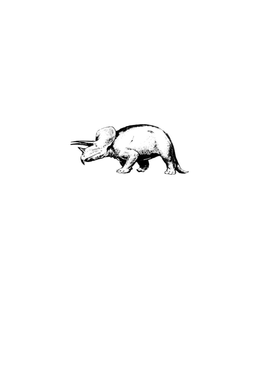
278
Cretaceous Period :- (130 to 65 million years ago)
The larger marine molluscs became extinct. The fossils of such or-
ganisms are available in places like Ariyaloor, of Tamil Nadu, today.
The Dinosaurs of the Mesozoic era abruptly became extinct during
this period. Several reasons are given for the extinction of the dinosaurs. Fos-
sils of dinosaurs were not obtained from later periods.
III. Cenozoic Era :
- (65 million years ago till date)
Plenty of fossil of organisms belonging to this era had been obtained.
All modern animals and plants were represented in these fossils. This era is
subdivided into Tertiary and Quarternary periods. Further this era contains
seven epochs. Through fossils we can trace the origin and evolution of inde-
pendent groups of animals, camels and man.
1. Paleocene epoch :-
Modern placental mammals originated during this time.
2. Eocene Epoch :-
Ungulates originated. The ancestral form of modern horses lived
during this epoch.
3. Oligocene epoch :-
Several animals with ancient characteristics became extinct. Modern
mammalian families were established. The apes originated during this epoch.
4. Miocene epoch :-
Several varieties of grasses evolved in Europe and N. America. Thus
large Priaries were formed. These changes encouraged the evolution of fast
Fig.7.2.5 Triceratops - a horned dinosaur

279
running herbivorus mammals and their predators. Thus the carnivorous
mammals came to prominence.
5. Pliocene Epoch :-
The priaries enlarged still further in several regions. The rodents
became more successful. The mammals increased in number.
6. Pleistocene epoch :-
Several glaciations happened during this time. This epoch is
popularly called the ‘Ice age’. The evolution of horses and man reached the
final stages during this period. The melting of ice that happened 1,500 years
ago is considered as the last stage of this epoch. Today we are living in an
inter-glacial Period.
7. 3 Fossils
The fossils are the preserved remains of animals, plants or their parts
found in various strata of earth. Fossils may be of entire organisms or a part
which got buried, a mould or cast, foot prints or imprints of a leaf on a stone.
Fossilization
:- Fossils can result by several methods. However these meth-
ods are purely accidental. There are many methods of fossilization.
1. Petrifaction :- It is the commonest method. In this method dead and buried
organisms turn into stones. This is due to formation of sedimentary rocks un-
der water. While the soft parts disappear due to decaying, hard parts get pre-
served due to mineralisation. The preservation happens in the original strata.
2. Petrifaction of soft parts :- Under certain conditions muscles and other
soft organs may get mineralised and form rocky fossils. Several such plant
fossils had been obtained.
3. Preservation of foot prints :- Moving animals on soft mud can leave foot
prints. These prints, if left undisturbed, can get hardened and form rocky fos-
sils. A study of such imprints can provide clues regarding the body form and
characteristics of the extinct animal.
4. Moulds and casts :- Fossilized moulds are found in volcanic ashes. Sev-
eral invertebrate fossils had been obtained as moulds. They provide details
about the exact physical features of the animal.
5. Fossilization in resins and amber :-
Normally, insects get entangled in soft sticky secretions of trees called
resin. The dried material can get fossilized. These fossils can even reveal the
colour of the organism.

280
6. Preservation in ice :- Entire animals can get frozen and may be pre-
served in ice. In such fossils the body parts remain intact without change.
Fossils of woolly mammoth from Siberia are classical fossils of this nature.
7. Dating of fossils :- The age of the fossils to a large extent can be assessed
accurately, using radioisotope method. It is known that all elements exist as
isotopes. Isotopes are atoms having slightly different atomic weights. e.g c
12
and c
14
. Some isotopes remain unstable. They undergo decay and release sub
atomic œ and ß particles till they become more stable. Examples are
Uranium
238
lead
206
Potassium
40
Calcium
40
+argon
39
Carbon
14
Carbon
12
The time required for the breakdown of half the given quantity of
unstable isotope is called the half-life. The half-life of many isotopes is
useful to assess the age of the rock accurately by measuring the ratio of
unstable : stable nuclides. Eg. c
14
: c
12
of the rock in which the fossil was
collected.
Evolutionary significance of fossils
1. Fossils tell us the full story of evolution. Fossil studies reveal the course of
evolution.
2. Through fossils the origin and evolution of specific groups of organisms can
be understood e.g. Horse evolution.
3. Fossils provide us clues regarding climatic conditions of various prehistoric
periods.
4. Study of fossils simplifies phylogenetic discussions.
5. Some fossils like woolly mammoth can provide vital clues regarding geneti-
cal make up.
Important fossils
Ichthyostega - interconnecting link between fishes and amphibians.
Seymouria - Interconnecting link between Amphibians and Reptiles.
Archaeopteryx - Ancestral form of birds
Dinosaurs - Extinct group of reptiles.
Hyracotherium - Early ancestor of horses.

281
7. 3 Extinct animals-mass extinction
Extinction may be defined as the termination of a lineage without is-
sue or abrupt disappearance of specific groups of organisms without leaving
descendents. Extinctions are of two types namely true extinctions and pseudo
extinction. In true extinctions a particular lineage totally disappears without
any progeny or evolutionary descendents.
Pseudo extinction may also be called as phyletic extinction or
phyletic transformation In this type a group may disappear leaving descen-
dents with evolutionary modifications. In horse evolution while the earliest
ancestor Eohippus became extinct its desecendent survived to produce the
modern ‘Equus’. Extinctions can happen for the taxonomic groups such as a
family or genus. Thus the extinction of dinosaurs as a group is a case of true
extinction. Similar extinctions happened to trilobites.
The process of extinction is not always predetermined. It happens due
to abrupt changes in evironmental conditions or other biological factors. Through
the use of computer models evolutionists can now examine the probalility of
extinction for large as well as small groups of organims. From the fossil record
it becomes evident that extinctions have occured at regular intervals of time.
Pattern of extiniction :-
Fossil record reveals a few patterns for extinction. Major groups of
herbivorous vertebrates are more susceptible than the carnivorous vertebrates
for extinction. Larger organisms easily became extinct. VanValen(1973)
recorded a constancy in the rate of extinction in a number of groups. He
explained this using ‘Mac Arthor’s law’. According to this law as every new
adaptation encourages the survival of a possessor it also decreases a fitness
of other related species of that area.
Causes of extinction :-
Even though extinctions are regular events in the history of earth they
are caused due to specific reasons (1) A mass extinction may be due to dras-
tic changes in the environmental conditions. (2) Any adaptive
advance in one species decreases the fitness of all other species. Thus
according to Red Queen’s hypothesis you have to keep running pretty fast,
just in order to stay in the same place. (3) Over specialistion to a specific
situation may cause extinction.(ex. Antlers.) (4) The spread of an epidemic
disease without any control can cause extinction. (5) An increase in the popu-

282
lation strength of herbivorus animals can cause rapid food shortage and cause
extinction for several inter-related groups. (6) A sudden cosmic radiation can
cause the death of large organisms. (7) A dust storm formed due to falling of
a meteorite is commonly mentioned as a cause for the disappearance of
dinosaurs.
In the recorded history of earth, extinctions of major groups of
organisms were due to natural causes. By the end of permian period of the
Paleozoic Era, nearly 60% of the varieties then existed, became extinct.
Similar large scale extinctions have been observed by the end of Mesozoic era
and during Cenozoic time.
However the extintion of animals and plants during our time is mostly
due to human interference. Thus the cause for the modern extinctions is
invariably human acitivities. The realisation of a such a cause lead to starting
of several international voluntary agencies to monitor and control extinctions.
“The red-data book brought out regularly by W. W. F (World
Wide Fund for
nature,
Formerly IUCN - International union for the conservation of nature
and natural resources) provides a list of animals and plants that are endan-
gered or have become extinct.
7.4 Evidences for evolution
Various theories of evolution explain that the life originated on earth,
as simple organisms (like ameobae), from organic substances. Later these
simple forms got specialized and diversified to suit their
diversified habitats and habits. Fossil records support the occurrence of
organic evolution to a large extent. Apart from these direct evidences many
fields of biology like comparative anatomy, embryology, physiology and
zoogeography also give positive evidences in support of evolution.
(A) The evidences from comparative anatomy :
General similarities in structures are seen between groups of
organisms. These similarities prove that the groups are related to each other.
The degree of relationship is measured by the degree of similarities they
possess. For example, a reptile is more closely related to mammal than an
insect. These comparisons also show the different plans of organisation in
various groups.
283
A basic plan of organisation could be seen when organs of different
vertebrates are compared. The fore limb of a frog, a reptile, a bird and
several mammals like bat, man, horse, cow and whale have striking
similarities in structure with modifications to suit their mode of life.
The fore limbs of these vertebrates are made up of similar bones
such as humerus, radius, ulna, metacarpels and phalanges. These
components show only little variations in these organisms. Thus, they help
frogs in hopping, the reptiles in crawling the birds and the bats in flying, the
whales in swimming, the horses in running and so on. Similarities are seen
not only in the construction of bones but also in the muscles and nerves.
Such structures with similar fundamental plan and different
functions are termed as homologous organs. Thus homology effects
divergent evolution. The brain of man resembles that of other vertebrates.
The olfactory region, the cerebral hemispheres, pineal body, optic
chiasma, optic lobes, cerebellum, medulla oblongate and spinal cord
are found in all vertebrates.
Organs like heart, eye, ear and kidney of various vertebrates, also
show homology. Such comparisons lead us to conclude that they could have
descended from a common ancestor.
Man
Horse
Bat
Bird
Whale
Fig.7.4.1
284
Vestiges:
Some organs which are well developed and functional in the
ancestral types have lost their functions in their descendants. Such organs
are found to be in the process of disappearance. Hence they are reduced
and remain as remnants. This might be because they were not used for a long
time.
A change in the environment has made them useless in the present
environment conditions. Such organs which are apparently of no use to
the possessor and hence not necessary for their existence are called as
vestigial organs. These vestigial organs serve as evidences for the process
of evolution in organisms.
Fig.7.4.2 Vestigial organs
horny termination
vent
of hind limbs
Hind limbs of python
rudimentary hind limbs of python
ilium
femur
muscles to move ears
nictitating membrane
hair on body
mammae on male
vermiform appendix
coccyx
Vestiges in
human body

285
Nearly 180 such vestigial organs can be cited in man. Some of them
are, reduced caecum, vermiform appendix, coccyx wisdom teeth,
nictating membrane of the eye, body hair and the muscles to move the
ears, lobes etc.,
Human vestigeal organs
a.Vermiform appendix
This is the remnant of a blind pouch, the caecum which is large and functional
in the digestive system of the herbivorous animals like rabbits. Herbivorous
food constituents like cellulose requires a long duration of time for digestion.
Hence it is stored in the caecum for a long time. The process of digestion is
carried out with the help of the intestinal bacteria that are lodged in the
caecum of these herbivores. A gradual change in the diet,
containing more meat and less cellulose has made caecum less
important. There by, it gradually became smaller and appears as a vestige,
in man.
b. Coccyx
The tail which is so useful and characteristic of lower vertebrates has lost its
function in human. It is represented as coccyx. The coccyx vertebrae are
originally meant for attachment tail muscles.
This is because of the change in
our mode of life from arboreal to terrestrial.
c. Wisdom tooth :
The last molars of upper and lower jaws are called as wisdom teeth.
This molar tooth is functional in primates. In humans, it differs from others in
their shape, size and the time of eruption. In some it may not erupt at all.
Many vestigial structures are much more fully developed in the
embryos for example the embryos of the whale is densly clothed with hair. In
their adults, they are represented only near the mouth and the eyes
(B) Evidences from embryology
Embryological studies are of considerable importance as they give
ample proof for the theory of organic evolution. The different stages in the

286
development of individual organs are collectively known as ontogeny. The
study of the evolutionary history of species or race is called as phylogeny.
The first stage in the ontogeny of an organism is the egg stage which
may be considered corresponding to the protozoan ancestors. In all the
vertebrates we are able to notice that cleavage starts only after fertilization.
Then it develops into a mass of cells called the blastula. The blastula is
converted into a gastrula by the process called the gastrulation.
During gastrulation different germ layers ectoderm, endoderm and
mesoderm get segregated to attain a triploblastic nature. In further devel-
opment the embryos of different vertebrates greatly resemble each other. All
of them including the human embryo possess tails and gill slits. In the earlier
days of the development, it is difficult to distinguish a human embryo from
that of a bird or an amphibian. These differences are less in the case of
groups which are very close to each other. A fish embryo very much
resembles an amphibian embryo than a mammalian embryo. The pattern of
further development of various organs in the embryos of various vertebrates
show striking similarities.
a). Development of kidneys
In all vertebrates the kidney develops as a set of tubules or
nephrons. The embryonic kidney of all vertebrates consists of pronephric
tubules. How ever, they do not form the adult kidney. The mesoanephros
appear and form the adult functional kidney in the fishes and amphibians.
Still another set namely the metanephros develops posterior to the
mesonephros in reptiles, birds and mammals and constitute the functional
kidney . This similarity in origin of the kidneys, in the various vertebrate groups
has led to infer that these three sets of tubules probably represent the single
kidney of the common vertebrate ancestor.
b). The development of heart:
In all the vertebrates, the heart begins as a pair of tubular structures,
the endocardial tubes which later fuse to form the primitive tubular heart.
This simple straight tubular heart becomes ‘S’ shaped. Later, by the
development of a partition it becomes two chambered as in the case of the

287
adult fishes. The two chambered heart becomes three chambered by the
development of the interauricular septum a condition found in the adult
amphibians. The three chambered heart later gives rise to the incipient
four chambered heart as in adult reptiles. This is effected by the formation
of an incomplete interventricular septum. Finally at the level of the birds and
mammals the heart becomes completely four chambered. This course of
development can be traced very clearly during the development of the heart
in a mammal. This type of development indicates a common ancestry for all
the vertebrates.
c). The development of tooth:
In the same way the appearance of tooth buds in the embryos of
birds indicates their descent from the ancestors (reptile like forms) which
had teeth.
The German Biologist Ernest Von Heackel was the first to notice the
stricking similarities between the stages of ontogeny and those of
phylogeny of a particular race. He propounded the famous law known as
the ‘Biogentic law’ or the theory of Recapitulation. This law states that
the ontogeny briefly repeats or recapitulates phylogeny or the evolutionary
history of the race. In other word ontogeny repeats phylogeny, which mans
the ancestral characters reappear in the developmental stages of an
individual. Hence this law tries to explain why a tadpole of a frog resembles
a fish, the caterpillar of a butterfly resembles a worm.
Though Hackel’s theory is accepted by many scientists a few raised
objections against his law (1). It is found that the order in which the
characters appear in phylogeny are not faithfully reproduced in ontogeny.
For example in animal phylogeny the teeth evolved before the tongue but in
the mammalian ontogeny the tongue appears earlier than the teeth
(2). Embryological development is not a precise record of ancestral evolu-
tion. An young mammal never looks like an adult fish or reptile. But it
resembles only the embryos of the reptiles or fishes. (3) The developmental
history is too much precised when compared with the evolutionary history.
Hence it is concluded that an animal does not recaptulate the adult
stages of any of its ancestors. An amphibian has not evolved from an adult
288
fish by proceeding beyond that stage. But it diverges as an embryo along
new lines during which stage the limbs are well developed and gills
degenerate. Similarly the human embryo recapitulates the embryonic history
and not the adult history.
(C) Evidences from physiology:
The studies on physiology and biochemistry prove that the process
of evolution has occurred biochemically. The protoplasm which is the basic
unit of life has the same quantitative and qualitative attributes in all living
organisms.
a) Similarity of biomollecules
The nucleic acid which forms the basic hereditory material for all
the diversified groups strongly support the common ancestry.
Fig.7.4.3 Comparison of embryos of vertebrates
Fish
Chick
Human
Tortoise
Salamander

289
The various enzymes and the hormones of the diversified groups
support the same view point. For example, trypsin, the proteolytic enzyme is
found through out the animal kingdom. The enzyme amylase could be seen
from poriferans up to the human.
The hormones of different vertebrates are also wide spread in
animals. The hormones seem to be simliar among the invertebrates. This could
observed among the vertebrates too.
The non-specific nature of the hormones could be proved by an
experiment done on amphibians. When the thyroid glands of a tadpole are
removed it remains as a tadpole and never metamorphos into an adult frog.
When these tadpoles are fed upon mammalian thyroid tissue they
metamorphose in to a frog. Like wise the hormones from the thyroid gand of
a cattle can be effectively used in the treatment of thyroid deficiencies.
The haemoglobin crystals of various vertebrates show variation. But
the haemoglobin crystals belonging to animals of the same genus are more or
less identical.
b) Similarity in Host parasite relationsip:
The protozoan parasite that causes malaria in birds cannot cause the
fever in human as their physiology is different.
The virus that causes polio in human can cause the same disease in
monkeys and apes but not in other vertebrates.
c) Similarity in blood group:
The anthropoid apes and man have A, B, O and AB blood groups.
A disease called gout occurs in both because of the accumulation of uric
acid in the blood vessels of the joints.
d). The serological tests:
The serological tests are conducted in closely related organisms. It is
found that they have similar type of antigen. For instance, the blood of a dog
was injected into a rabbit. The antigens in the rabbit stimulated it to produce
antibody. Sufficient time was given for the antibodies in the rabbit to attain a
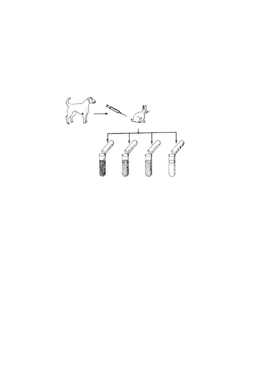
290
high level of concentration. After that the serum of the rabbit (anit dog
serum) was extracted. When it was mixed with the serum of the dog, the
antigen-antibody reactions took place. This made the serum cloudy. This
reaction will not happen if the rabbit is not immunized.
If the antidog serum is mixed with the serum of the wolf, precipitation
occurs but not to that extent as in the case of the dog When the same, test is
conducted with the fox and the cat (all coming under the order carnivora) the
same degree of relationship could be shown with the degree of precipitation.
When it is done with the cat no precipitation occurs showing that cow is very
distant from the carnivores.
Such biochemical tests apart from providing a sound evidence for
evolution they solve many taxonomical problems too.
(D) Zoogeographical evidence:
Geographical distribution of animals and plants provide ample
evidence for the fact of organic evolution. Biogeography is the spatial
distribution of organisms upon the earth’s surface at various periods. The
organisms belonging to the same species when happen to grow in
different areas with different climatic conditions may undergo adaptations to
suit their habitat. In course of time they may be modified so as to call them as
different species.
Dog
Rabbit
blood serum
wolf
fox
cow
dog
blood serum
serum
serum
serum
serum
Fig.7.4.4 Serological tests
291
This could be explained with the camels. The Arabian one humped
camel living in the vast deserts is highly adapted for the desert life by storing
water in the (pouches of the) stomach, food in the hump and having broad
heavily padded feet to hold grip in the loose sand.
On the other hand the two humped camel in the colder regions of
central Asia, has a coat of shaggy hair which insulates its body against the
extremely cold climate. Its feet are not padded but quite hard enabling the
animal to move about more freely on the rocky terrains of central Asia. From
these two examples we can understand that the differences are due to
different environments for several generations.
Charles Darwin during the course of his voyage in the ship ‘HMs
Beagle’ observed the flora and fauna of Galapagos islands which is 600
miles away from the west coast of south America, The inhabitants of each
island differed from those of other islands as well those of the main land.
Each one of these islands had their own type of giant tortoise, which was
different from those of the main land as well as from those of others. We may
suggest that these are the migrants from the main land or from other
landmasses.
The famous Darwin’s finches (13 species of finches) inhabiting
different islands exhibit special adaptations for their particular modes of life.
Those that fed on insects had long slender beaks by which they could pick
Fig.7.4.5 Darwin’s finches

292
up small insects from small crevices and pierce them. On the other hand
plant eaters had short strong beaks, which were useful in breaking open hard
nuts. Those living on islands covered with remains of volcanic eruptions
possessed black feathers, which merged with the surroundings. Those living
among green, had green feathers, which blended well with the background.
Those inhabiting pebbly beaches possessed speckled gray plumage. And
hence they had a wider range of adaptive radiation.
Self Evaluation
Part - A
1. Oparin’s theory is called as
(a) the Biogenesis theory
(b) the Primary abiogenesis
(c) Cosmozoic theory
(d) Special creation theory
2. The resistant spores that led to origin of life on earth are named as
a) protozoans
b) cosmozoa
c) viruses
d) bacteria
3. The proposal that living forms are animated forms of non-living matter was
provided by
a) Empedocles
b) Thales
c) Lamarck
d) Aristotle
4. The protocell model was formed of
a) coacervates
b) proteins
c) ozone
d) methane
5. Mesozoic era is commonly refered to as
a) age of mammals
b) age of fishes
c) golden age of reptiles
d) cradle of ancient life
6. Cenozoic era is called as
(a) Cradle of ancient life
(b) Age of mammals
(c) golden age of fishes
(d) All the above.
7. The first vertebrates were included in the group
a) Amphibia
b) Agnatha
c) Carinata
d) Aves

293
8. An earliest Jawless fish was
(a) Sword fish
(b) Cat fish
(c) Mullet
`
(d) Jamoytius
9. The duration of cenozoic era was
a) 210 to 65m years ago
b) 65m year ago to till date
c) 600 to 440 m years ago
d) 210m year ago to till date
10. The coal and petroleum are obtained from the forests of
a) Devonian period
b) Mesozoic era
c) Cretaceous
d) Tertiary period
11. The earliest ancestor of horses were
a) Eohippus
b) Equus
c) Seymouria
d) Dinosaurs
12. Archaeopteryx was a connecting link in between
(a) Amphibians and reptiles
(b) Fishes and amphibians
(c) Birds and reptiles
(d) Birds and mammals
13. Fossils of woolly mammoths were obtained from
a) Siberia
b) Sahara
c) Europe
d) Bavaria
14. Identify the early ancestor of horses
a) Dinosaurs
b) Seymouria
c) Archaeopteryx
d) Hyracotherium
15. Fossilized moulds are found
(a) in rocks
(b) in volcanic ashes
(c) in sand
(d) all the above
Part - II
1. What was the nature of the primordial earth according to J.B.S Haldane ?
2. Explain Cosmozoic theory.
3. Why do we call the palaeozoic era as the cradle of ancient life.
4. Write a short note on the significance of Palaeozoic era.
5. What was Archceopteryx ?

294
6. What is ‘ice age’ ?
7. What are Placoderms? Give example.
8. What is dating of fossils ?
9. What is the significance of Seymouria ?
10. Name the various methods of fossilization.
11. What is Ichthyostega and Hyracotherium.
12. What is Mac Author’s law ?
13. Write any two causes for extinction of animals.
14. What is precambrian period ?
15. What is meant by divergent evolution?
16. What is vestige.
Part III
1. What is abiogenesis ?
2. Give an account of Urey-Miller hypothesis.
3. Give an account on significant events of Mississippian Period.
4. Explain J.B.S. Haldane’s hypothesis.
5. Give an account on Coacervation theory.
6. Explain the six epochs of Cenozoic era.
7. What is the evolutionary significance of fossils?
8. Give an account of the significant events of the Palaeozoic era.
9. Give an account of various epochs included in Cenozoic era.
10. Explain the statement ontogeny recapitulates phylogeny
Part IV
1. Tabulate the geological time scale.
2. Give an account of the Mesozoic era.
3. What is mass extinction ? Provide the causes for such extinctions.
4. Write an essay on fossils and methods of fossilization.
5. Give an account on Extinct animals.
6. How do the serological tests help in relating the animals?
7. Write an essy on the evidences for evoltion from embryology.

295
Reference books
1. Manual of Zoology Vol. I.
Part. I.(Invertebrata), M. Ekambaranatha
Ayyar and T.N. Ananthakrishnan, Reprint 2003.
2. Manual of Zoology Vol. I.
Part. II.(Invertebrata), M. Ekambaranatha
Ayyar and T.N. Ananthakrishnan, Reprint 2003.
3. Manual of Zoology Vol. II. Chordata M. Ekambaranatha Ayyar and
T.N. Ananthakrishnan, Reprint 2003. S. Viswanathan
(Printers andPublishers) Pvt. Ltd. 38, Mcnichols Rd,
Chetput, Chennai - 600031.
4. Chordate Zoology E. L. Jordan and P. S. Verma. Reprint 2003.
S. Chand .and Company Ltd, Ram nagar,
New Delhi - 110 055.
5. A Text book of Zoology R. D. Vidyarthi and P. N. Pandey S. Chand and
Company Ltd, Ram nagar, New Delhi - 110 055.
6. Concept of Cell Biology P. S. Verma and V. K. Agarwal 1/e Reprint
2002. S. Chand and Company Ltd, Ram nagar,
New Delhi - 110 055.
7. Cell Biology N. Arumugam Reprint. 1999. Saras Publication
A R P Camp Rd, Peria vilai, Kottar,
Nagercoil - 629 002.
8. Genetics P. S. Verma and V. K. Agarwal Reprint 2003. S. Chand and
Company Ltd, Ram nagar, New Delhi - 110 055.
9. A Text book of Human Anatomy T. S. Ranganathan 6/e Rev. 2002.
S. Chandand Company Ltd, Ram nagar,
New Delhi - 110 055.

296
10. Chordate Embryology P. S. Verma and V. K. Agarwal Reprint 2003.
S. Chand and Company Ltd, Ram nagar,
New Delhi - 110 055.
11. A Text book of Embryology
N. Arumugam Reprint. 1999. Saras
Publication A R P Camp Rd, Peria vilai, Kottar,
Nagercoil - 629 002.
12. Economic Zoology G. S. Shukla and Upadhyay 4/e 1998. Rastogi
Publications, Shivaji Rd, Meerut - 250 002.
13. A Hand Book on Economic Zoology Jawaid Ahsan and Subhas Prasad
Sinha. Reprint 2003. S. Chand and Company Ltd,
Ramnagar, New Delhi - 110 055.
14. Concept of Evolution P. S. Verma and V. K. Agarwal Reprint 1999.
S. Chand and Company Ltd, Ram nagar,
New Delhi - 110 055.
15. Organic Evolution N. Arumugam 9/e.1999. Saras Publication
A R P Camp Rd, Peria vilai, Kottar,
Nagercoil - 629 002.
Visit websites
http://home.pcisys.net/~dlblanc/taxonomy.html
http://www.utm.edu/~riwin/b120lab.htm
http://can-do.com/uci/lessons 98/Invertebrates.html
http://www.student.loretto.org/zoology/chordates.htm
http://cellbio.utmb.edu/cellbio/cancer biology
http://dir.yahoo.com/Health/Medicine/Oncology
http://www.bartleby.com/107/
http://www.mhhe.com/biosci/ap/seeleyap/