
Lec.3
Dr. Inas Khalifa Al-Sharquie
2012-2013
1
Bacteriology
Cell wall Growth
Microbial Nutrition & Growth
Learning Outcomes:
At the end of this lecture, the students should be able to:
1. Describe the process of synthesis bacterial cell wall
2. Define L forms
3. Identify the chemical and environmental factors affecting bacterial growth
4. Explain how microbes are classified on the basis of O
2
needs
5. Explain the bacterial growth curve
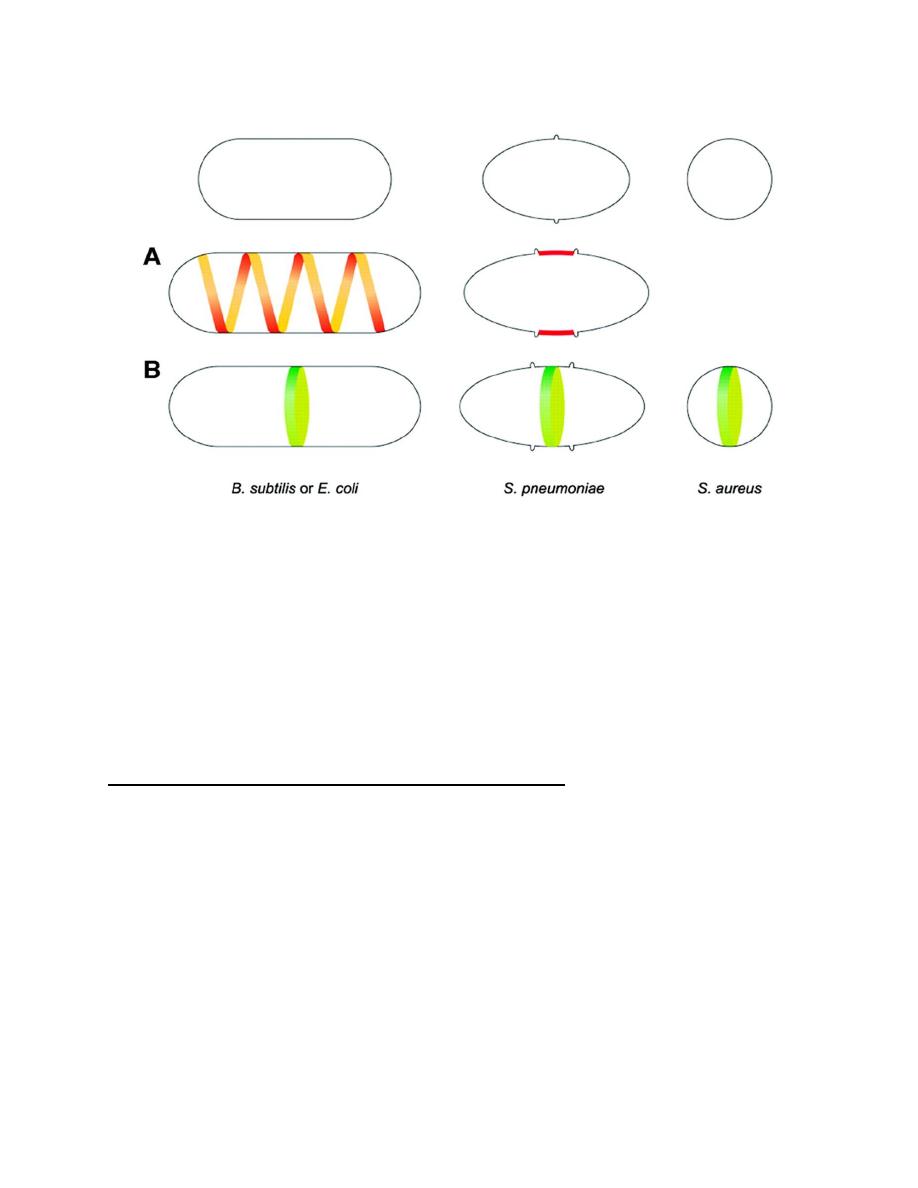
Cell wall growth
Cell wall synthesis is necessary for cell division; however, the incorporation of
new cell wall material varies with the shape of the bacterium. Rod-shaped bacteria
(eg, E coli, Bacillus subtilis) have two modes of cell wall synthesis; new
peptidoglycan is inserted along a helical path leading to elongation of the cell, and
is inserted in a closing ring around the future division site, leading to the formation
of the division septum. Coccoid cells such as S aureus do not seem to have an
elongation mode of cell wall synthesis. Instead, new peptidoglycan is inserted only
at the division site. A third form of cell wall growth is exemplified by S
pneumoniae, which are not true cocci, as their shape is not totally round, but
instead have the shape of a rugby ball. S pneumoniae synthesize cell wall not only
at the septum but also at the so-called equatorial rings (Figure 1).

Lec.3
Dr. Inas Khalifa Al-Sharquie
2012-2013
2
Protoplasts, Spheroplasts, & L Forms
Removal of the bacterial wall may be accomplished by hydrolysis with lysozyme
or by blocking peptidoglycan synthesis with an antibiotic such as penicillin. In
osmotically protective media, such treatments liberate protoplasts from gram-
positive cells and spheroplasts (which retain outer membrane and entrapped
peptidoglycan) from gram-negative cells.
If such cells are able to grow and divide, they are called L forms. L forms are
difficult to cultivate and usually require a medium that is solidified with agar as
well as having the right osmotic strength. L forms are produced more readily with
penicillin than with lysozyme, suggesting the need for residual peptidoglycan.
Some L forms can revert to the normal bacillary form upon removal of the
inducing stimulus. Thus, they are able to resume normal cell wall synthesis. Others
are stable and never revert. The factor that determines their capacity to revert may
again be the presence of residual peptidoglycan, which normally acts as a primer in
its own biosynthesis.
Some bacterial species produce L forms spontaneously. Lead to chronic infection
and they resist antibiotic treatment and give us a great problem in healing the focus
of infection.
Lec.3
Dr. Inas Khalifa Al-Sharquie
2012-2013
3
Figure 1:
Incorporation of new cell wall in differently shaped bacteria. Rod-shaped bacteria such as
Bacillus subtilis or Escherichia coli have two modes of cell wall synthesis: new peptidoglycan is inserted
along a helical path (A), leading to elongation of the lateral wall, and is inserted in a closing ring around
the future division site, leading to the formation of the division septum (B). Streptococcus pneumoniae
cells have the shape of a rugby ball and elongate by inserting new cell wall material at the so-called
equatorial rings (A), which correspond to an outgrowth of the cell wall that encircles the cell. An initial
ring is duplicated, and the two resultant rings are progressively separated, marking the future division
sites of the daughter cells. The division septum is then synthesized in the middle of the cell (B). Round
ce
o
lls such as Staphylococcus aureus do not seem to have an elongation mode of cell wall synthesis.
Instead, new peptidoglycan is inserted only at the division septum (B).
Nutritional requirement for microorganisms
The essential elements for the bacterial growth in the body of the host or on culture
media are:
1-Carbon source: Chemolithotrophs, organisms that use an inorganic substrate
such as hydrogen or thiosulfate as a reductant and carbon dioxide as a carbon
source. Heterotrophs require organic carbon for growth, and the organic carbon
must be in a form that can be assimilated.

Lec.3
Dr. Inas Khalifa Al-Sharquie
2012-2013
4
2-Nitrogen source: Most microorganisms can use NH
3
as a sole nitrogen source,
and many organisms possess the ability to produce NH
3
from amines (R—NH
2
) or
from amino acids. Many microorganisms possess the ability to assimilate nitrate
(NO
3
–
) and nitrite (NO
2
–
) reductively by conversion of these ions into NH
3
. A few
bacteria use N
2
in nitrogen fixation, a process by which nitrogen (N
2
) in
the atmosphere is converted into ammonia (NH
3
).
3- Sulfur Source: Sulfur in its elemental form cannot be used by plants or animals.
However, some autotrophic bacteria can oxidize it to sulfate (SO
4
2–
). Most
microorganisms can use sulfate as a sulfur source, reducing the sulfate to the level
of hydrogen sulfide (H
2
S). Some microorganisms can assimilate H
2
S directly from
the grwth medium, but this compound can be toxic to many organisms.
4- Phosphorus Source:
Phosphate (PO
4
3–
) is required as a component of ATP,
nucleic acids, and such coenzymes as NAD and NADP. In addition, many
metabolites, lipids (phospholipids, lipid A), cell wall components (teichoic acid),
some capsular polysaccharides, and some proteins are phosphorylated. Phosphate
is always assimilated as free inorganic phosphate (P
i
).
5-Mineral Sources: Numerous minerals are required for enzyme function.
Mg
2+
and K
+
are both essential for the function and integrity of ribosomes. Ca
2+
is
required as a constituent of gram-positive cell walls, though it is dispensable for
gram-negative bacteria. Many marine organisms require Na
+
for growth.
6-Growth Factors:
are organic compounds obtained from the environment, where
cells must have them in order to grow, but unable to synthesize them, like
vitamins.

Lec.3
Dr. Inas Khalifa Al-Sharquie
2012-2013
5
Environmental factors affecting growth
1. Nutrients
2. pH: each organism need a certain PH for its growth.
Neutrophiles 6-8,
Acidophiles 3
Alkalophiles 10.5
Majority of microorganisms grow at a pH between 6-8
3. Temperature: Different microbial species vary widely in their optimal
temperature ranges for growth: Psychrophilic (15-20
0
C), Mesophilic (30-
37
0
C), Thermophilic (50-60
0
C).
Some organisms are hyperthermophilic and can grow at well above the
temperature of boiling water, which exists under high pressure in the depths of
the ocean. Most organisms are mesophilic; 30°C is optimal for many free-living
forms, and the body temperature of the host is optimal for symbionts of warm-
blooded animals.
4. Oxygen (O2): most bacteria require O2 necessary (strict aerobes).Others can
live with or without O2: Facultative bacteria as E.coil. Other requires a small
amount of O2: (microaerophilic). The rest cannot grow in presence of O2:
anaerobic bacteria e.g., Clostridium.
5. Osmotic Pressure:
cytoplasmic membrane and cell wall protect bacteria cell
from varying contents of salts in the medium .For most species the maximum
conc. of salts is (5-12%), some other species can grow in high conc.
(osmophilic bacteria) e.g: Staph. aureus. Organisms requiring high salt
concentrations are called halophilic; those requiring high osmotic pressures
are called osmophilic.

Lec.3
Dr. Inas Khalifa Al-Sharquie
2012-2013
6
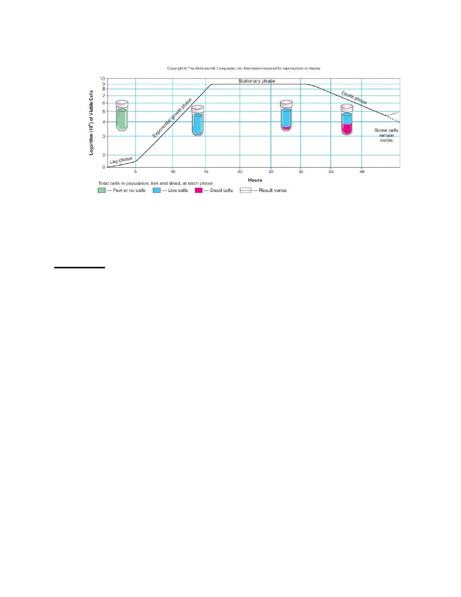
Bacterial Growth
Bacteria reproduce by binary fission, a process by which one parent cell divides to
form two progeny cells.
When a small number of bacteria are inoculated into a liquid nutrient medium and
the bacteria are counted at frequent intervals, the typical phases of a standard
growth curve can be demonstrated.
The number of bacteria in a medium could be:
1- A total cell count curve is based on the number of cells present that are alive or
dead.
2-A viable cell count curve measures only living (viable) cells (capable of growing
and producing a colony on a suitable growth medium).
The typical phases of a standard growth curve are (Figure 2):
1- Lag phase: during vigorous metabolic activity occurs but cells do not
divide. This can last for a few minutes up to many hours.
2- Log (logarithmic) or exponential phase: is when rapid cell division occurs.
There is liner relationship between time and log of number of cells. Β-
Lactam drugs, such as penicillin, act during this phase because the drugs are
effective when cells are making peptidoglycan (i.e., when they are dividing).
3- Stationary phase: occurs when nutrient depletion or toxic products cause
growth to slow until the number of new cells produced balances the number
of cells that die, resulting in a steady state.
4- Death or decline phase: decline in the number of viable bacteria.
Lec.3
Dr. Inas Khalifa Al-Sharquie
2012-2013
7
Figure 2:
The typical phases of a standard Growth Curve
Summary
1. L-forms are bacterial variants that lack a cell wall
2. Nutrients : Macronutrients/micronutrients
3. Generation time: Time required for cell to divide/for population to double
4. The growth cycle of a culture of bacteria is divided into four phases: lag
phase, exponential phase, stationary phase, decline phase
Main References:
1. Jawetz, Melnick and Adelberg’s Medical Microbiology (Brooks,
Butel,Morse). 25
th
ed. Copyright 2010.
2. Levinson W. Review of Medical Microbiology and Immunology.12
th
ed.
Copyright 2012, McGraw-Hill.