Lecture 3
ORGANS OF IMMUNE SYS.
Antigen Presenting Cells

OBJECTIVES
Primary (central) Lymphoid organs
Secondary (peripheral) lymphoid organs
T cell educations
Antigen Presenting Cells (APC)
Stem cells originate from the
yalk sac
in the 1
st
6 wks of gestation
Then the
LIVER
for the next few month
Then the
BONE MARROW
will be resp. for origination &
proliferation of stem cells under control of diff, hormones
, enzymes & interleukin like IL3 ,
IL7
,MG CSF Macrophage
granulocyte colony stimulating factor
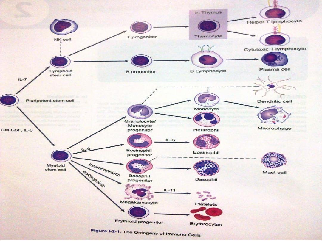
Stem cell
Lymphoid series
(Lymphocyte & Nk
cell)
MYELOID SERIE
(RBC Granulocyte
Monocyte)
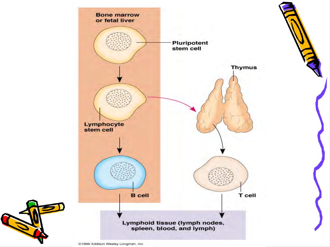

Organs of Immune System
Primary (central)
Thymus
for T cells &
bone
marrow
for B cells
Site where lymphocyte
maturation
Secondary (peripheral) encapsulated (spleen &
lymph node) and Unencapsulated (Mucosa
associated lymphoid tissue M.A.L.T.)
Site
where Lymphocyte interact with Antigens &
other cells
Naïve (virgin ) lymphocyte
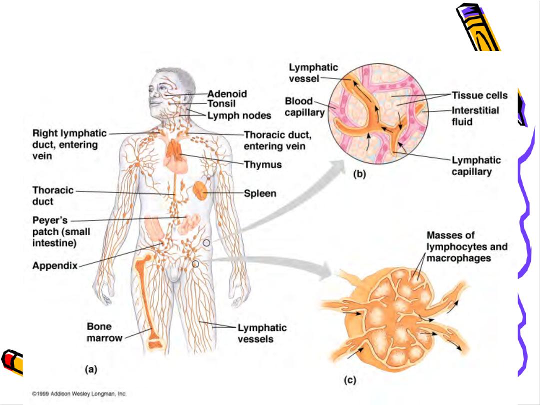
Components of Human Immune System

Thymus
2 lobs each with Cortex & medulla
T Lymphocyte Education
Cortex: T cells are Immature Highly dividing
,Highly dying (95%)die by Apoptosis because
Auto-reactive cells (
Negative selection
)
Medulla less dividing less dying cells , only that
recognize self MHC ( by CD4 or CD8 ) will
expand(
positive selection
)


•
Cells of the thymus
Epithelial cells
secrete thymic hormones as
thymopoietin ,Thymoline &Thymosine also
Enzymes as ADA(adenosine deaminase) & PNP
(purine necleoside phosphorylase) which help
in differentiation & maturation of Tcells
Inter –digitating dendritic cells
they are rich
in class II Ag & teach T cells how to deal
with an Ag

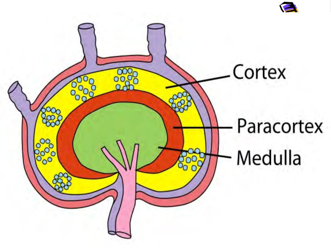
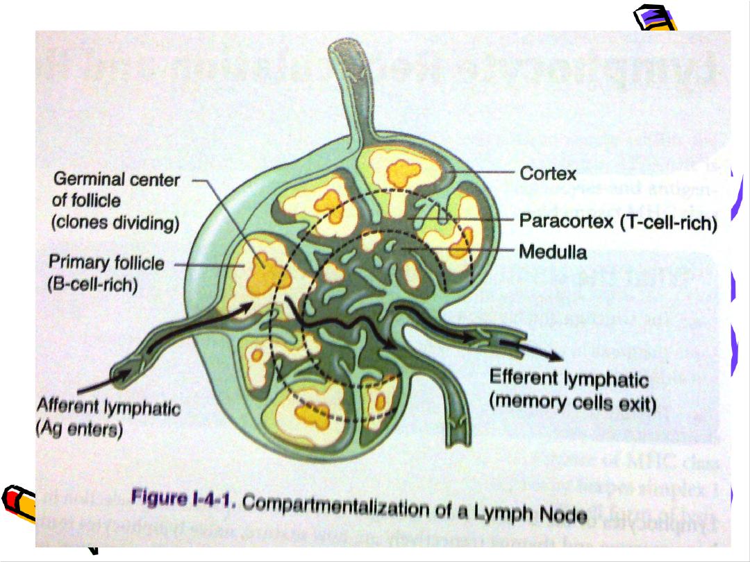
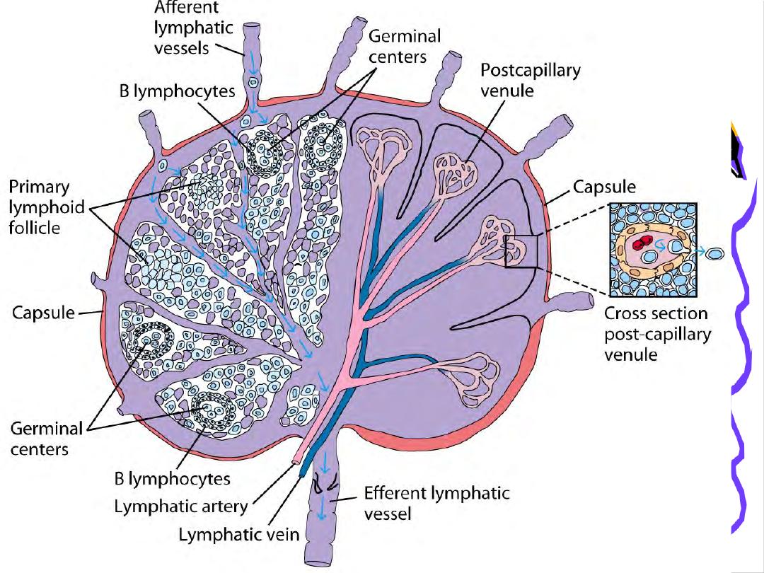
Lymph node
•
Filters Ag from the lymph
Cortex containing aggregations of B
lymphocytes as a primary follicles after Ag
stimulation become secondary follicles
containing large dividing B lymphocytes ( blast
cells & plasma cells
Para cortex contains T lymphocytes

spleen
Red palp site where old RBCs are destroyed
White palp surrounds the spleenic arteries
forming Peri-arteriolar lymphoid sheath (
area
for T lymphocytes)
Between red & white palp the Marginal Zone
which is(
area for B lymphocytes)
Inter-digitating cells will take the
Blood born Ag
to the peri arteriolar
lymphoid sheath
ANTIGEN PRESENTIG CELL
(APC)
•
Monocyte in the blood(1-6% of WBC) circulate
for 3 days Tissue as a Macrophage
Like alveolar cell,kupffer cell in the liver & glail
cell in the Brain where they live for months &
when activated they become APC where they
have B7 molecule & Class II MHC
Activation by
phagocytosis, Gamma-interferon &
cytokines from T helper cells as IL2,IL12
While IL 8 is a chemotactic

APC
Include(any cell have B7 mol.& class II MHC)
•
Langerhans cell
•
Dendritic cells
•
B lymphocytes
•
Macrophages
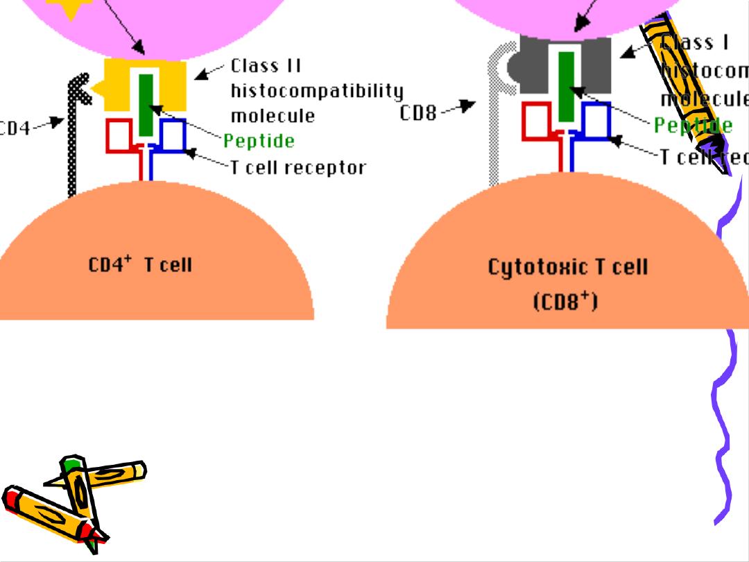
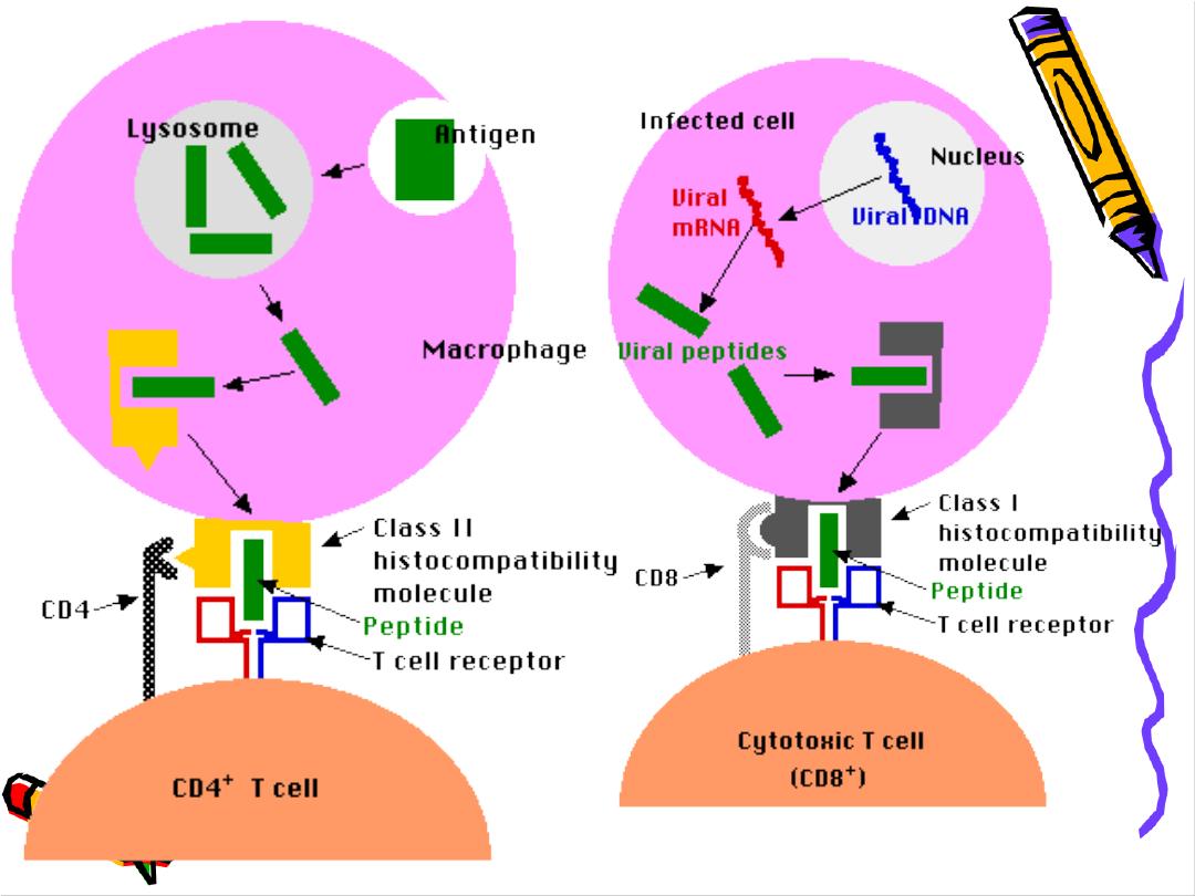
They process the Ag & present it to T lymphocytes
with class I for CD8+ cells or Class II for CD4+ cells
Also they deliver B7 Mol. To react with CD28 on T
helper cells

APC
•
APC secretes IL1, TNF, (both are endogenous
•
pyrogen) also IL12 which activate T cells
•
IFN α ( Anti-virus)
•
Hydrolytic Enz., nitric oxide H2O2 , Super oxide
•
Lysozyme
•
APC has many Receptors as
•
CR 1,2,3 For C3b
•
FC receptor
•
ICAM (inter cellular adhesion Mol.
)

Functions of APC
-Identifications of Microbes (Ag) by
recognition
Recptor as TL R
-Engulfment (
Phagocytosis
) into phagosomes
-
Lysosomes
which are filled with digestive enz. Fuse
with phagosome to form
phagolysosome
Which will digest the Ag with
preserving
the
Epitope
-
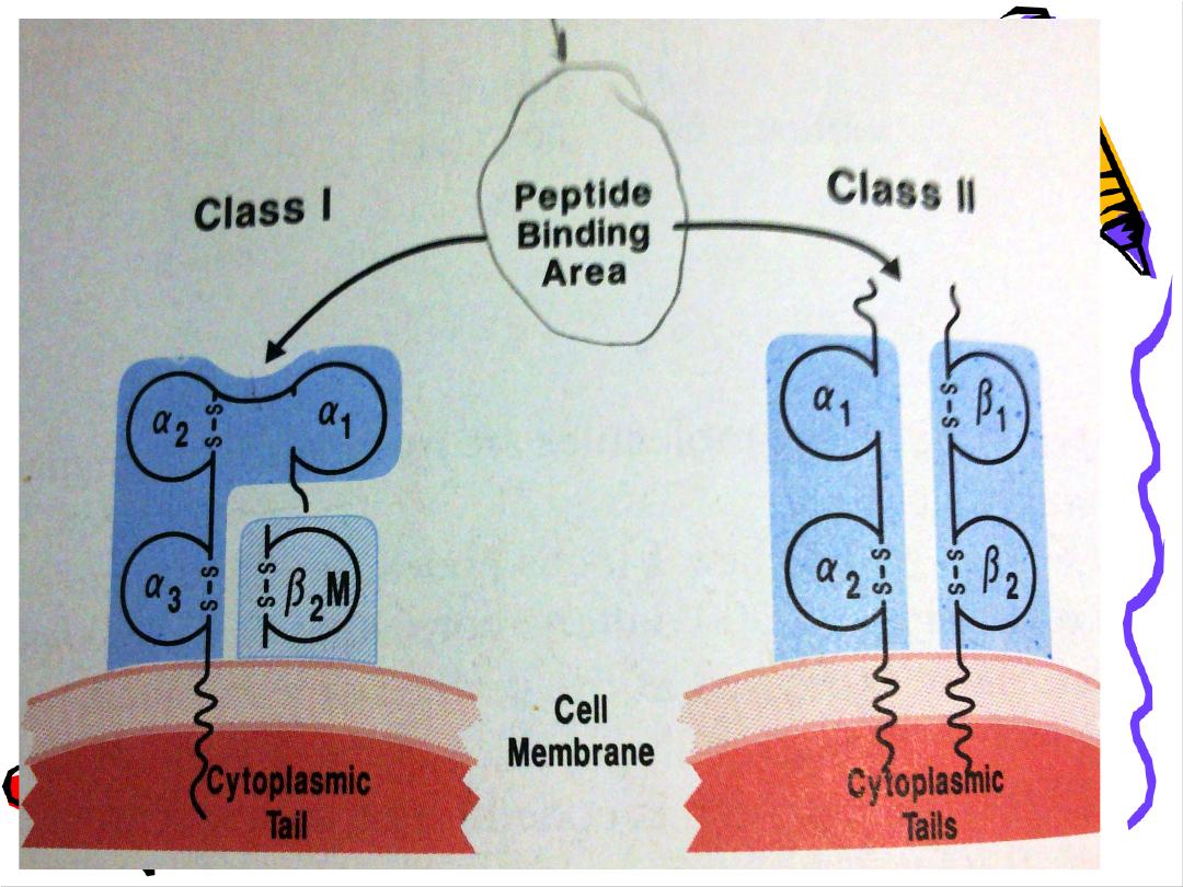
Presents
the epitope in the groove of MHC class I
or class II on their surface for CD8 &CD4
respectively in association with
B7
molecule
.
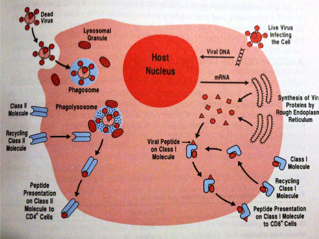
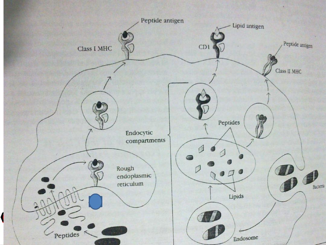
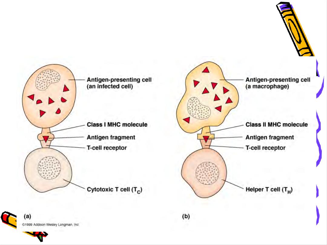
T Cells Only Recognize Antigen Associated
with MHC Molecules on Cell Surfaces
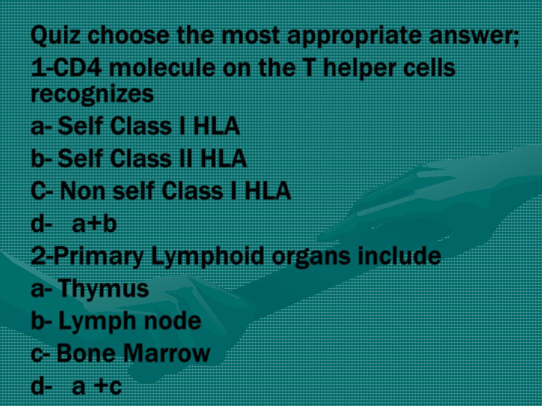
Quiz choose the most appropriate answer;
1-CD4 molecule on the T helper cells
recognizes
a- Self Class I HLA
b- Self Class II HLA
C- Non self Class I HLA
d- a+b
2-Primary Lymphoid organs include
a- Thymus
b- Lymph node
c- Bone Marrow
d- a +c