Physiology
Dr. Basim Mohammed Alwan Lecture (15)MUSCLE SENSORY RECEPTORS AND SPINAL REFLEXES
Proper control of muscle function requires not only excitation of the muscle by the anterior motor neurons but also continuous sensory feedback information from each muscle to the spinal cord giving the status of the muscle at each instant, that is:What is the length of the muscle?
What is its tension?How rapidly is its length or tension changing?
To provide this information, the muscle and its tendon are supplied abundantly with 2 special types of sensory receptors "muscle spindles and Golgi tendon organs".
LENGTH-MONITORING SYSTEM AND THE STRETCH REFLEX
Absolute muscle length and changes in muscle length are monitored by stretch receptors embedded within the muscle.There are 2 kinds of stretch receptors:
One respond best to how much the muscle has been stretched.
Other responds to both the magnitude of the stretch and the speed with which it occurs.These receptors consist of afferent nerve fiber endings that are wrapped around modified muscle fibers. Several of which are enclosed in a connective tissue capsule. The entire structure is called "muscle spindle".
Each spindle is 3-10 mm long.
It is build around 3-12 very small modified intrafusal fibers that are pointed at their ends and attached to glycocalyx of the surrounding large "extrafusal fiber".
Intrafusal skeletal muscle fibers
They are small muscle fiberThe central region of which has either no or few actin and myosin filaments. Therefore, this central portion does not contract when the ends do. Instead they function as sensory receptors.
The end portions that do contract are excited by small gamma motor neurons.
They are of 2 types:
Nuclear-bag fibers
They are 1-3 in each spindle.There is large number of nuclei congregated in an expanded bag in the central portion of the receptors.
They are innervated by primary nerve endings
Nuclear-chain fibers
They are 3-9 in each spindle.They are about 1/2 in diameter and 1/2 as long as the nuclear-bag fibers.
Their nuclei aligned in a chain throughout the receptor area.
They are innervated by primary and secondary nerve endings.
Their ends are connected to the nuclear-bag fibers.
The sensory fibers which originate in the central portion and stimulated by stretching of this mid portion of the spindle are of 2 types:
The Primary Endings (Annulospiral) Fibers
They are large sensory fibers encircles the very center of the receptor area.
They are termination of type Ia afferent fibers.
They have 17 µm diameters.
They transmit signals to the spinal cord at a velocity of 70-120 m/second.
They discharge most rapidly while the muscle being stretched and less rapidly during sustained stretch. Thus, respond to changes in length and change in the rate of stretch.
The Secondary (Flowerspray) Fibers
They are the termination of usually one and sometimes two smaller type II sensory nerve fibers.Their diameter is 8 µm.
They innervate the receptor region on one side of the primary ending.
They discharge at increasing rate throughout period when the muscle is stretched.
They respond to change in the length alone.
Note: Both primary and secondary nerve endings are stimulated when the spindle is stretched but the pattern of response is different.
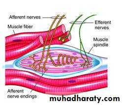
Fig. 15-1 muscle spindle
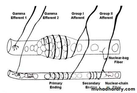
Fig. 15-2 muscle spindle
The muscle spindles are parallel to the extrafusal fibers, such that stretch of the muscle by external force pulls on the intrafusal fibers, stretching them and activating their receptor endings with reflex contraction of the extrafusal fibers of the muscle.
The more the muscle is stretched or the faster it stretched, the greater the rate of the receptor firing.
In contrast, contraction of the extrafusal fibers and the resultant shortening of the muscle remove tension on the spindle and slow the rate of firing of the stretch receptor.
When the afferent fibers form the muscle spindle enter the CNS, they divide into branches that take several different paths:
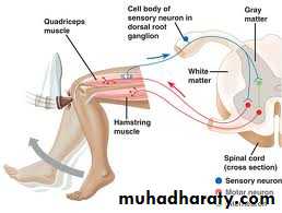
First Path:
Directly stimulates motor neuron going back to the muscle that was stretched, thereby completing a reflex arc known as the "stretch reflex".This reflex is probably the most familiar in the form of the knee jerk and it is the probably the only momosynaptic reflex.
The examiner taps the pattelar tendon which passes over the knee and connects extensor muscles in the thigh to the tibia in the lower leg.
As the tendon is pushed in, and thereby stretched by tapping.
The thigh muscles to which it attached are stretched and all the stretched receptors within these muscles are activated.
More action potentials are generated in the afferent nerve fibers from the stretched receptors.
They are transmitted to the motor neurons that control these muscles.
The motor units are stimulated
The thigh muscles are shortens
The patient's lower leg is extended to give the knee jerk.
Note: In contrast to the knee jerk, during normal movement, stretch receptors are rarely all activated at the same time, nor are they activated so strongly. Moreover, movement occurs in response to the integration of many types of local descending controls.
Second Path
The afferent nerve fibers from stretch receptors end on interneuron.
When activated, inhibit the motor neuron controlling the antagonistic muscles (in the knee jerk, the flexor muscles of the knee are inhibited).
Reciprocal Innervations: The activation of one muscle with simultaneous inhibition of its antagonistic muscle.
The only reflex that does not obey the principle of reciprocal innervations is the positive supporting reflex which is occurring by the pressure exerted by the body weight during standing.
Third Path
Afferent nerve fibers activate motor neurons of synergistic muscles, that is, muscles whose contraction assists the intended motion (in the knee jerk, other extensor muscles).The muscles activated are on the same side of the body as the receptors and the response therefore is ipsilateral.
Fourth Path
Afferent nerve fibers continue to the brain stem.They synapse there with interneuron which forms the next link in the pathway that conveys information about the muscle length to areas of the brain dealing with motor control.
(NOTE: It is the dynamic type of stretch reflex)
ALPHA-GAMMA COACTIVATION
Whenever the alpha motor neurons are activated, the gamma motor neurons are activated at the same time. The role of gamma efferent coactivation is to prevent relaxation of the muscle spindle during extrafusal muscle contraction and to maintain them capable of reflexly adjusting the alpha motor neuron discharge thought the movement.
This is important for both the damping as well as the servo-assistant functions of the stretch reflex.
TENSION-MONITORING SYSTEM AND GOLGI TENDON REFLEX
Any given set of inputs to a given set of motor neurons can lead to various degrees of tension in the muscles they innervate depending on:Muscle length
Load on the muscle
Degree of muscle fatigue
Therefore, feedback is necessary to inform the motor control systems of the tension actually achieved.
Such feedback is provided by Golgi tendon organs which monitor how much tension is being exerted by the contracting motor units.
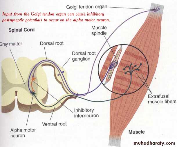
Fig. (15-5) Golgi tendon organ
Golgi tendon organThey are encapsulated sensory receptors through which a small bundle of muscle tendon fibers pass.
They are located in the tendons near their junction with the muscle.
About 10-15 muscle fibers are usually connected in series with each Golgi tendon organ.
Endings of the afferent nerve fibers are wrapped around collagen bundles in the tendon.
When he attached extrafusal muscle fibers contract, they pull on the tendon, which straightens the collagen bundles and distorts the receptor endings, and activating them.
Thus, the Golgi tendon organs discharge in response to the tension generated by the contraction and initiates action potentials that are transmitted to the CNS via large rapidly conducting type IIb nerve fibers.
Branches of the afferent neurons cause widespread inhibition via interneurons, of the motor neurons to the contracting muscle and its synergists.
They also stimulate the motor neurons of the antagonistic muscles.
Importance
Prevent tearing of the muscle or avulsion of the tendon from its attachments to the bone.Equalize the tension of the separate muscle fibers (that is fibers which exert excess tension become inhibited by the reflex and the reverse is true. This will spread the muscle load over all fibers and would prevent damage in isolated areas of a muscle where small numbers of fibers might be overloaded.
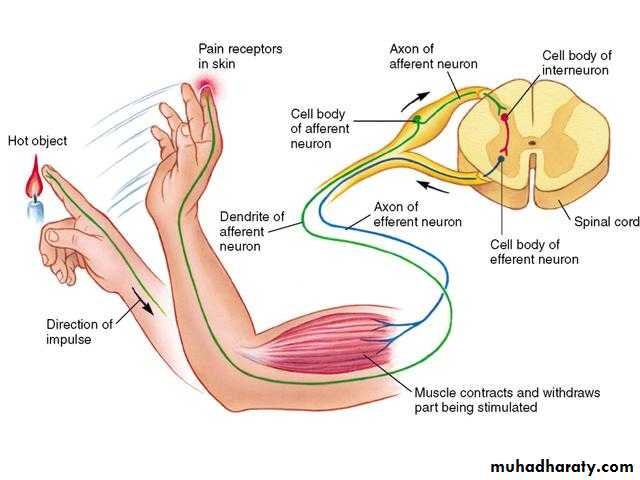
Fig. 15-6 Withdrawal reflex
WITHDRAWAL REFLEX (fig.13-6)Cutaneous sensory stimuli on a limb are likely to cause the flexor muscles of the limb to contract, thereby stimulating withdrawing the limb from the stimulating object. This is called "flexor reflex" or the "nociceptive reflex" or simply "pain reflex".
It is polysynaptic reflex
The response includes flexor muscles contraction and inhibition of the extensor muscles, so that the part stimulated is flexed and withdrawn from the stimulus.
When a strong stimulus is applied to a limb, the response includes not only flexion and withdrawal of that limb but also extension of the opposite limb. This is called crossed extensor reflex (fig.15-7) which has an even longer period of afterdischarge than does the flexor reflex.
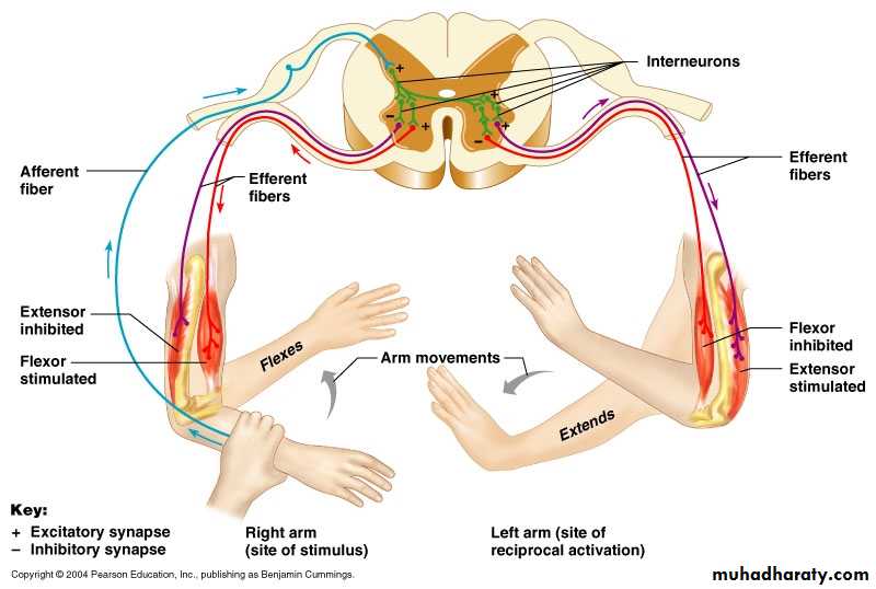
Fig. 15-7 Crossed extensor reflex
THE SKELETAL MUSCLE TONEDEFINITION: This is a state of continuous mild contraction of skeletal muscle during rest.
MECHANISM: It is a static type of stretch reflex.DISTRIBUTION: It is present in all skeletal muscles, but specially in the antigravity muscles. These muscles include:
Extensors of the lower limbs
Flexors of the upper limbs.
The muscle of the back and back of neck.
The anterior abdominal wall.
FUNCTIONS OF THE SKELETAL MUSCLE TONE
Maintenance of the erect posture.
Help both venous and lymph flow.
Prevent abdominal visceral ptosis.
Important source of heat production.
THE TENDON JERKS
It is the dynamic type of stretch reflex, that is consists of a rapid contraction of the muscle followed by a rapid relaxation.Reinforcement of the tendon jerks
The tendon jerks can be reinforced by facilitating the spinal centers. In lower limb jerks facilitation can be produced by asking the patient to grasp his hands together and exert maximal effort to pull them apart. In upper limb jerks, facilitation can be produced by asking the the patient to clench his teeth.This reinforcement is called Jendrassik maneuver.
This maneuver acts by the following mechanisms:
Contracted muscles send signals to stimulate the gamma efferent neurons.
Distracting the patient's attention, which prevent any voluntary inhibition of the reflex?