The ear:
The external ear constitutes:
1. The auricle (pinna).
2. The external auditory (acoustic)
meatus.
3. The tympanic membrane (eardrum).
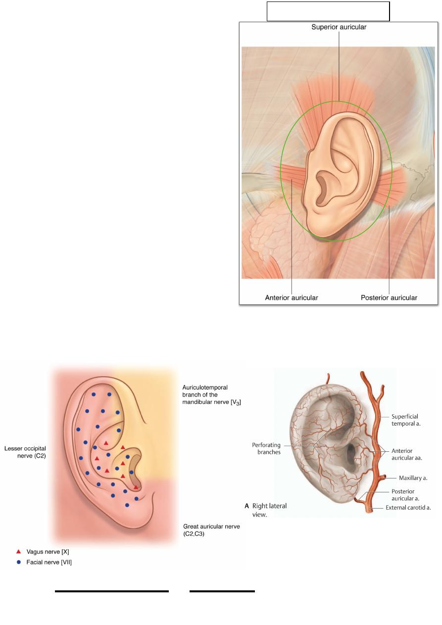
The auricle:
• Is an oval cartilage attached to the
side of the skull by anterior &
posterior auricular ligaments.
• The skin is thin & well attached to the
u n d e r l y i n g p e r i c h o n d r i u m &
prolonged inward into the external
auditory meatus as far as the tympanic
membrane.
• The cartilage is prolonged inward to
be continuous with the external 1/3 of
the EAM.
• Three auricular muscles are present to
move the auricle but their function is
negligible in human.
• Vessels; posterior auricular &
superficial temporal vessels.
• Nerves; great auricular, vagus and auriculotemporal nerves share the supply of
the auricle as mentioned.
!
111
Head & Neck Dr. Nawfal K. Al-Hadithi
Undergoes ossification with age
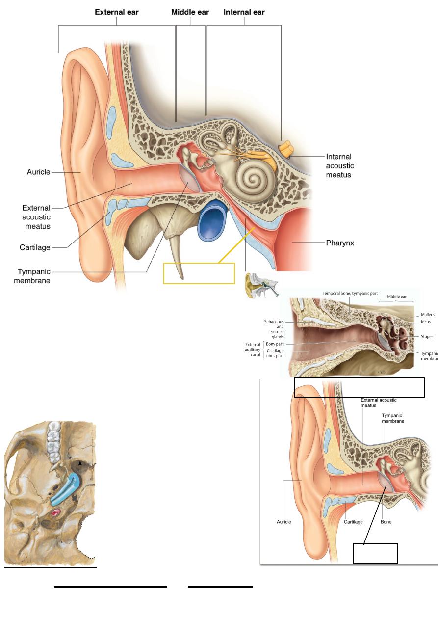
The eam:
• Is 24 mm in length
• Its lateral 1/3 is cartilagenous &
directed upward & backward as it goes
medially
• Its medial 2/3 is bony & directed downward
& forward as it goes medially
•The bony canal is narrower than
the cartilaginous
•The skin is adherent to the
underlying bone & cartilage
•The narrowest part of the canal is
the isthmus which is the junction
between its two parts
•The skin of the cartilagenous part
contains hair with sebaceous &
ceruminous glands
•The EAM is bounded anteriorly
by the TM joint & parotid gland &
posteriorly by the mastoid process
!
112
Head & Neck Dr. Nawfal K. Al-Hadithi
Ear drum
Eaustachian tube
Cartilaginous part Secretes wax
• The inferior wall of the canal is 5 mm longer than the superior one due to the
obliquity of the eardrum
• Vessels; as the auricle & deep auricular vessels.
• Nerves; auriculotemporal & auricular branch of the vagus nerves.
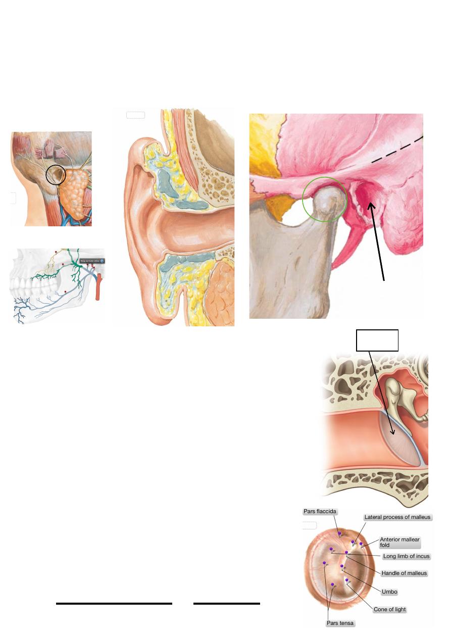
The eardrum:
• A nearly oval membrane 8X10 mm, set in a cone like
shape whose concavity faces outward & its most
concave point is its center “umbo”
• It is semitransparent, pearly grey in color
• It is applied in an oblique manner in the EAM so that
its lateral surface faces downward, forward & laterally
making 55O angle with the floor
• Its circumference is a fibrocartilagenous ring set in the
tympanic sulcus
• It is composed of three layers:
a. Outer layer of modified skin continuous with that of the
EAM
b. Inner layer of m.m continuous with that of the
middle ear
c. Intermediate fibrous layer formed of circular &
radial fibers which is responsible for the strength of
the membrane
• The upper 1/6 of the membrane lacks the
intermediate layer so it is lax & called pars flaccida,
!
113
Head & Neck Dr. Nawfal K. Al-Hadithi
Tm
Eam
Eardrum
the rest of the membrane is called pars tensa
• The handle of the malleus is fused with the upper part of the membrane
Applied anatomy:
• In order to straighten the EAM in examination of the ear it should be pulled
upward, backward & laterally in adults and downward, backward & laterally in
children
• Wax of the external ear mostly affects the lateral 1/3 of the EAM
• In ear syringing, the nozzle should be directed forward at first then backward,
upward & medially to avoid injury to the tympanic membrane
• Normal tympanic membrane is semitransparent & pearly grey with a cone of
light in its antero-inferior part, a diseased membrane looses its shiny
appearance & the cone of light`
The middle ear:
Is a small, six-walled cavity in the temporal bone where the sound waves are
converted into mechanical waves
• The cavity is 15 mm in height, 15 mm in AP dimension but narrow from side
to side where it is narrowest in
the center (2 mm)but wider
a b o v e & b e l o w ( l i k e a
biconcave lens)
• The cavity communicates
anteriorly with the nasopharynx
via the Eustachian tube &
posteriorly with the mastoid air
cells through the aditus, their
mucosal lining is continuous
with each other & is respiratory
in type
!
114
Head & Neck Dr. Nawfal K. Al-Hadithi
1
2
3
4
5
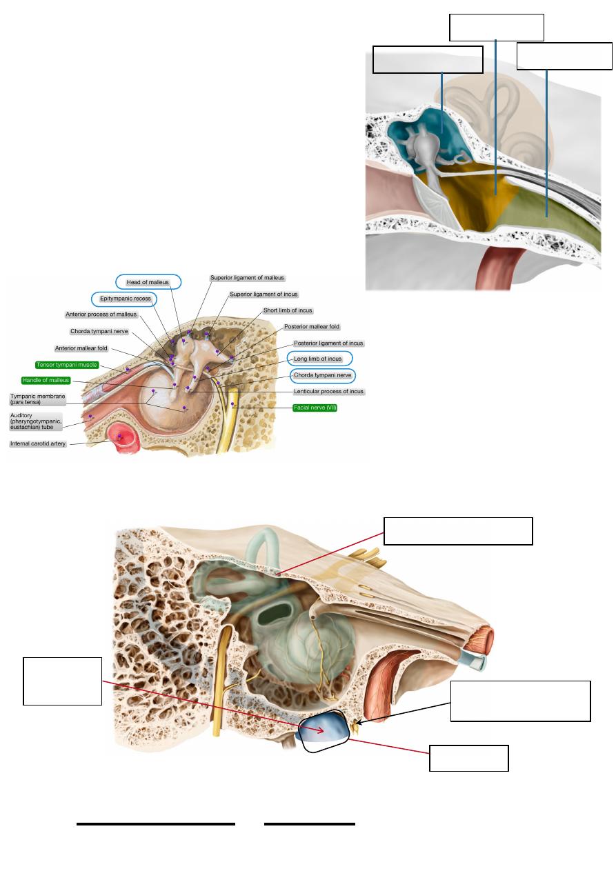
Parts:
a. Epitympanum “epitympanic recess OR
attic”; the part of the cavity which extends
above the level of the tympanic membrane
b. Mesotympanum; the part of the cavity
opposite to the eardrum
c. Hypotympanum; the part below the level of
the eardrum
• Contents:
a. ONE nerve; chorda tympanip
b. TWO muscles; tensor tympani & stapedius
c. THREE bones; incus, malleus & stapes
Walls of the middle ear:
The roof:
The roof of the tympanic
cavity is formed by the thin
plate of tegmen tympani
which separates the ear from
the cranial cavity
The floor:
•The floor of the middle ear
separates the ear from the
jugular fossa
• It is perforated by the tympanic branch of glossopharyngeal nerve
The lateral wall:(discussed)
!
115
Head & Neck Dr. Nawfal K. Al-Hadithi
Epitympanic recess
Hypotempanum
Mesotympanum
Tegmen tympani (the roof)
Jugular fossa
Tympanic nerve (from
glossopharyngeal nerve )
Internal
jugular vein
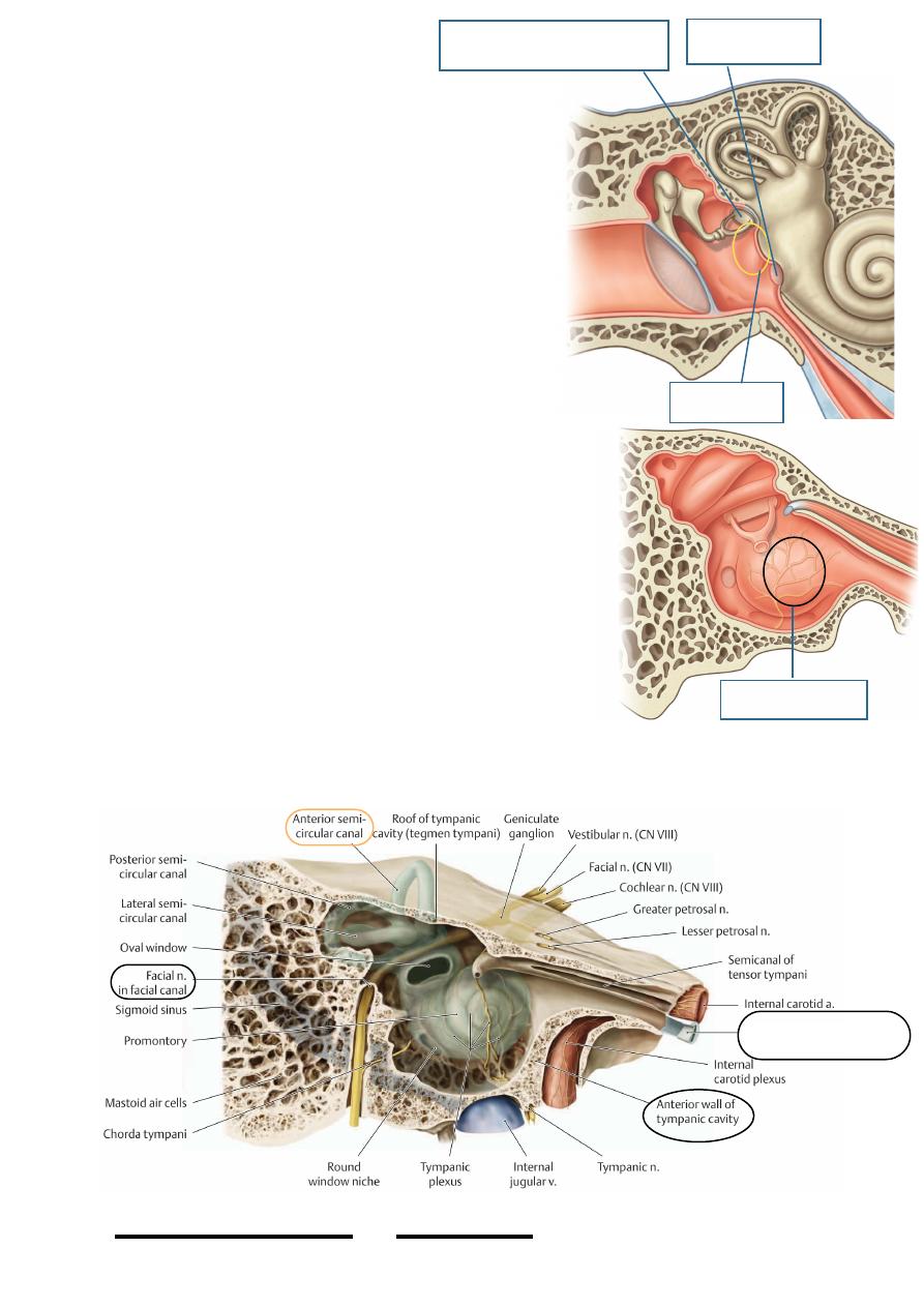
The medial wall:
* The most prominent feature of the medial wall is
the promontory of the internal ear which is the
basal turn of the cochlea
* The promontory is grooved by branches of the
tympanic plexus
* The oval window “fenestra vestibuli”: is an oval
opening above the promontory whose long axis is
horizontal & maximum convexity is superior, it is
closed in life by the footplate of the stapes
* The round window “fenestra cochleae”; lies
below & behind the promontory & closed in life by
the secondary tympanic membrane
* The facial canal seen in the medial wall as a
prominence of bone above the oval window which
then curves inferiorly & nearly vertically behind the
promontory, the bone may be so thin in this area
* The prominence of the lateral semicircular canal
sometimes seen as a prominent ridge above the facial
canal
The anterior wall:
• This wall separates the middle ear from the
carotid canal
• It is perforated by the coraticotympanic nerves
which leaves the carotid plexus around the ICA
to enter the tympanic plexus over the promontory
• In the upper end of the anterior wall lies the
opening of the Eustachian tube
!
116
Head & Neck Dr. Nawfal K. Al-Hadithi
Promontary
Round window
Tympanic plexus
Oval window closed by F.P
Eustachian tube
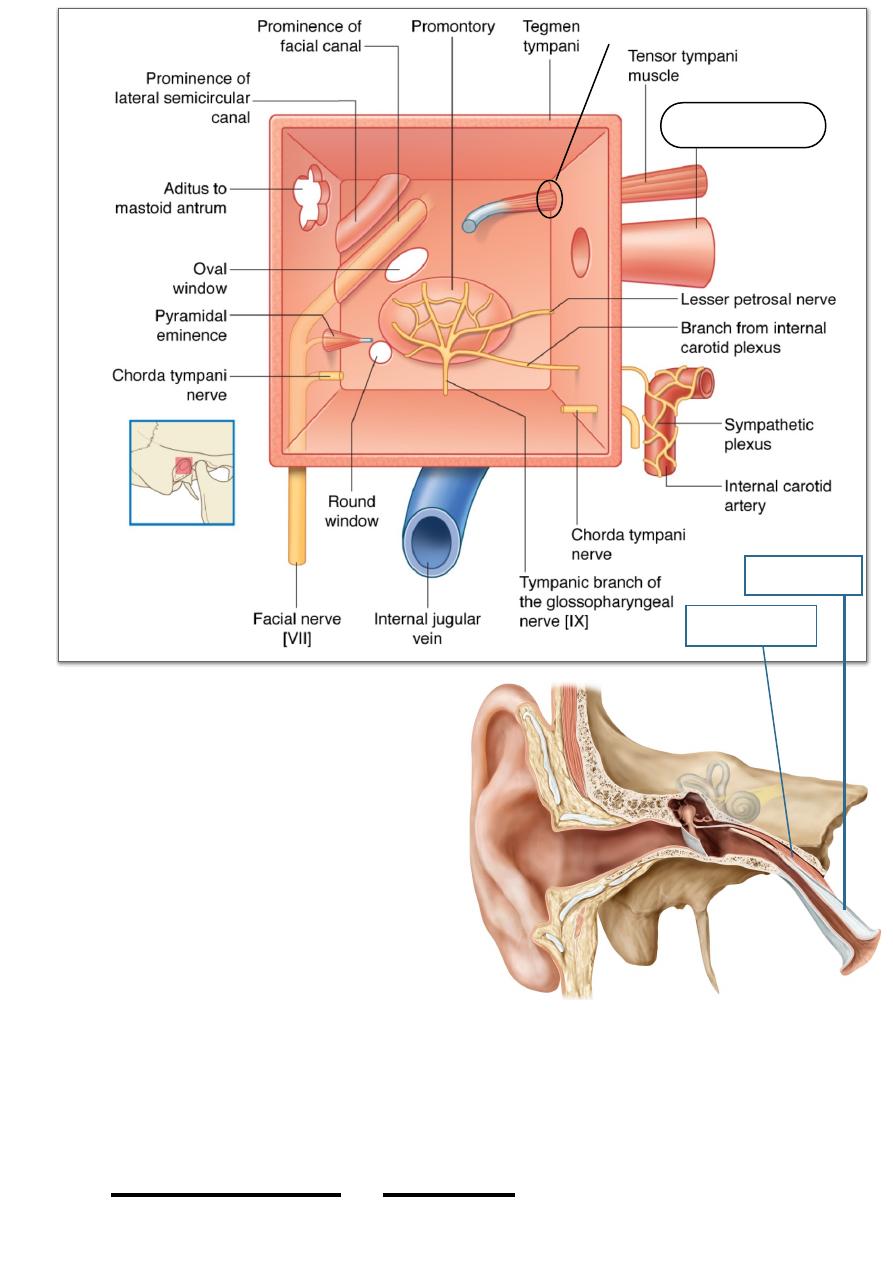
• Above the auditory tube opening
lies the semicanal for tensor
tympani muscle
• Tensor tympani, a 2cm long muscle
which arises from the septum
between the auditory tube & its
canal & from the cartilagenouos
part of the tube gives rise to a
slender tendon which hooks around
the processus & then directed
laterally to insert into the handle of
the malleus, its contraction tenses
the tympanic membrane by pulling
the handle of the malleus medially
resulting in dampening of its
vibrations
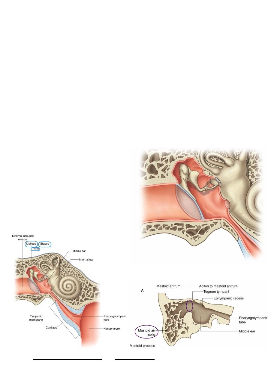
The auditory (Eustachian) tube:
• This 3.5 cm long tube connects the cavities of the middle ear & nasopharynx
• Its tympanic 1/3 is osseous & pharyngeal 2/3 is cartilagenous
!
117
Head & Neck Dr. Nawfal K. Al-Hadithi
Eustachian tube
Semicanal
Tensor tympani
Auditory tube
• The direction of the tube from the ear to
the nasopharynx is downward, forward
& medially making 45O angle with the
sagittal plane & 35O angle with the
horizontal plane
• The mucosa of the middle ear therefore
is continuous with that of the
nasopharynx through the tube
• Mucous glands are present in the
cartilagenous part whose pharyngeal end
is surrounded by the tubal tonsils
• The tube is shorter, wider & more
horizontal in children
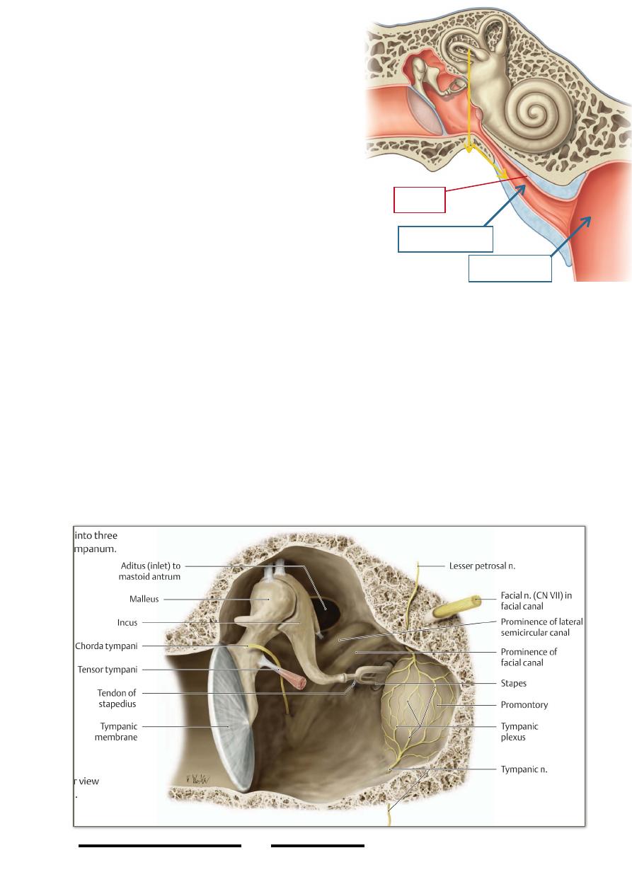
The posterior wall:
• The upper part of the posterior wall is open to the mastoid antrum through the
aditus ad antrum which is a large irregular opening leading from the middle ear
to the mastoid antrum
• Fossa incudis lies below the opening of the mastoid antrum, it lodges the short
process of the incus
• The vertical facial canal lies medially in the posterior wall
• The pyramidal eminence projects from the posterior wall in front of the facial
canal, it is hollow structure whose walls give rise to stapedius muscle
• Stapedius arises from the pyramidal eminence, its tendon is inserted into the
posterior part of the neck of the stapes, its contraction tilts the footplate of the
stapes resulting in dampening of its effect on the internal ears (protective
function)
!
118
Head & Neck Dr. Nawfal K. Al-Hadithi
Eustachian tube
Nasopharynx
Mucosa
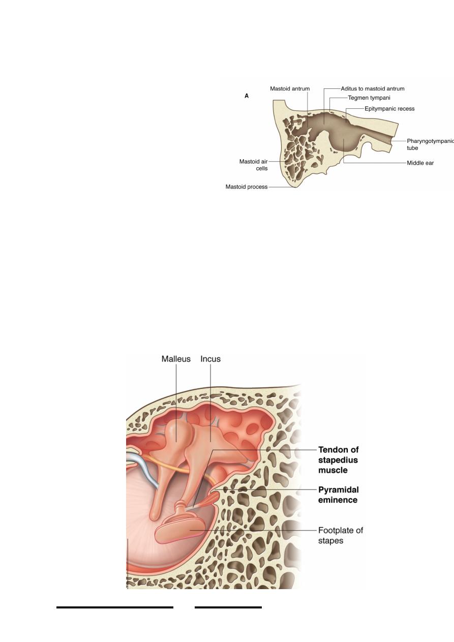
The mastoid air cells:
• These are small bony cavities communicating with each other located within
the mastoid process
• The first cell is the largest &
called mastoid antrum which lies
immediately behind the attic
with which it communicates
through the aditus ad antrum
• The size & number of mastoid
air cells vary considerably,
sometimes only few small cells
are present within the mastoid &
called sclerotic mastoid
• The mastoid process develop into a definite elevation only at the age of 2 years
• The lining mucosa is continuous with that of the tympanic cavity
The auditory ossicles:
• Three bones, the incus, malleus & stapes united by true synovial joints form a
lever system which convert the vibrations of the tympanic membrane into
mechanical energy represented by the pressure of the footplate of stapes on the
oval window
• The fixation of these bones in the tympanic cavity is provided by:
1. The attachment of the malleus handle to the eardrum
2. The attachment of the stapedial footplate to the oval window
3. The anterior & posterior ligaments of the bones
!
119
Head & Neck Dr. Nawfal K. Al-Hadithi
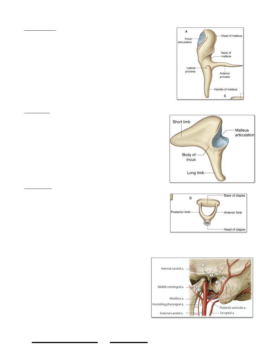
The malleus:
- The bone’s name is derived from its resemblance to a
hammer
- The rounded head of the bone lies in the epitympanic
recess
- The long handle is fused with the upper half of the
tympanic membrane
- The head shows a posterior oval concavity which
receives the incus in the incudo-mallear joint which is
of the saddle variety
- The short anterior process is connected to the petro-
tympanic fissure of the anterior wall by a ligament
The incus:
- The anterior part of the body of the incus has a
concavo-convex facet for articulation with the
mallear head
- The short process (posterior crus) extends
posteriorly to lie in the fossa incudis
- The long process (inferior crus) descends vertically
parallel to the handle of the malleus to end in a
rounded structure, the lenticular process which is
received by the head of stapes in the incudo-
stapedial joint which is of ball & socket variety
The stapes:
- The head of stapes is hollowed for reception of the
lenticular process of the incus
- The narrow neck receives posteriorly the insertion of
stapedius
- Two crura diverge from the neck to attach the footplate
- The footplate closes the oval window to which it is
attached by a ring like ligament
Blood supply of the middle ear:
The arterial supply:
- The main artery of the eardrum is the
anterior tympanic branch of maxillary
artery
- The main artery of the tympanic cavity,
mastoid antrum & mastoid air cells is the
stylomastoid branch of posterior auricular
branch of the ECA together with the
anterior tympanic branch of the maxillary
artery
- Smaller branches from the ascending
pharyngeal artery, middle meningeal artery,
artery of pterygoid canal share in the supply of the middle ear
!
120
Head & Neck Dr. Nawfal K. Al-Hadithi
The veins:
- Are parallel to arteries & drain to:
1. Superior petrosal sinus
2. Pterygoid plexus
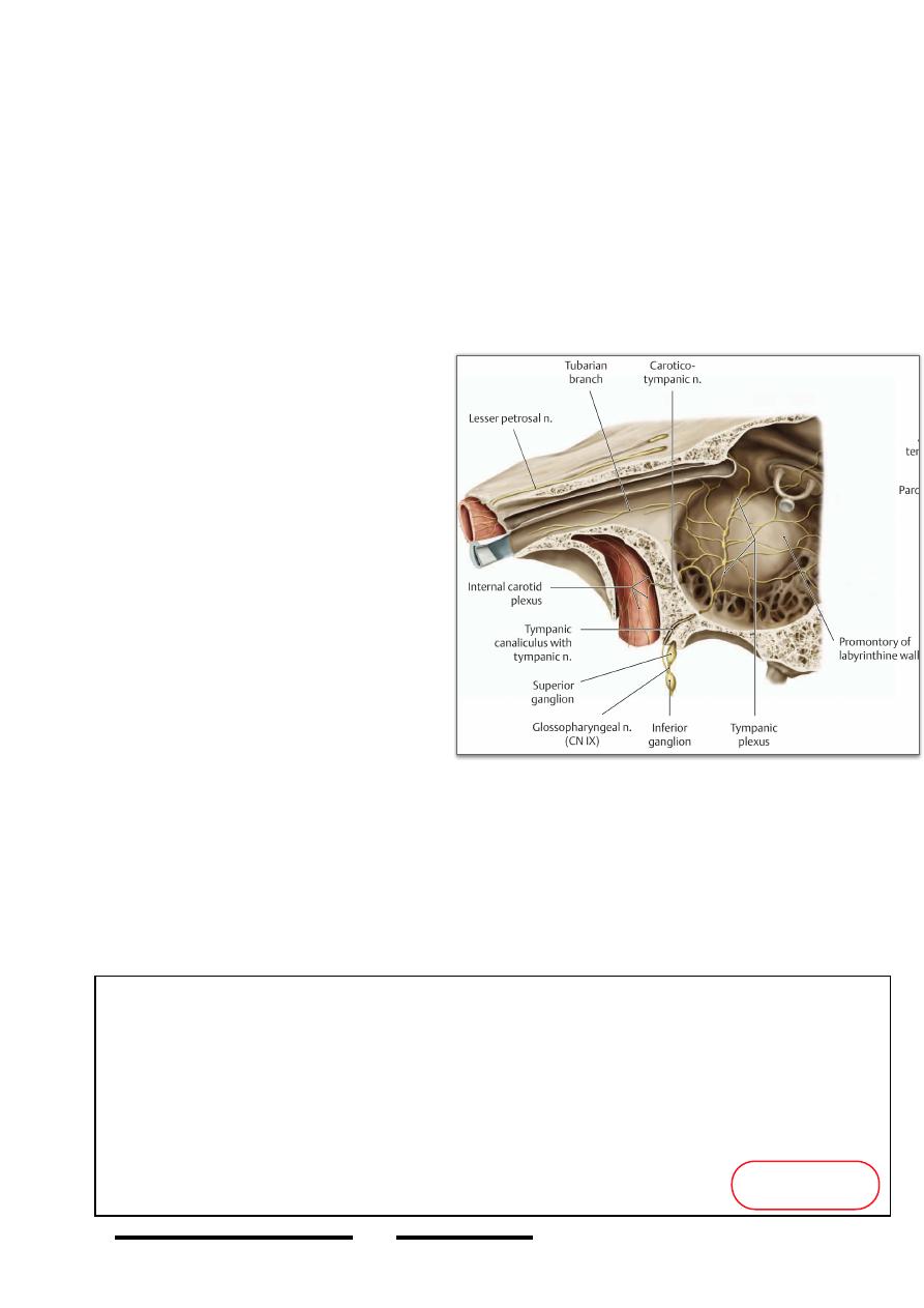
Nerve supply of the middle ear:
The tympanic cavity, the deep surface of the tympanic membrane & mastoid air cells
are supplied by the tympanic plexus.
Tympanic plexus:
An autonomic plexus formed at the promontory of the internal ear by contribution of:
1. The tympanic branch of glossopharyngeal nerve
2. Coraticotympanic branches of
the carotid sympathetic plexus
Nerves in the middle ear:
1. Facial nerve has part of its course
in the medial & posterior walls of
the tympanic cavity but this part
does not contribute to ear supply
2. Chorda tympani traverses the
tympanic cavity between its
bones but also give no branch to
the ear.
Applied anatomy:
• Communication between the
nasopharynx & middle ear results
in transmission of infections
from the nose & pharynx to the
ear so otitis media is one of the
c o m p l i c a t i o n s o f u p p e r
respiratory tract infection which complicates children infections more than
adults due to the shape of their tubes & the possibility of associated adenoids
• Communication between the middle ear & the mastoid air cells results in
transmission of infection from the middle ear to the mastoid resulting in acute
or chronic mastoiditis
• Facial nerve involvement may be associated with diseases of the middle ear
(LMND), Bell’s palsy
!
121
Head & Neck Dr. Nawfal K. Al-Hadithi
*The oblique position and concavity of eardrum is protective because it was vertical it will be more susceptible to damage
*The sound waves are transmitted across the footplate of the stapes to the perilymph so it moves a membrane covered by
nerve fibers leading to stimulation of the nerves and hearing
*Pars flaccida is more liable to damage than the rest
*When the footplate transmit the waves to the perilymph the excess pressure produced will be absorbed by secondary
tympanic membrane
*Geniculate nucleus lies at the angle of loop of facial nerve
*Chorda tympni at first run with facial n then it leaves it and pass through the cavity and leave it through the anterior wall
*Tendon of tensor tympani hooks ninety degrees from anterior wall to lateral wall of middle ear
*High intensity sound lead to reflex contraction of tensor tymapni and elevation of footplate of stapes by contraction of
sntapideus
*Tensor tympani protects middle ear
*Stapedius protects inner ear
!"#$.د ت'()*+