
The
Diencephalon
consists of the:
1-Thalamus,
2-Hypothalamus,
3-Subthalamus,
4-Epithalamus.
Prof. Dr. F. AL-Khafaji

Diencephalon & Basal Ganglia
•Learning objectives:
1- To become familiar with the
four
major anatomical
divisions of the diencephalon.
2- To understand the
main
structure, connections and
functions of the various parts of the diencephalon.
3- To become familiar with the
major
anatomical
divisions of the Basal ganglia.
4- To understand the
main
structure, connections and
functions of the various parts of the Corpus Striatum.
Prof. Dr. F. AL-Khafaji

Borders of the Diencephalon
Rostral
: plane through the optic chiasm and anterior
commissure.
Caudal
: plane through the posterior commissure and
the caudal edge of the mammillary bodies.
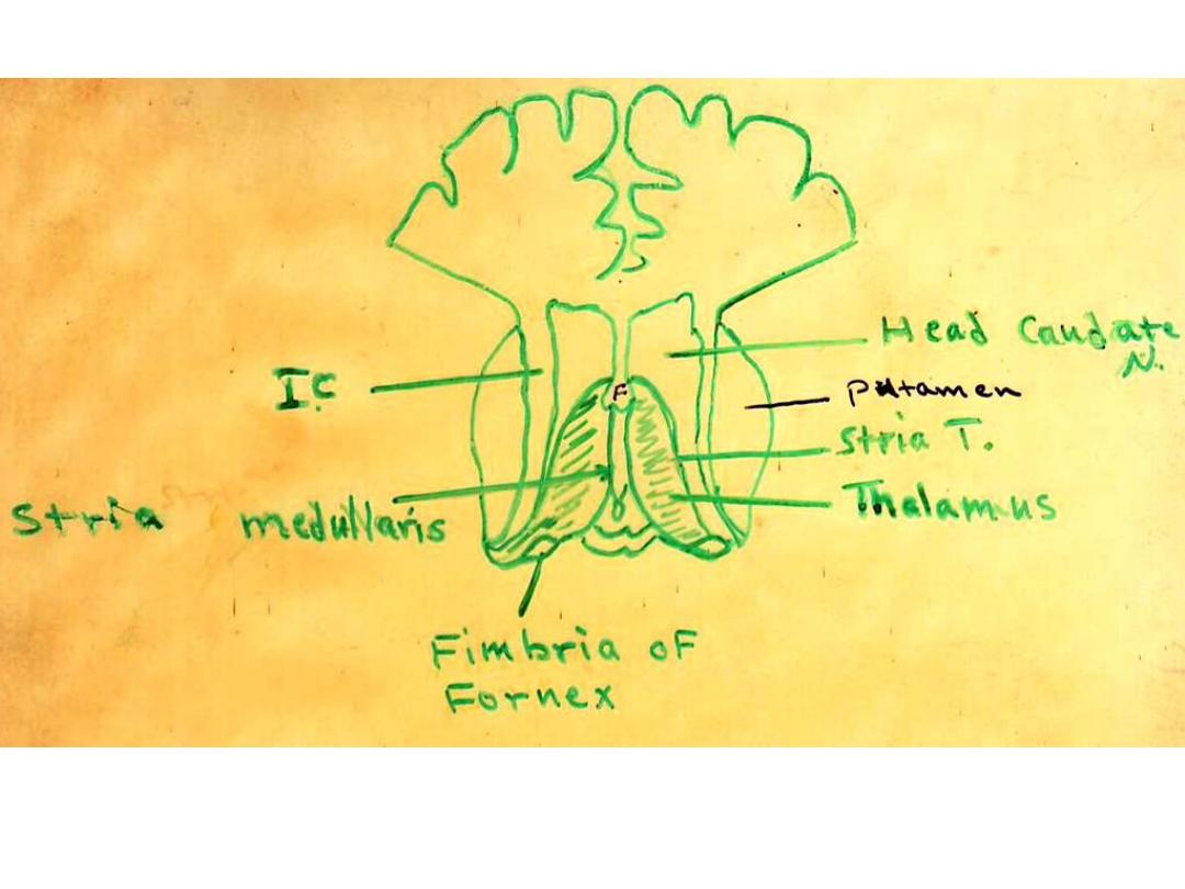
Medial
: wall of third ventricle, stria medullaris thalami
and massa intermedia.
Lateral
: the internal capsule, tail of caudate nucl. and stria
terminalis.
Dorsal
: the fornix and floor of the lateral ventricles.
Prof. Dr. F. AL-Khafaji

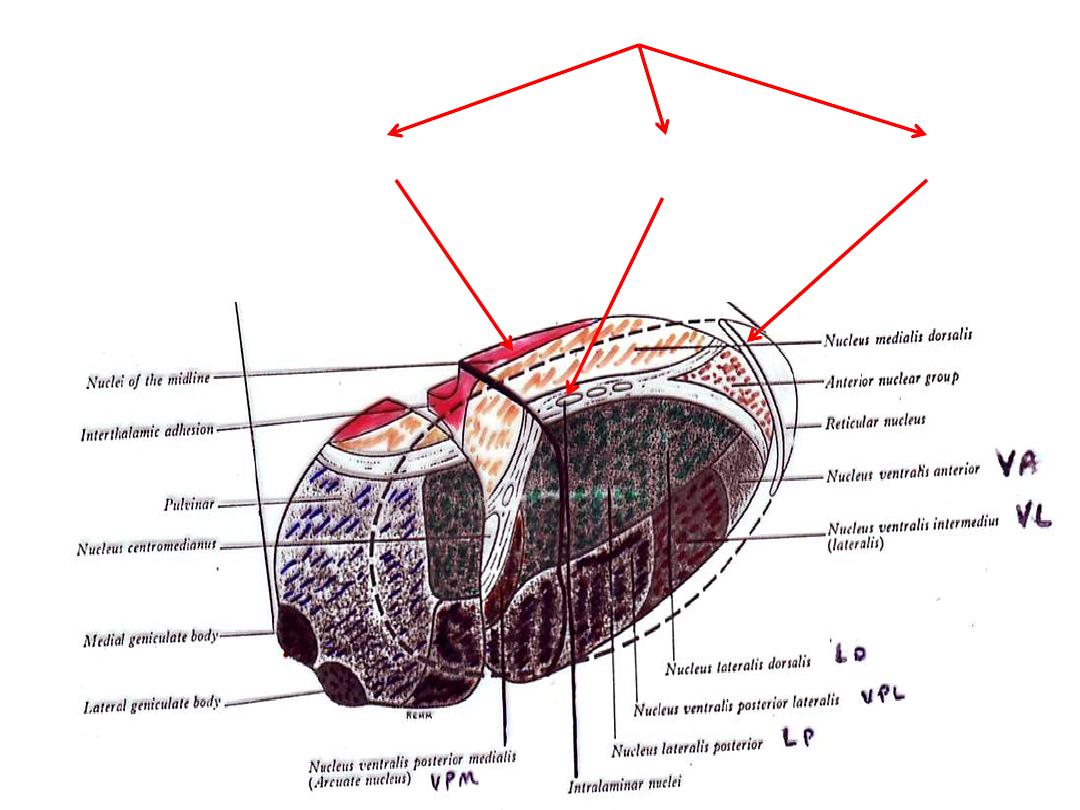
The Thalamus
•Large, egg-shaped (ovoid), 4 cm in length nuclear mass.
•It makes up about 80% of the mass of the diencephalon.
•Consists mainly of grey matter, but its sup. & lat. Surfaces are covered
by thin layers of white matter termed respectively the stratum zonale &
the external medullary lamina
•The grey matter is incompletely divided into anterior, medial &
ventrolateral nuclei by a Y-shaped lamina of white matter called the
internal medullary lamina
• Has
two
ends (Ant. & Post.) &
four
surfaces (Sup., Inf., Med. & Lat.).
•It extends:
1.Anteriorly
to the interventricular foramen;
2.Superiorly
to the transverse cerebral fissure (bet. Corpus callosum &
fornix)
3.Inferiorly
to the hypothalamic sulcus
4.Posteriorly
it overlaps the midbrain (Pulvinar).
Prof. Dr. F. AL-Khafaji

Ant. End:
Smaller than post. End
Lies behind interventricular foramen which connects the lat. Ventricle
& 3
rd
. Ventricles
Post. End:
Large & project backwards & laterally over sup. Colliculus of midbrain &
is called the pulvinar ( )وسادة
There are two small swellings on inf. Surface of pulvinar called the med.
& lat. Geniculate bodies.
Sup. Surface:
Not clearly demarcated from lat. Surface.
Stria medullaris thalami
mark the junction between the sup. & med.
Surfaces.
Separated from the ventricular surface of caudate nucl. By the
stria
terminalis & thalamostriate vein
Divided into two areas by an impression produced by the lat. Margin of
fornix
the lat. Area is coverd by ependema & forms part of the floor of
the body of lat. Ventricle( lamina affixa) the median area is coverd by
the tela choroidea of 3
rd
. Ventricle (double fold of pia mater)
Prof. Dr. F. AL-Khafaji

Inf. Surface:
Lies upon the subthalamic tegmental region (ie: hypothalamus,
subthalamus & midbrain (from before backwards)
Med. Surface:
Forms part of lat. Wall of 3
rd
. Ventricle
Separated from corresponding surface of opposite thalamus by a
narrow interval
The two thalami are connected by a short band called the interthalamic
adhession
Lat. Surface:
Separated from
lentiform nucl.
By post. Limb of
internal capsule
Many fibers stream out of this surface & enter internal capsule en route
for cerebral cortex (they Form the thalamic radiation, which form a
Stratum on lat. surface called the external medullary lamina
Prof. Dr. F. AL-Khafaji
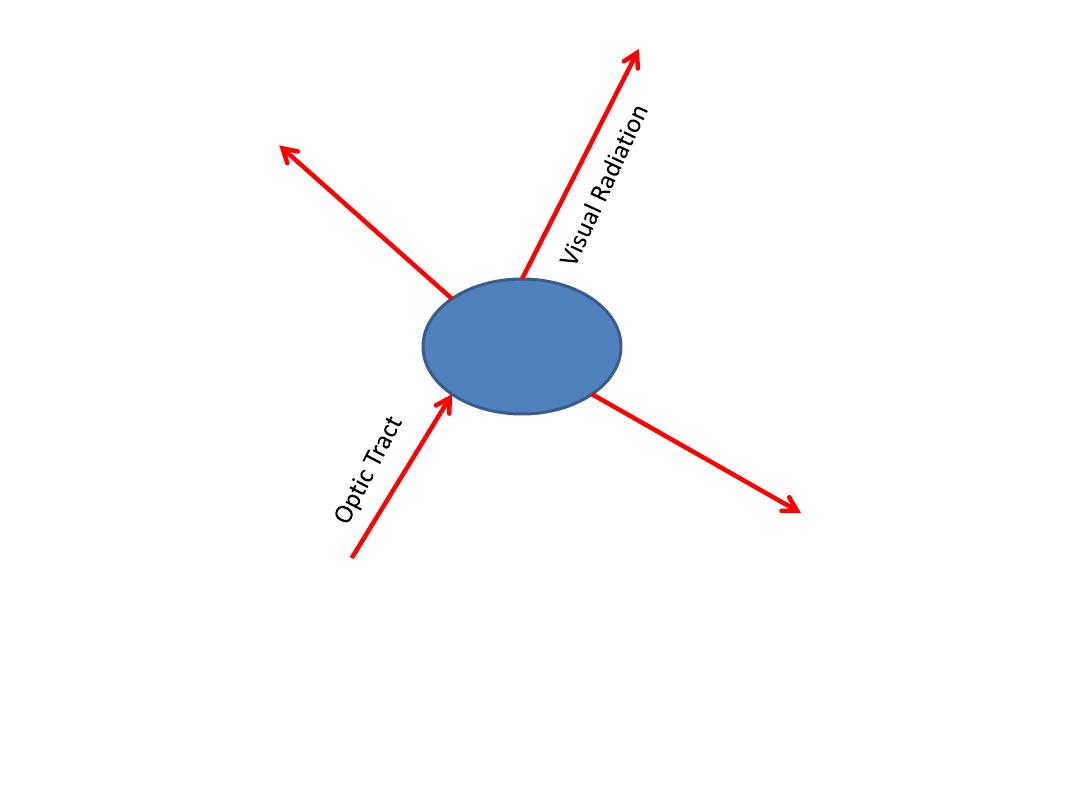
Prof. Dr. F. AL-Khafaji

Functions of Thalamus
1.Relay station
A. Most somatic sensory pathways except olfaction.
B. few motor pathways (eg. Cerebellar)
2.Integrating center
For impulses from many sources (eg. Somatic sensory, visual,
visceral, Some motor eg. Cerebellar, corpus straital)
3.Maintenance & regulation of state of:
consciousness, alertness, attention (through influence upon
cerebral cortex)
4.Emotional connotations
(
المحتوى العاطفي
)
(which accompany most sensory experiences)
5.Crude sensations
(eg. Pain which may reach consciousness at this level even
when all connections between thalamus
& cortex are
destroyed
).
Prof. Dr. F. AL-Khafaji
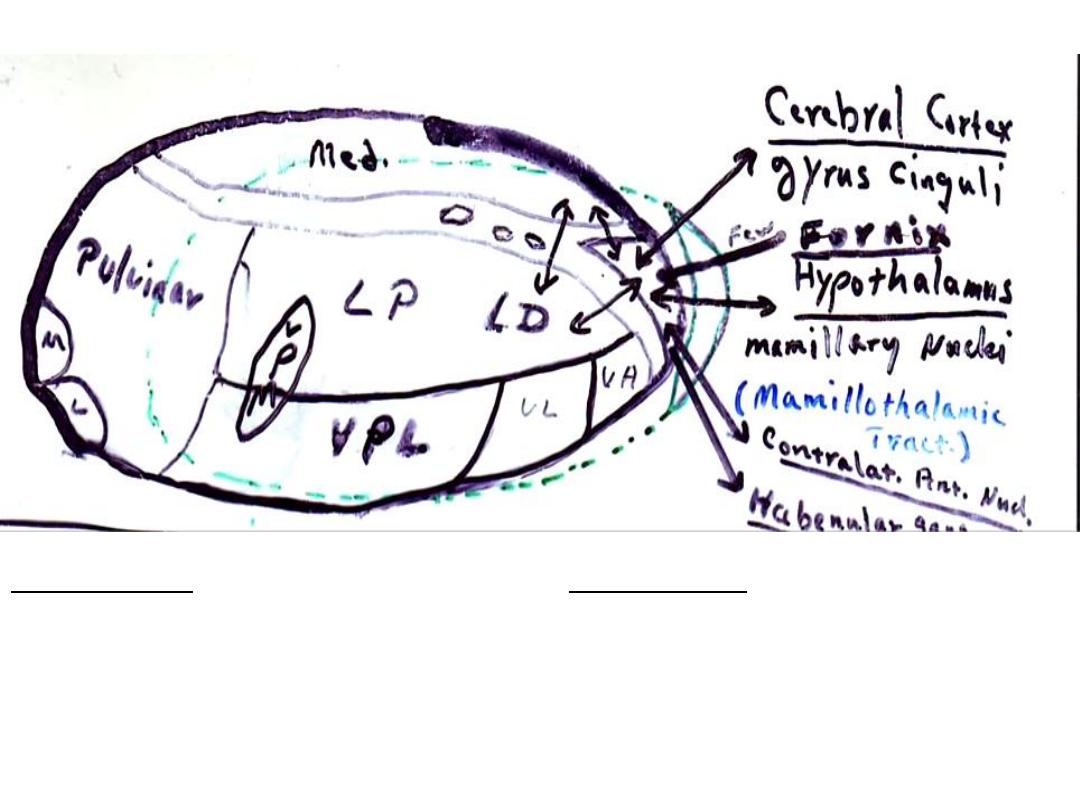
Connections anterior nucleus
Aff. Connections
1.Cerebral cortex (gyrus cinguli)
2.Hypothalamus (mammillary nuclei)
3.Contalateral ant. nucl.
4. Other thalamic nuclei
5.Fornix
Eff. Connections
1.Cerebral cortex (gyrus cinguli)
2.Hypothalamus (mammillary nuclei)
3.Contalateral ant. nucl.
4.Other thalamic nuclei
5.Habenular nuclei
Prof. Dr. F. AL-Khafaji
Ant. Nucl.
*Closely associated with the limbic system.
*Concerned with :
1-Emotional tone.
2-Mechanisms of recent memory.
*Stimulation or ablation of mammillothalamic tract causes:
1-Alteration in autonomic control.
2-Memory loss for recent events.
The Limbic Loop
Hippocampus Fornix Mammillary body Ant. Thalamic Nucl.
Parahipocample gyrus Cingulate gyrus Cingulum
-----------------------------------------------------------------------------------------------
NB.: Limbic system: centers that effect emotional & visceral aspects of
behavior & memory processing
Prof. Dr. F. AL-Khafaji

Limbic System (
ّوفيُحلا ُزاهِجلا
)
centers that effect emotional & visceral aspects of
behavior & memory processing .
I.
Structures with close olfactory connections
1.Paraterminal gyrus
2.Septal nuclei
3.Piriform cortex
4.Amygdaloid body
II.
Others
1.Hippocampus, fornix, hypothalamus,
cingulate gyrus, parahippocampal gyrus
Prof. Dr. F. AL-Khafaji
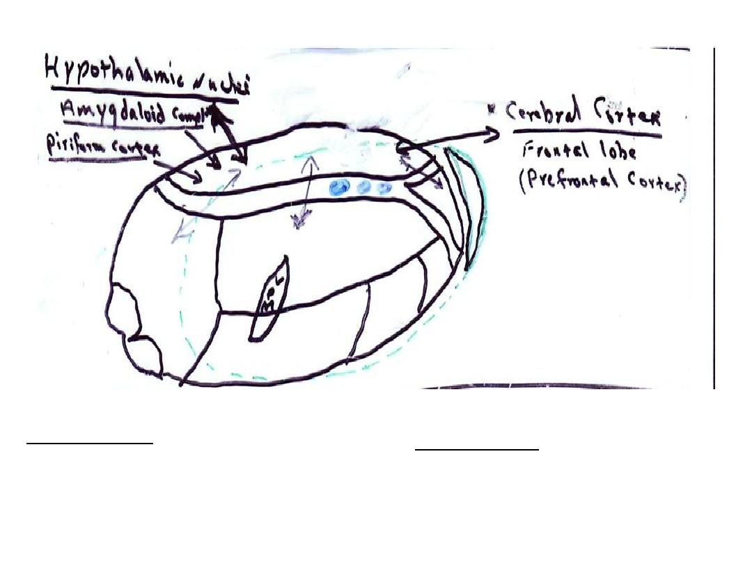
Connections medial nucleus (dorsomedial nucl.)
Aff. Connections
1.Cerebral cortex (prefrontal cortex)
2.Hypothalamus
3.Other thalamic nuclei
4.Amygdaloid complex
5.Piriform cortex
Eff. Connections
1.Cerebral cortex (prefrontal cortex)
2.Hypothalamus
3.Other thalamic nuclei
Prof. Dr. F. AL-Khafaji

Medial Nucl.
*
Provides mechanisms for the
integration
of certain
Somato-visceral impulses projecting to prefrontal cortex.
*
Mediate impulses of an
affective nature
which
contributes to the formation of personality.
*
Stimulation, disease, or surgical ablation of nucl. in man
results in
changes
in:
1- Motivational drive.
2- Ability to solve problems.
3- Consciousness level.
4- General personality.
5- Subjective feeling status (affective tone).
6- Pain perception (indifference to pain).
7- Emotional content.
Prof. Dr. F. AL-Khafaji

Types of thalamic nuclei
R
= Relay nucl.
A
= Association nucl.
SC
= Subcortical projections
DC
= Diffuse cortical projections
Prof. Dr. F. AL-Khafaji
Prof. Dr. F. AL-Khafaji

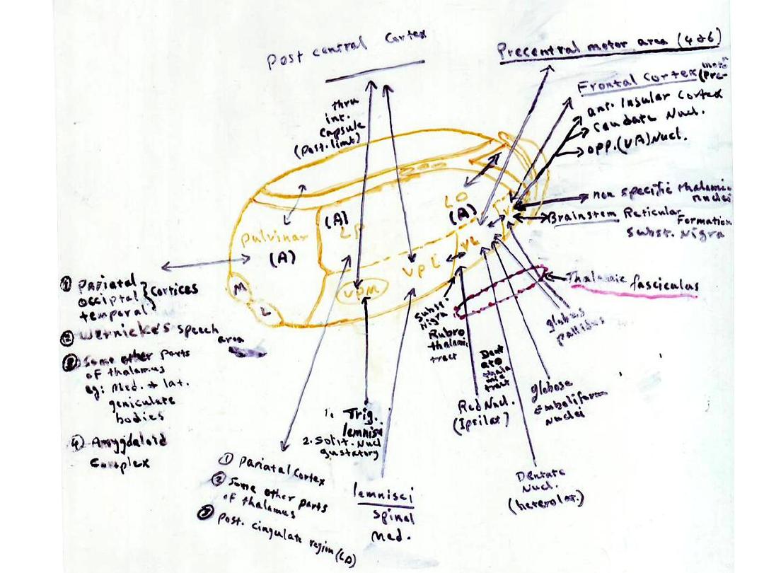
Nucleus Ventralis Lateralis
*Related to
motor control system
.
* Makes important contribution to initiation of movements,
control of muscle tone, regulation of cortical reflexus.
* The
cerebello-Thalamo-Cortical relay system
may play an
important role in unconscious regulation of muscle tone.
(interruption of this system , at thalamic level, may be
responsible for the reduction in muscle tone following
stereotaxic surgery in Parkinsonism.
* Lesions in (VL) ameliorate some aspects of dyskinesia (due
to both cerebellar or corpus striatal lesions) by reducing
output from (VL) to motor cortex.
Prof. Dr. F. AL-Khafaji

Nucleus Ventralis Anterior
*
Related to
motor control system
& therefore affects
activities of motor cortex.
*
Regarded as important link in the final stages of the
ascending activating system (arousal reaction).
*
Stimulation increases Parkinsonian rigidity &
tremor.
*
Abalation reduces or abolish this tremor.
Prof. Dr. F. AL-Khafaji
VPI
Nucleus Ventralis
Intermedius
(R)
Vestibular Nerve
Postcentral Gyrus
(caudal portion close to junction of
Bradman's area 2,5)
(Primary vestibular area for conscious
vestibular perception)
Prof. Dr. F. AL-Khafaji
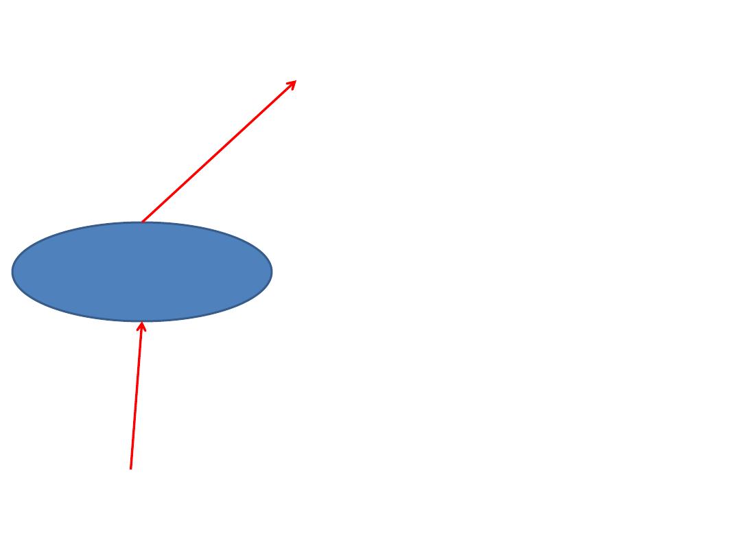
MGB
Lower auditory
center
Lat. Lemniscus
Inf. Colliculus
1-Inf. Colliculus
2-Brainstem auditory relay nuclei
Sup. Temporal gyrus (41)
(Transv. gyrus of Heschl)
1-Ventrolat. Nuclei of thalamus
2-Pulvinar
P.N: Asc. Fibs. Are paralled by desc. Fibs. Which may serve as regulatory feed-back mechanism.
Prof. Dr. F. AL-Khafaji
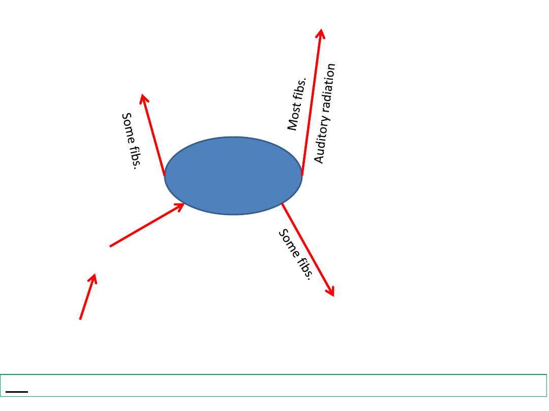
LGB
Lower visual
centre
Ganglion cells of retina
1-Ventrolat. Nuclei of thalamus
2-Pulvinar
Visual Cortex (Striate, 17)
1-Pretectum
2-Sup. Colliculus
3-Suprachiasmatic Nucleus
4-Zona Incerta
P.N.: LGB is the main end station of optic tract.
Prof. Dr. F. AL-Khafaji
None specific thalamic nuclei
Nuclei of midline
Intralaminar nuclei
Reticular nucl.

(SC)
Reticular Nucleus
*
Important final link in the diffuse Thalamo-Cortical radiation.
*
Produces wide spread effects on cortical activity (cortical
arousal).
*
Integrates intra-thalamic activities.
Afferents:
1- Cerebral Cortex.
2-Other thalamic nuclei.
3-Brainstem reticular formation.
4-Globus pallidus.
Efferents:
1-Other thalamic nuclei.
2-Mid-brain reticular formation.
3-Corpus striatum.
Prof. Dr. F. AL-Khafaji

(SC, DC)
Centromedians
Nucleus
*Intra-thalamic integrating mechanism bringing the activities of various
other thalamic nuclei into functional relation with each other.
Afferents
1-Asc. Reticular formation.
2-Other thalamic nuclei.
3-Globus pallidus.
4-Spinal,medial & trigeminal lemnisci (collaterals).
5-Sup. Cerebellar peduncle (contribution)
Efferents
1-Cerebral cortex (not directly) via collaterals to corpus straitum.
2-Corpus striatum.
3- Other none-specific thalamic nuclei.
4-VA nucleus of thalamus
Nuclei of the mid-line
*Lie in periventricular grey matter of dorsal 1/2 of ventricular wall &
inter-thalamic adhesion.
*Poorly developed in man (but well developed in many mammals).
*Concerned with visceral functions & connected to hypothalamus
Prof. Dr. F. AL-Khafaji

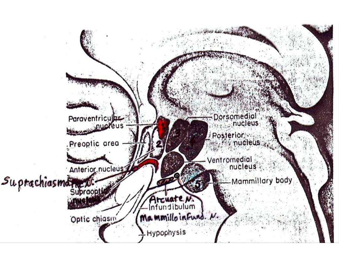
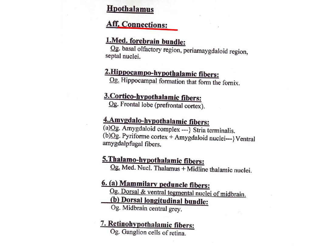
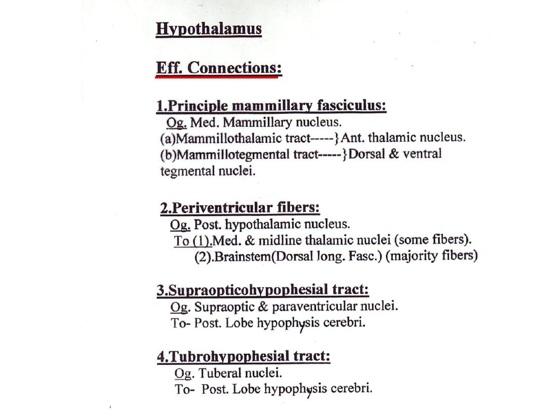
Hypothalamic Nuclei
Zone Nuclei
1.Supraoptic (Ant.) 1.Supraoptic nucl.
2.Paraventricular nucl.
3.Suprachiasmatic nucl..
2.Infundibluotubular (middle) 1.Dorsomedial nucl.
2.Ventromedial nucl.
3.Arcuate nucl.(
Infundibular
)
3.Mammillary (caudal) 1.Posterior nucl.
2.Mammillary nucl.
3.Mammilloinfundibular nucl.
Prof. Dr. F. AL-Khafaji
Prof. Dr. F. AL-Khafaji
Functions of Hypothalamus
1.Endocrine control:
*Release factors
Anterior pituitary
*Release inhibiting factors
2.Neurosecrtion:
*Vasopressin (supraoptic nucleus)
Posterior pituitary
*Oxytocin (paraventricular nucleus)
3.Autonomic control:
Higher center for control of lower autonomic centers in brainstem & spinal cord
*Ant. Region
Influence parasypathetic activity
*Preoptic region
*Post. Region
Influence sympathetic activity
*Lat. Region
4.Tempereture regulation:
*Ant. Region controls dissipation of heat
*Post. Region controls conservation of heat
Prof. Dr. F. AL-Khafaji

5.Food intake regulation:
*Lat. Region (Hunger centre) initiates eating & increase food intake
*Med. Region (Satiety centre) inhibits eating & reduces food intake
6.Water intake & balance:
*Lat. Region (Thirst center) increase water intake
PN:
Vasopressin effect on distal convoluted tubules & collecting
tubules of kidney
7.Emotion & behavior:
Function of hypothalamus, limbic system & prefrontal cortex
It generates behaviors involved in rage, aggression, escape, etc.
8.Circadian rhythms
(daily rhythm of a biological function)
eg.:
Body temperature, Adrenocortical activity,
Sleep & wakefulness (ant. Region)
PN:
Suprachiasmatic nucleus (aff. From retina) play a role in control of
circadian rhythms (variation in intensity of light are transmitted by this
nucl. To many hypothalamic nuclei.
Prof. Dr. F. AL-Khafaji
Prof. Dr. F. AL-Khafaji
Prof. Dr. F. AL-Khafaji
Prof. Dr. F. AL-Khafaji
Prof. Dr. F. AL-Khafaji
Prof. Dr. F. AL-Khafaji

Epithalamus
*
Composed of :
1.Pineal body.
2.. Habenula
نانِعلا
(
بنية تشريحية تشبه لجام الفرس
[ )
ج
:
أعنة
]
3.Posterior & Habenular commissures
*
Most dorsal, smallest, and oldest part of Diencephalon
*
Functionally and anatomically linked to the limbic system
*
It is implicated in:
1.
Autonomic
functions (e.g. respiratory, cardiovascular….etc.)
2.
Endocrine
functions (e.g. Thyroid functions)
3.
Reproductive
functions ( e.g. mating behavior)
Prof. Dr. F. AL-Khafaji

Habenular Nucleus
*
Center for integration of olfactory, visceral & somatic afferent
pathways (correlation of olfactory & somatic afferent impulses)
*
Ablation of this nucl. Produce changes in metabolism, endocrine
regulation & thermoregulation
Aff. Fibs.:
1. Amygdaloid complex (via. Stria terminalis)
2. Hippocampal formation (via. Fornix)
3. Olfactory tubercle
4. Pre-optic & septal areas (via. Stia medullaris thalami)
5. Ant. Perforated substance
6. Various hypothalamic Nuclei
7. Globus Pallidus
Eff. Fibs.:
1. Interpeduncular nucl. (via. FR)
2. Medial nucl. Of thalamus
3. Tectum & reticular formation of mid-brain
Prof. Dr. F. AL-Khafaji

PN.:The Stria Medullaris Thalami, Habenula, & Fasciculus
Retroflexes form segments of Visceral Eff. Pathways which
carry impulses to parts of brainstem & spinal cord
(eg. Tectotegmentospinal Tracts & Dosal Longitudenal
Fasciculus which connect with autonomic preganglionic
centers
Prof. Dr. F. AL-Khafaji
Nucleus Subthalamicus
Aff. Fibs.:
1.Globus Pallidus
2.Motor cortex (precentral)
3.Pedunclopontine Nucl.
Eff. Fibs.:
1.Globus Pallidus
2.Contrlateral Globus pallidus
3.Substantia Nigra
4.Opposite Subthalamic nucl.
Zona Incerta
Aff. Fibs.:
1.Motor cortex (precentral )
Eff. Fibs.:
1.Mid-brain reticular formation
Nucleus Subthalamicus
*Lies medial to internal capsule
*Continuous with substantia nigra
Function
Important site for integration of a
number of motor control centers
Especially through its connections with
the corpus striatum & midbrain
Tegmentum
Lesions of one subthalamic nucl.
Results in condition called Hemiballismus
(Subthalamic Dyskinesia)
Zona Incerta
*Thin strip of gray matter
*Lies between thalamic & lenticular fasciculei
*Continuous with thalamic reticular nucl.
Functionally associated with the Zona Incerta are
the Nucl. of prerubral field & Entopeduncular nucl.
Prof. Dr. F. AL-Khafaji
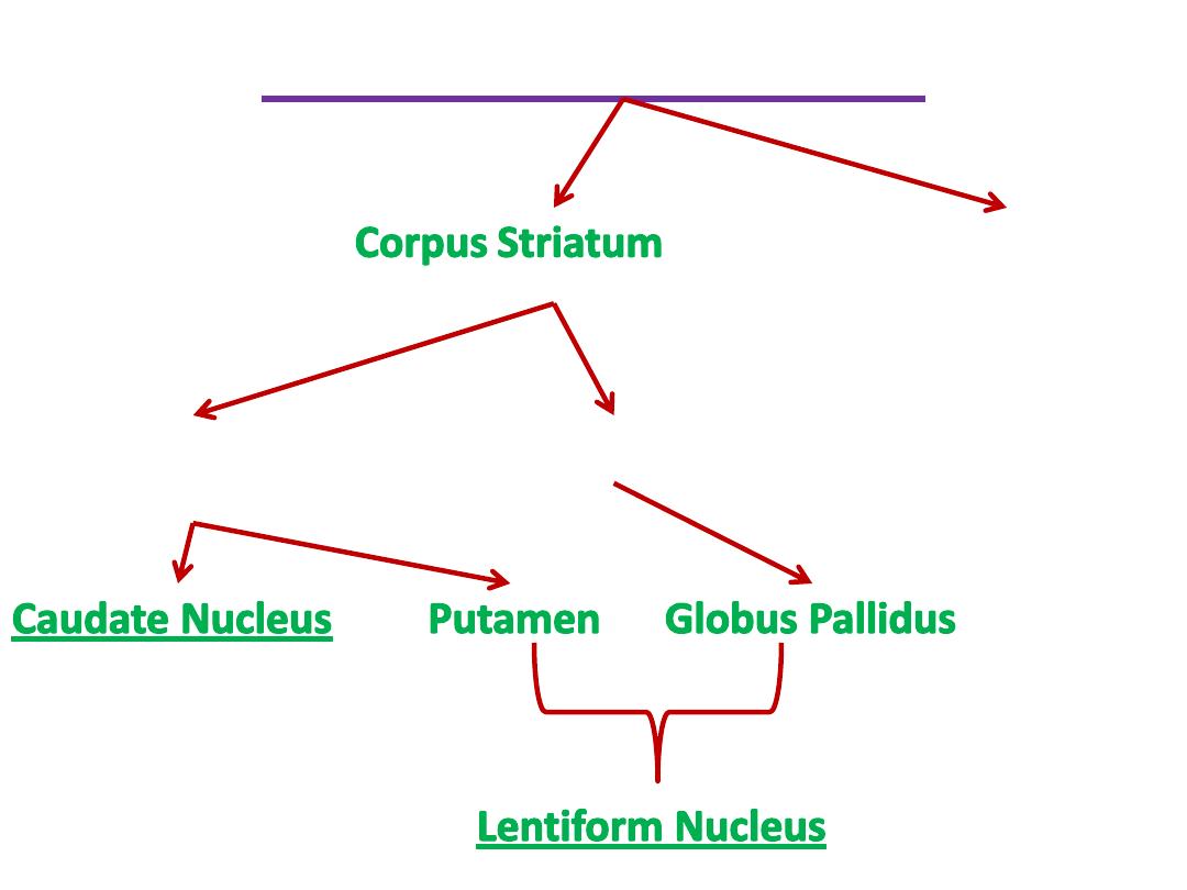
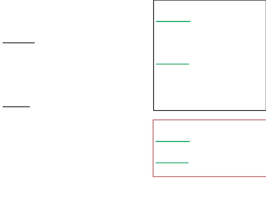
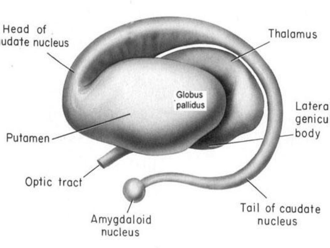
Basal Ganglia (Nuclei)
(Concerned with somatic motor function)
Amygdaloid nuclear
complex
(Archistriatum) (oldest)
(Component of Limbic system)
Ie. visceral, endocrine, behavioral
Neostriatum
(Striatum)
Paleostiatum
Prof. Dr. F. AL-Khafaji

Caudate Nucleus
*
C-shped structure, related to lateral ventricle.
*
The
head
lies rostral to thalamus & protrude into ant. Horn of lat. Vent.
*
The
body
is slender & elongated & arches along dorsolateral border of
thalamus (lat. To stria terminalis)
*
The
tail
lies caudal to thalamus in roof of inferior horn of lat. Vent.
(in relation to amygdaloid nuclear complex)
Putamen
*
Largest & most lateral part of corpus striatum
*
Medial to external capsule
*
Traversed by striopallidal fibers
*
Rostrally & ventrally is continuous with head of caudate nucl.
*
But in more dorsal regions, is connected to caudate nucl. by strial
bridges (passing between fibers of internal capsule)
Prof. Dr. F. AL-Khafaji

Globus Pallidus
*
Phylogentically older than striatum
*
Smaller inner part of lentiform nucl.
*
Lies med. To putamen
*
Dorsomedial margin borders the post. Limb of internal
capsule
*
Has a thin
lat. Medullary Lamina
(between it & putamen)
*
Has
Med. Medullary Lamina
which divide globus pallidus
into med. & lat. Segments
*
Has less distinct
Accessory Medullary Lamina
which divide
med. segment into inner & outer portions
Prof. Dr. F. AL-Khafaji
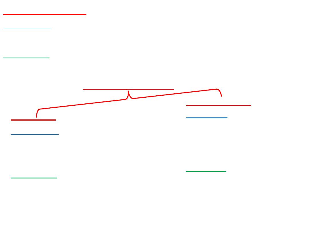
Caudate Nucleus
Afferents:
1- Cerebral Cortex
Efferents:
1- Globus Pallidus (mainly)
Putamen
Afferents:
1- Cerebral Cortex
2- Caudate Nucl.
Efferents:
1- Globus Pallidus
Globus Pallidus
Afferents:
1- Caudate Nucl.
2- Putamen
3- Subustantia Nigra
4- Subthalamic Nucl.
Efferents:
1- Thalamus (VL,VA,Centomedian)
2- Subthalamus
3- Substantia Nigra
4- Hypothalamus
5- Reticular formation
6- Habenular nuclei
Lentiform Nucleus
Prof. Dr. F. AL-Khafaji
Diseases of corpus striatum
Various types of abnormal involuntary
movements (
Dyskinesia ةَك
َ رَحلا ُلَل َخ
) eg.
Tremor, athetosis, chorea, ballism
Disturbances of muscle tone
(usually increased muscle tone)
eg. Rigidity of Parkinson disease
PN:
Cerebral cortex must play an important role in dyskinesia
Evidence:
1- All forms of abnormal involuntary movements cease during sleep
2- Abolished by general anesthesia
3- Exaggerated by excitement & anxiety
4- Ablation of motor cortex & interruption of corticospinal tract at
various levels abolishes dyskinesia (may suggests that impulses from
centers responsible for dyskinesia must be transmitted to segmental
levels via the corticospinal tract
Prof. Dr. F. AL-Khafaji