1
Fifth stage
ENT
Lec-14
د.سعد
24/3/2016
The Ear
Anatomy and physiology
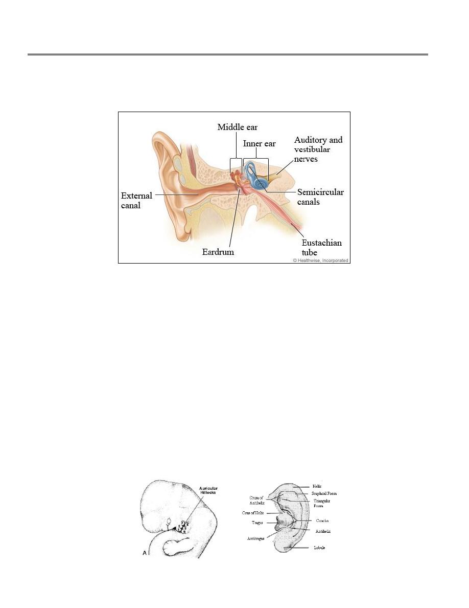
The ear can be divided anatomically and clinically into three parts – The external ear, the
middle ear and the inner ear. The external and middle ear are concerned particularly with
transmission of sound. The inner ear functions both as the organ of hearing and as part of the
balance system of the body.
The External Ear
consists of;
1. The auricle (pinna).
2. The external canal (external auditory meatus).
The Auricle;
Is developed from 6 tubercles (Hillocks) that originate from 1
st
and 2
nd
brancheal
arches, and is fully formed by the 12
th
week of fetal life.. The underlying skeleton of the auricle
consists of yellow elastic cartilage, except for the lobule which is composed only of fat and
fibroalveolar tissue. The skin on the lateral surface is closely adherent to the perichondrium. The
auricle is attached to the side of the head by ligaments and functionless muscles.

2
The external auditory meatus;
In adults it measure about 24mm from the introitus to the tympanic membrane, the outer third
is cartilaginous and the inner two thirds are bony. The sebaceous glands, hair follicles and
ceruminous glands (which secrete wax) present only in the cartilaginous portion
.
(The
epithelium lining the bony canal is thinner and devoid of these structures).
Owing to the tight union of the cartilage and skin any inflammatory process (such as boil in the
cartilaginous meatus) can be extremely painful.
In the adult there is an angle in the meatus: the outer part runs upwards and backwards as it
passes inwards; the inner part runs more horizontally. Accordingly the pinna must be pulled
upwards, backwards and outwards when using a speculum to examine the ear drum. In children,
on the other hand, the meatus is shorter and straighter and the drum can often be examined
without this maneuver.
The auricle and external meatus are supplied by branches of the 5
th
, 9
th
and 10
th
cranial nerves.
The medial surface of the auricle is supplied by fibers of the great auricular nerve (C2 and C3)
and the lesser occipital nerve (C2).
Relations of external auditory meatus;
1. Anteriorly; temporomandibular joint.
2. Posteriorly; mastoid antrum and mastoid air cells.
3. Superiorly; the middle cranial fossa.
4. Inferiorly and anteriorly; the parotid gland.
The tympanic membrane (drumhead or eardrum); separate the external meatus from the
middle ear and consist of three layers: an outer epithelial layer continuous with that of external
auditory meatus, a middle layer of fibrous tissue and an inner layer of mucous membrane
continuous with the lining of the tympanic cavity. It has elliptic, funnel shape, about 8mm wide,
9-10 mm high and about 0.1 mm thick. The ear drum is supported around its periphery by a
fibrous thickening, the annulus.
The tympanic membrane is divided into two parts:
1. The pars tensa; represent the lower portion of the tympanic membrane.
3
2. The pars flaccida (Shrapnell`s membrane); this part of the drum is small and comprises
the upper most part . The fibrous tissue layer is deficient in this area, hence the flaccidity.
It is frequently referred to as the attic. Perforations in this area are potentially unsafe.
Pars tensa
Pars flaccida
Lower portion of the tympanic
membrane
Upper portion
Larger (⅔)
Smaller (⅓)
Thicker
Thinner
In the middle layer there is fibrous
tissue.
Deficient of fibrous tissue.
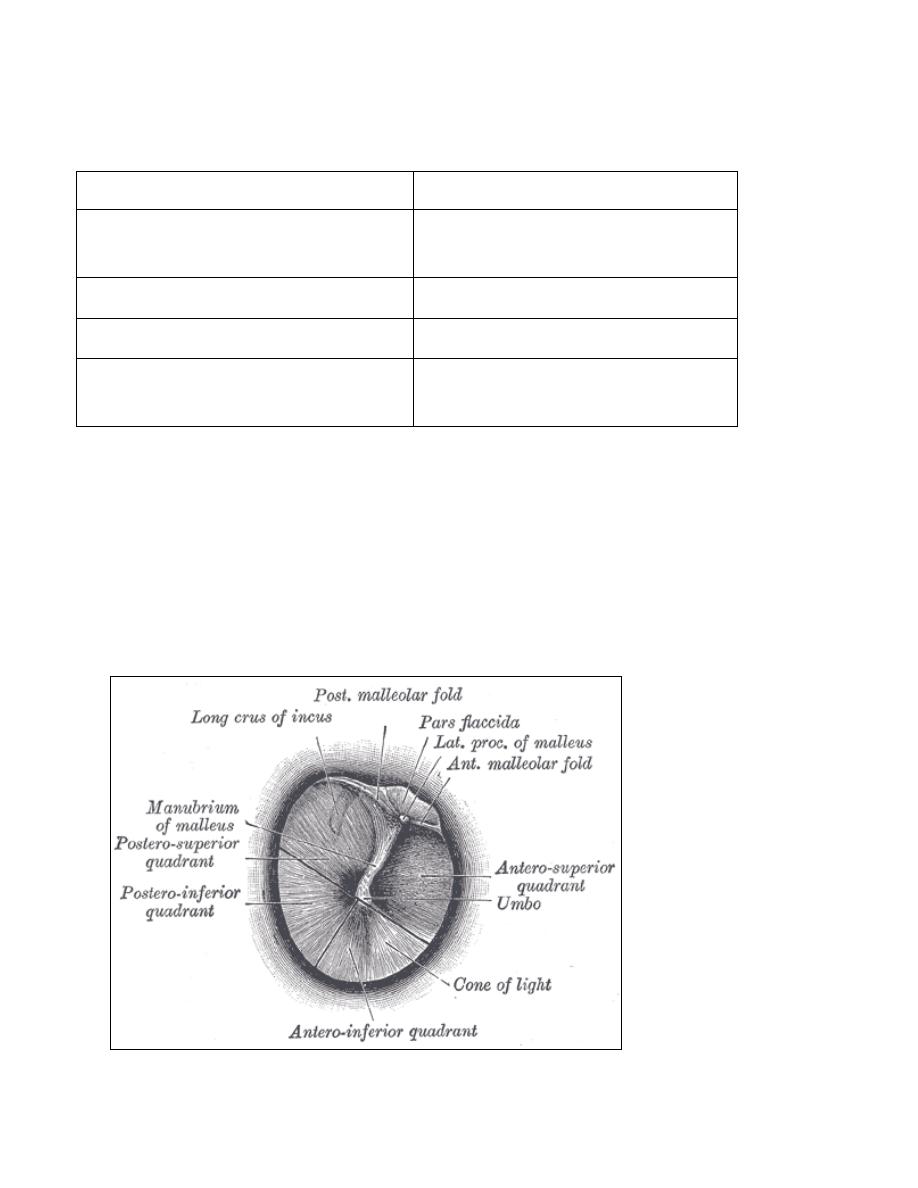
Landmarks of the eardrum;
1. The handle of malleus, running down to the approximate center, the umbo.
2. The lateral process of the malleus.
3. The anterior and posterior malleolar folds, separate pars tensa from pars flaccida.
4. It has a pearly grey colour with a triangular bright area, the cone of light, extending
downwards and forwards from the umbo.
4
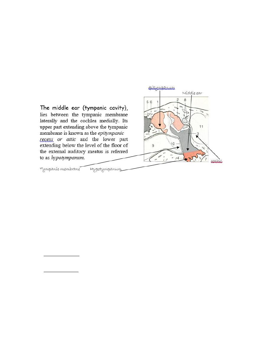
The Middle Ear
The
middle ear cleft in the temporal bone (which develops from the Eustachian tube
early in life) includes;
1. The middle ear (tympanic cavity).
2. The Eustachian tube ( opens anteriorly into the nasopharynx)
3. The aditus which leads posteriorly to the mastoid antrum and air cells.
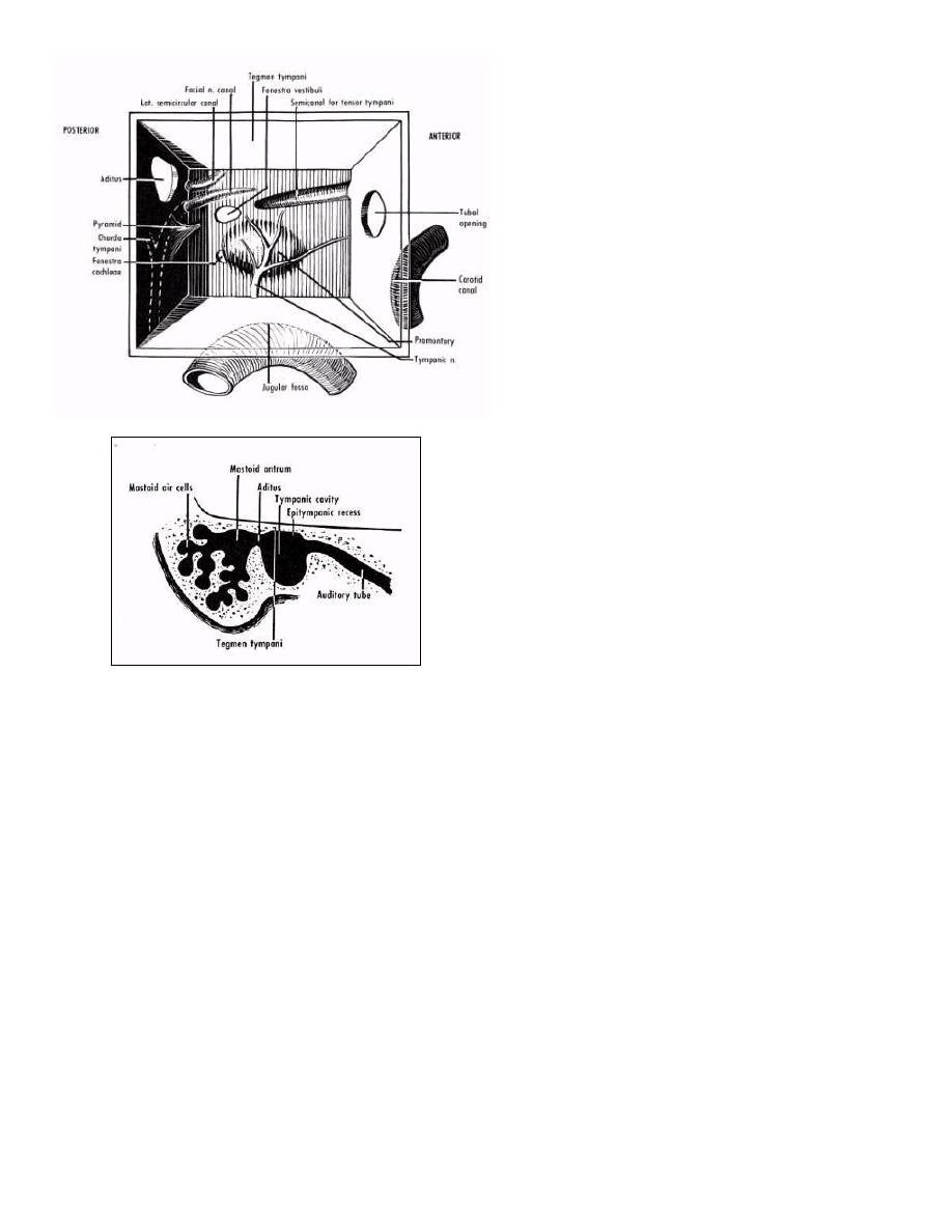
This cavity may be described as six-sided box frequently linked in shape to a match box.
Roof; is formed by a thin plate of bone (tegmen tympani). This plate separates the tympanic
cavity and mastoid antrum from the middle cranial fossa. Injury of tegmen tympani leads to CSF
leak.
Floor; a thin plate of bone separates the cavity from the bulb of internal jugular vein.
Anterior wall:
The lower portion is formed by a thin plate of bone separating the cavity from the
internal carotid artery.
The upper portion has two openings, the lower one being the Eustachian tube and above
it lies the
canal for tensor tympani muscle.
5
Posterior wall;
High up in the posterior wall there is
an opening (the aditus), which leads
to the mastoid antrum.
Just below the entrance to the aditus
is a small pyramid of bone which
contains the stapedius muscle.
Medial wall;
The promontory: is round bony swelling covering the basal turn of the cochlea.
Facial nerve: which run approximately horizontally superior to the promontory.
The round window niche: is a deep recess posteroinferior to the promontory. The round
window itself is closed by a thin membrane, deep to which is the scala tympani of the
cochlea.
Oval window: This is occupied by the footplate of the stapes bone and leads into the scala
vestibuli of the cochlea. It lies posterosuperior to the promontory.
Lateral wall; is mostly formed by the tympanic membrane,
with the lateral bony walls of the
epitympanum above and hypotympanum below
.
6
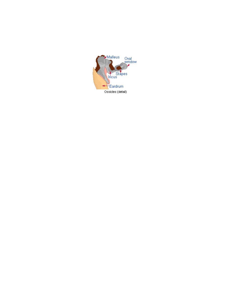
Contents of middle ear;
1. Air.
2. Ossicles; Malleus, incus and stapes. The handle of malleus is firmly embedded in the middle
layer of the tympanic membrane and its head articulates with the body of the incus. The
long process of the incus articulates with the stapes, which in turn occupies the oval
window through its foot plate.
3. Muscles; Tensor tympani and stapedius. The latter comes into action reflexly in response
to loud noise to protect the ear against damage.
4. Nerves;
a) Chorda tympani; arise from the facial nerve in the Fallopian canal.
b) Tympanic plexus; Lies on the promontory. It is formed by the tympanic branch of the 9
th
cranial nerve (Jacobson) and branches from the sympathetic plexus. This plexus supplies
the lining mucosa of the Eustachian tube, tympanic cavity and mastoid air cells.
The Eustachian tube; Connects the tympanic cavity with the nasopharynx and in adult is
about 36mm in length. In infant the tube is shorter and wider and its course is more horizontal
than in adults. It is lined by columnar ciliated epithelium. The tube has 2 parts:
1) Pharyngeal cartilaginous part (2/3 of its length)
2) Tympanic bony portion.
On swallowing or yawning, the tube is opened by contraction of tensor palati muscle which is
attached to the lateral wall of the tube and thus equality of air pressure is maintained on both
sides of the tympanic membrane
The Inner Ear:
The inner ear, or labyrinth, consists of a bony capsule (otic capsule) that is almost embedded in
the petrus bone. Within this capsule is the membranous labyrinth. The membranous labyrinth
contains fluid known as endolymph which is similar to the intracellular fluid (has high potassium
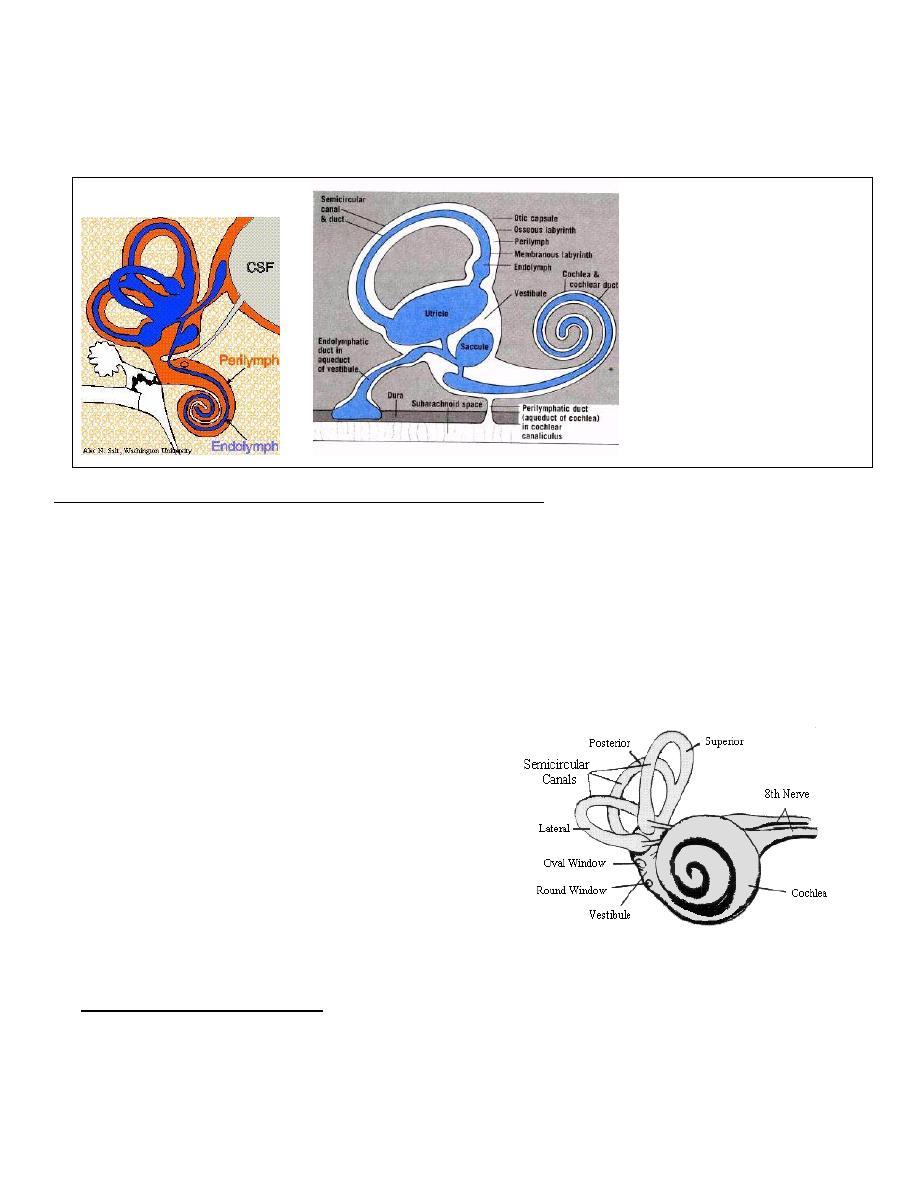
7
and low sodium concentration). The space within the bony labyrinth, between its wall and the
membranous labyrinth contains another fluid known as perilymph. The composition of the
perilymph is very similar to the extra cellular fluid and CSF (has low potassium and high sodium
concentration).
The Otic Capsule (bony labyrinth) consists of three parts;
1. The cochlea, a snail-like structure lies
anteriorly. It wounds two and a half
times round its central axis or
modiolus.
2. The vestibule, in the middle
3. Three semicircular canals, posteriorly.
The Membranous Labyrinth consists of;
1.
The membranous cochlear duct or
scala media;
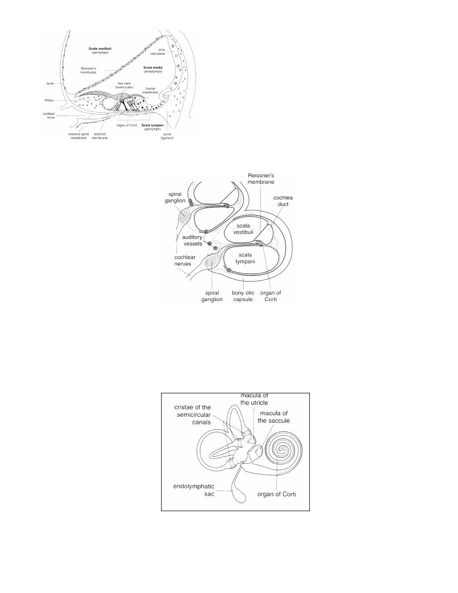
8
This is a blind tube, triangular in
section, situated in the bony cochlea. The
floor of this tube is formed by the basilar
membrane. On it lies a neuroepithelium
called the organ of Corti, which is the
sense organ of hearing. It consists of a
complex arrangement of supporting and
hair cells. The terminal fibers of the
cochlear division of the 8
th
cranial nerve
end in contact with these hair cells.
2.
Saccule and utricle; lie in the bony vestibule. They are stimulated by linear
acceleration. They are joined by Y – shaped endolymphatic duct which extends to the
endolymphatic sac. The sac is probably concerned with absorption of endolymph.
9
3.
The membranous semicircular ducts; occupy , but not filing the lumens of the
corresponding bony canals and are set at right angles to each other. They are stimulated by
angular acceleration.
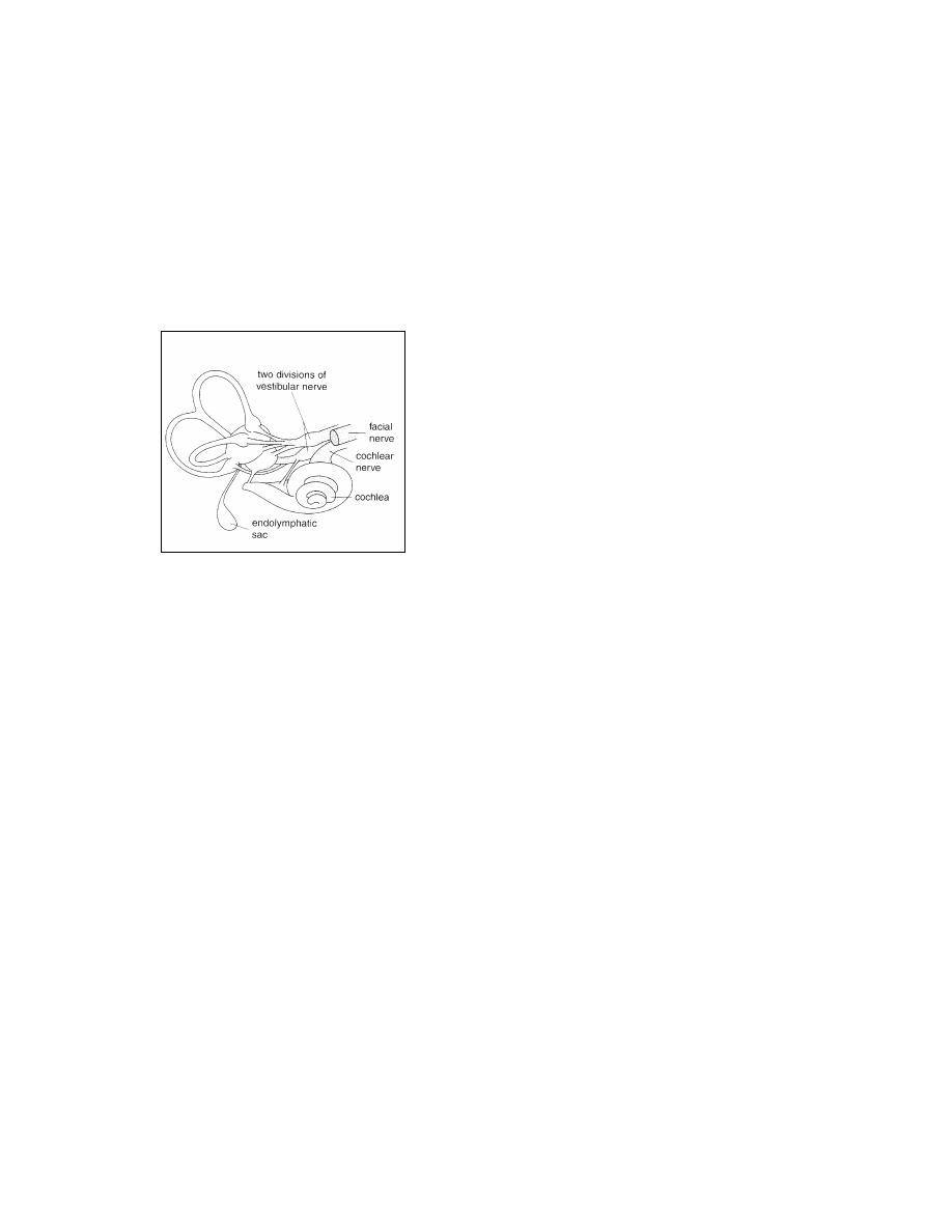
The ampullary*, utricular and saccular nerves unite to form the vestibular nerve. The vestibular
and cochlear nerves together constitute the 8
th
cranial nerve.
* Ampula is a dilated end in the semi circular duct
Blood Supply;
The main supply comes from the labrynthine artery (internal acoustic artery) which arises
from the basilar or anterior inferior cerebellar artery.
Facial Nerve
This nerve emerges from the pons and
after crossing the cerebellpontine angle it
enters the temporal bone at the internal
auditory meatus. It passes over the labyrinth
until it reaches the medial wall of the
tympanic cavity. Hear it bends backwards at
right angle where the geniculate ganglion is
situated and passes almost horizontally,
enclosed in the fallopian canal, above the
oval window. When it reaches the aditus it
turns downwards behind the pyramid and
continues almost vertically till it merges
from the stylomastoid foramen.
The nerve to the stapedius is given off close
to the pyramid. The chorda tympani nerve
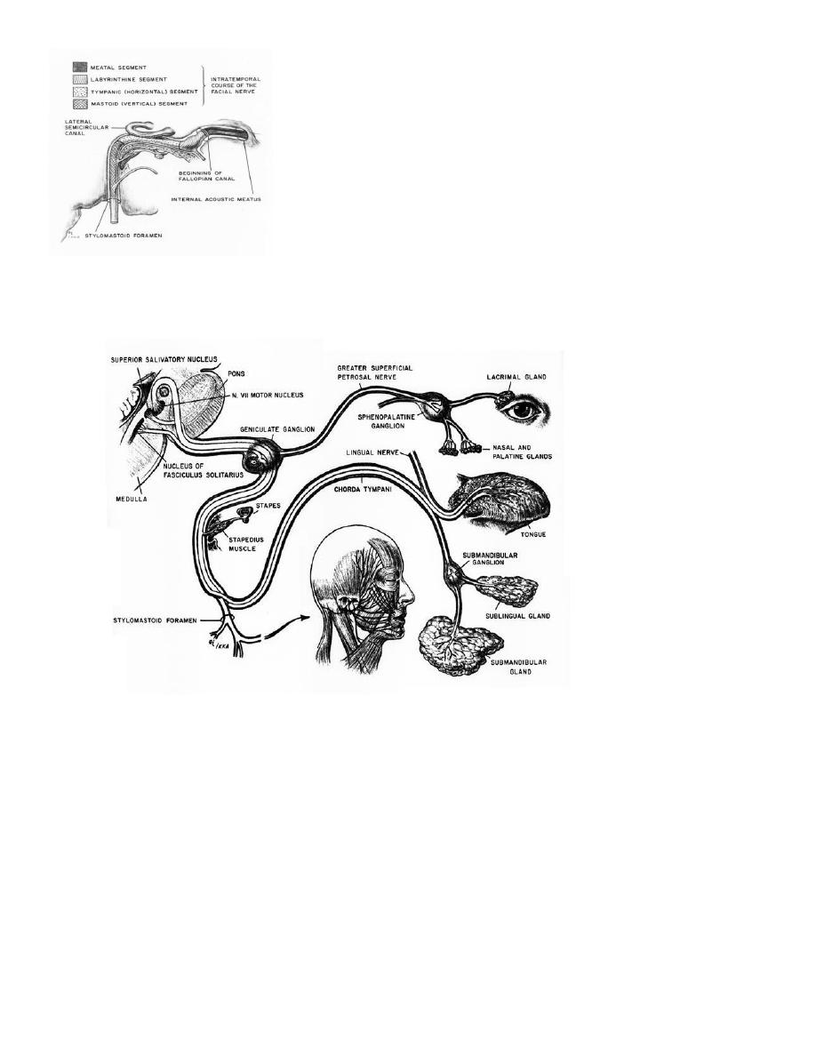
10
leaves the descending part of the facial
nerve and enters the tympanic cavity.

11