
AFTER MID
LEC: 1
DR. KHUDAIR
Oncology
Principles of Surgical
Oncology
TOTAL LEC: 1
Dr. Khudair


Principles of Surgical Oncology
Dr. Khdair Al-Rawaq
✴
Definitions
✴
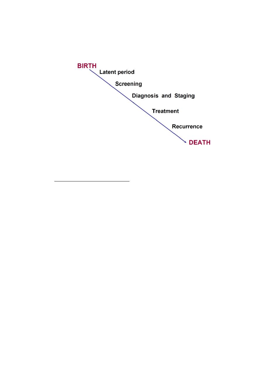
The history of surgical oncology
✴
The role of surgery in management of cancer, including :
1-Prevention
2-Screening
3-Diagnosis
4-Treatment
5-Rehabilitation
6-Palliative care
.Future of Surgical Oncology
Definitions
•
Surgical oncology, It is refered to the specific application of surgical principles to the
oncologic setting. These principles have been derived by adapting standard surgical
approaches to the unique situations that arise when treating cancer patients.
•
The surgeon is often the first specialist to see the patient with a solid malignancy, and,
in the course of therapy, he or she may be called upon to provide diagnostic,
therapeutic, palliative, and supportive care. In each of these areas, guiding paradigms
that are unique to surgical oncology are employed.
•
A surgical oncologist is a well-qualified surgeon who has obtained additional training
and experience in the multidisciplinary approach to the prevention, diagnosis,
treatment, and rehabilitation of cancer patients, and devotes a major portion of his or
her professional practice to these activities and cancer research.”
1

Ancient History of Surgery for Cancer Treatment
•
1600 BC First recorded description of the surgical treatment of cancer (in Egypt)
•
400 BC Hippocratesdescribes the stages of cancer and advises against surgery for
terminal disease; he coins the terms “carcinoma”(crab-leg tumor) and
“sarcoma”(fleshy tumor)
•
200 AD Galenidentifies cancer as a systemic disease (primary and metastasis).
•
Before 1850 Early heroic attempts to resect cancer
•
1850-1950 Development of standard surgical resection techniques
•
1950-1960 Development of extended radical surgical procedures
Historical Eras of Surgery to Treat Cancer
•
1960-1980 Exploration of combined-modality treatment
•
1980-2000 Multimodality therapy improves organ preservation and
survival
•
2000-present Surgical practice incorporates improved understanding of
the molecular basis of
Landmark Advances in Surgical Oncology:
•
1775 Etiologic basis of cancer Percival Pott
•
1809 Elective oophorectomy Ephraim McDowell
•
1829 Metastatic process Joseph Recamier
•
1846 Ether used as anesthesia John Collins Warren
•
1867 Carbolic acid used as antisepsis Joseph Lister
•
1873 Laryngectomy Albert Theodore Billroth
2

•
1878 Resection of rectal tumor Richard von Volkman
•
1880 Esophagectomy Albert Theodore Billroth
•
1881 Gastrectomy Albert Theodore Billroth
•
1890 Radical mastectomy William Stewart Halstead
•
1896 {Oophorectomy for breast cancer} G. T. Beatson
•
1904 Radical prostatectomy
Hugh H. Young
•
1906 Radical hysterectomy Ernest Wertheim
•
1908 Abdominoperineal resection
W. Ernest Miles
•
1909 Nobel prize for thyroid surgery
Theodore Emil Kocher
•
1910 Craniotomy
Harvey Cushing
•
1912 Cordotomyfor the treatment of pain
E. Martin
•
1913 Thoracic esophagectomy
Franz Torek
•
1927 Resection of pulmonary metastases
George Divis
•
1933 Pneumonectomy
Evarts Graham
•
1935 Pancreaticoduodenectomy
Allen O. Whipple
•
1945 Adrenalectomy for prostate cancer
Charles B. Huggins
•
1957 Isolated limb perfusion
Oliver Creech
•
1958 First multicenter clinical trial
Bernard Fisher
•
1965 Hormone therapy for cancer
3
Charles Huggins
•
1971 Microvascular free-tissue transfer
Harry Buncke
Surgery for Cancer Prevention
•
Role of Surgery in Cancer Prevention
Pre-cancerous lesions
–
Leukoplakia of the tongue
–
Thyroid gland in MENS II
–
Colon in FAP
•
•Organs at high risk of malignancy even where a pre-cancerous lesion has not been identified
–
Breast in cariers of deleterious BRCA mutations
–
Colon in HNPCC
4

Surgery for Cancer Screening
•Colonoscopy in colon cancer
•Digital rectal examination in prostate cancer
•Clinical breast examination
Surgery for Diagnosis
History:
•
Ascertain presence of risk factors
•
Evidence of metastases
•
Presence of co-morbid factors
•
Family and social history
•
Psychological assessment of patient
•
Ascertain patient’s social and economic resources
•
Ascertain patient’s expectation from therapy
•
Patient’s treatment preferences
•
Educate patient on diagnosis, treatment and follow-up, and correct mis-
information
Investigations:
•
Knowledge of all available modalities of investigating particular case
•
Microscopic diagnosis is compulsory
Biopsy:
•
Surgeon’s responsibilities:
•
Selection of appropriate biopsy method and site
5

•
Responsible that the tissue reach the pathologist timely and properly .
•
Communicate the results to the patient, family, other physicians
•
Provide initial prognosis and information on follow-up care
Types of Biopsy Methods:
• Transcutaneous
• Image-directed (with fine-needle aspiration or cutting needle)
– Ultrasonography
– Computerized tomography
– Magnetic resonance imaging
• Open incisional (A portion of the tumor)
• Open excisional (All tumor mass removed)
Types of tissue Biopsy
1. Lymph node biopsy
•
lymphoma
•
metastatic carcinoma.
2. Biopsy of a tissue-based mass.
6

Lymphoma:
•
The goal of biopsy in the patient with an abnormal lymph node and
suspected lymphoma is to make the general diagnosis and to establish the
lymphoma type and subtype. Additional analyses of the cells in the node, its
internal architecture, and the subpopulations of cells are critical for
subsequent treatment.
• Diagnosis of lymphoma should be made on a completely excised node that
has been minimally manipulated
•
The use of needle aspiration does not consistently allow for the complete
analyses. Eg. RS cell
•
can be performed as needed.
Carcinoma
•
The diagnosis of metastatic carcinoma often requires less tissue than is needed for
lymphoma. Fine-needle aspiration (FNA), core biopsy, or subtotal removal of a single
node will be adequate in this situation.
•
For metastatic disease, the surgeon will use a combination of factors, such as location
of the node, physical examination, and symptoms, to predict the site of primary
disease. When this information is communicated to the pathologist, the pathologic
evaluation can be focused on the most likely sites so as to obtain the highest
diagnostic yield. The use of immunocytochemical analyses can be successful in
defining the primary site, even on small amounts of tissue.
7

Sentinel node biopsy:
•
Technique
•
The node or nodes that preferentially drain a particular primary tumor”basins”
are identified by mapping and then surgically excised.
•
The mapping agents include radiolabeled materials and vital dyes that are
specifically taken up by, and transported in, the lymphatic drainage systems.
These mapping and localizing agents, used alone or in combination, are critical in
defining the unique flow patterns to specific lymph node(s) and ambiguous
drainage patterns (eg, a truncal melanoma that may drain to the axilla,
supraclavicular, or inguinal spaces).
Biopsy of a tissue-based mass:
–
Several principles must be considered
•
Mass in the aerodigestive tract In the aerodigestive tract, biopsy of a lesion should
include a representative amount of tissue taken preferably from the periphery of
the lesion, where the maximum amount of viable malignant cells will be present.
Because the treatment of in situ and invasive diseases varies greatly, the biopsy
must be of adequate depth to determine penetration of the tumors. This is
particularly true for carcinomas of the oral cavity, pharynx, and larynx.
•
Mass in the trunk or extremities For soft-tissue or bony masses of the trunk or
extremities, the biopsy technique should be selected on the basis of the planned
subsequent tumor resection. The incision should be made along anatomic lines in
the trunk or along the long axis of the extremity. When a sarcoma is suspected,
FNA can establish the diagnosis of malignancy, but a core biopsy will likely be
required to determine the histologic type and plan neoadjuvant therapy.
8
Breast mass
•
Although previously a common procedure, an open surgical biopsy of the breast is
rarely indicated today
•
A core biopsy/FNA, performed either under image guidance (ultrasonography or
mammography) or directly for palpable lesions, is the method of choice.
•
The core biopsy method establishes the histologic diagnosis, provides adequate
tissue for analyses of hormone-receptor levels and other risk factors, causes little
or no cosmetic damage, does not perturb sentinel node analyses, and does not
require extended healing prior to the initiation of therapy. In addition, a small
radioopaque clip can be placed in the tumor to guide the surgical extirpation.
Surgeon’s Tasks in Performing Biopsy:
•Orient the specimen
•Ensure the integrity of the tissue plane
•Ensure the adequacy of the tissue sample
•Be sure tissue reach the pathologist !
9

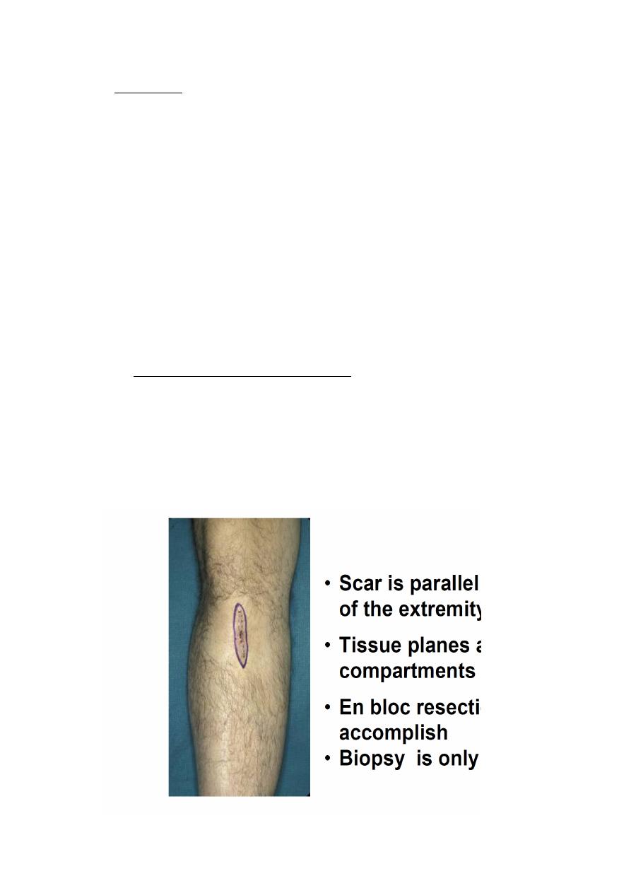
Appropriate Open Biopsy
•Scar is parallel to the long axis of the extremity
•Tissue planes and compartments are intact
•En bloc resection will be easy to accomplish
•Biopsy is only the first step
Surgery and Staging
•Classifies patients according to the degree of spread of cancer in order
•Guide selection of primary and adjuvant treatment
•Estimate prognosis
•Assist in evaluating result of treatment
•Facilitate exchange of information
•Contribute to continuing investigation of cancers
Types of staging:
•
TNM most common
•
AJCC head & neck
•
FIGO Gyenicological Malig.
•
Duke GIT
10

Surgery for cancer treatment:
•Surgery
Zero-order kinetics—100% of cells at risk are killed with a single treatment
•Radiotherapy/Chemotherapy
First-order kinetics—only a portion of cells at risk are killed during treatment, which is
followed by regrowth.
Preoperative Assessment and Preparation
•Surgeon’s responsibility to assess the risk-to-benefit ratio and identify and correct
underlying, relevanthealth problems .
–Nutritional status
–Co-morbid medical conditions
–Hypertension
–Diabetes
–Congestive heart failure
–Liver or renal insufficiency
–Immunosuppresion
Types of surgery:
•Local resection
•Radical resection with en-bloc resection of lymph nodes
•Supra-radical resections
•Surgery for metastasis
11

•Surgical management of complications
•Vascular access surgery
Treatment:
Principles of surgical resection of tumor
•Adequate margin of resection
•Prevention of tumor spillage
•Minimal manipulation
•Reconstruction
Metastasectomy:
This is done when:
• The primary tumor is controlled or can be controlled
• Metastasis is single or where multiple is localized
• Evidence that metastasectomy is associated clinical benefits
• Tumor doubling time is sufficiently long
• No significant co-morbid factor.
Surgery and rehabilitation
Role of surgery in the rehabilitation of cancer patients
•Restoration of form
•Restoration of function
•Care of ostomies
•Psychological treatment and support
12

•Maintenance and improvement of quality of life.
Surgery and palliative care:
Goals of Palliative Surgery
•Relieve symptoms for patients beyond cure when non-surgical measures are not feasible, not effective,
or not expedient
•Palliation means patient should be better at the completion of the procedure
“It is axiomatic that one cannot palliatively improve an asymptomatic patient using a scalpel.”
R. G. Martin, 1982
Palliative Improvement of Function and quality of life:
• Adequate control of pain
• Relief gastrointestinal and biliary obstruction
• Stop hemorrhage
• Supplement poor nutrition
• Airway obstruction
• Renal failure
• Rectal or urinary incontinence
Factors that influence outcome of treatment
1. •Patient related factors
2. •Health care provider related factors
3. •Environment related factors
Surgical Oncology in the Future:
•Preemptive surgery in populations at genetic risk for the development of cancer
•Tissue and function
-preserving improvements
13

–Minimally invasive and robotic surgery
–Implantable monitors
–Treatment sensitizers
–Tissue-engineered, implantable “spare parts”
•Refinements in surgical practice will be driven by the underlying molecular basis of tumor
biology
Thank you
واﺧريا ﺧﻠﺼﺖ ﻫﺬي اﳌﺤﺎﻇﺮة اﻟﻮﻏﻒ
Done by :Hussein Sadun Al-Nuaimy
14