
PERTUSSIS
Hasanein Ghali, MD - Department of Pediatrics
College of Medicine – University of Baghdad
December, 29
th
, 2015

EPIDEMIOLOGY
The vast majority of outbreaks of Pertussis are caused
by the bacterium
Bordetella pertussis
.
Pertussis syndrome
;paroxysmal coughing, can be
produced by related bacteria and several types of
adenovirurus.
Transmission requires
close contact
with a case.
healthy carriers are rare but adolescents and adults
who have pertussis may have relatively
minor
symptoms
and be
contagious
.
The incubation period is
7
to
10
days.

SYMPTOMS AND SIGNS
Pertussis is an infection of the respiratory tract that,
in the typical case, progresses through a sequence of
symptoms over a period of
4
to
6
weeks.
1-
Catarrhal Stage:
The initial symptoms are Coryza,
mild cough, lacrimation, and low-grade fever.
Patients are most
contagious
at this time.
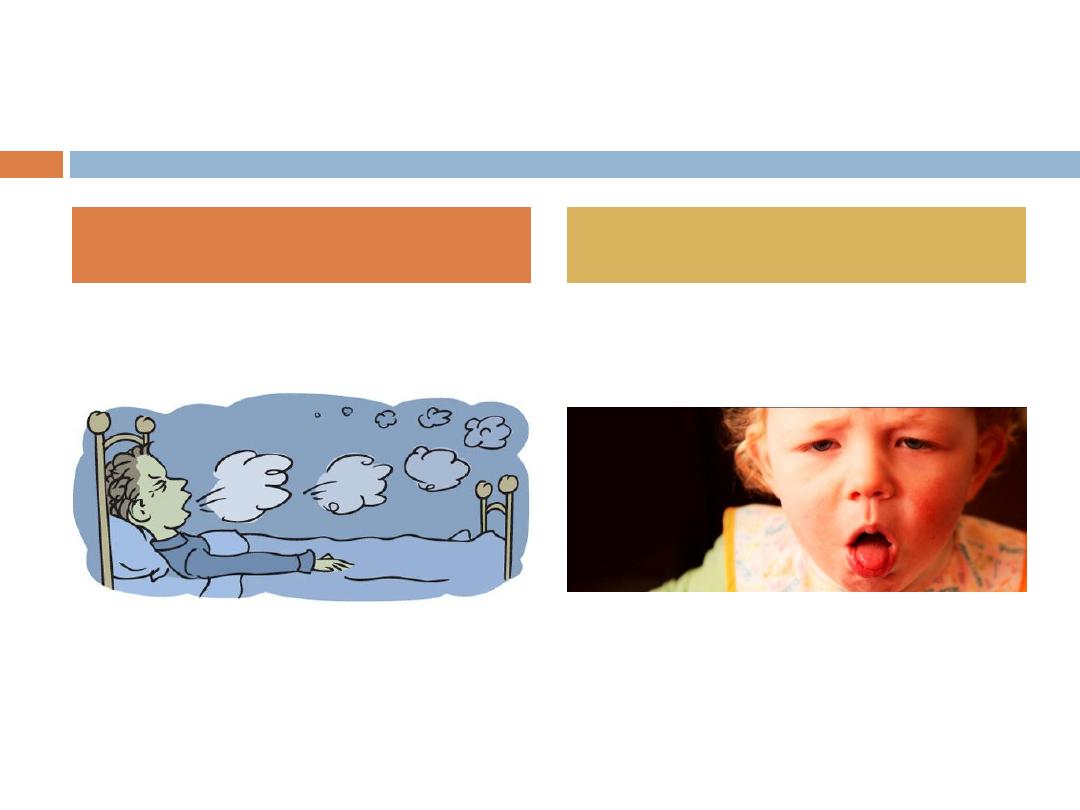
2-
Paroxysmal Stage:
The coughing increases in
frequency as the young patient works at trying to keep
the tracheobronchial tree open.
Various stimuli, such as feeding provoke a series of
several coughs during a single expiration, followed by a
deep inspiration, often with a characteristic
whoop
.

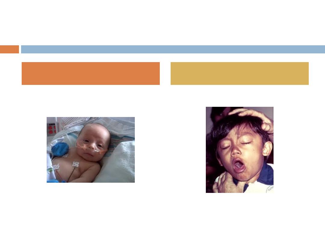
SYMPTOMS AND SIGNS
(cont.)
These bouts of coughing gradually increase in severity,
requiring hospitalization of infants and younger
children.
The child may become cyanotic during the struggle to
take a breath and clear thick mucus secretions.
Vomiting
is common at this time.
Gentle suctioning and positioning will help air exchange.
3-
Convalescent Stage:
Symptoms gradually diminish
over 2 to 4 weeks, and the patient returns to normal.
The clinical course is not altered dramatically by the
administration of antibiotics or other medications.

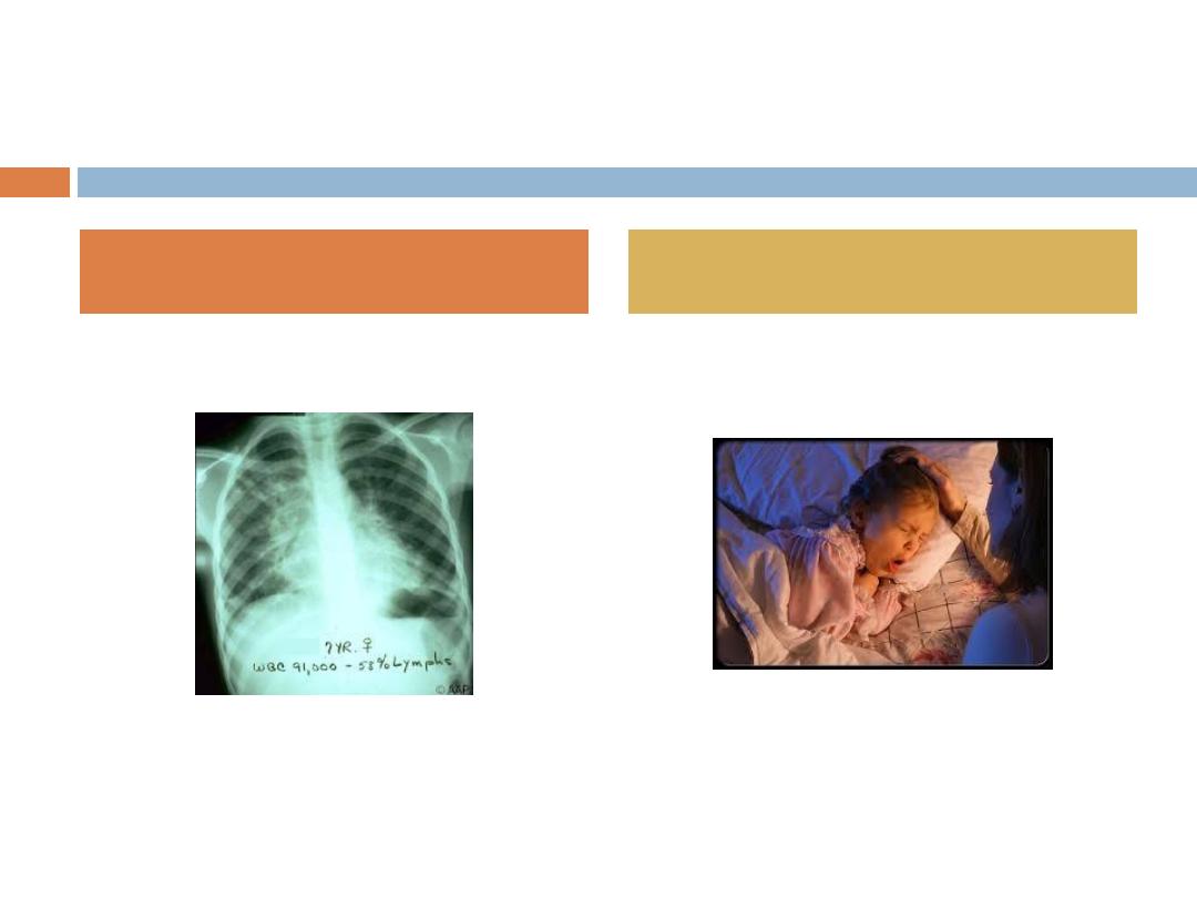
COMPLICATIONS
Pneumonia
due to the Pertussis organism or,
more
frequently
, to secondary bacterial invasion is a
common complication and a major contributor to the
death of infants and young children who become
hypoxic and may have
intracranial hemorrhages
.
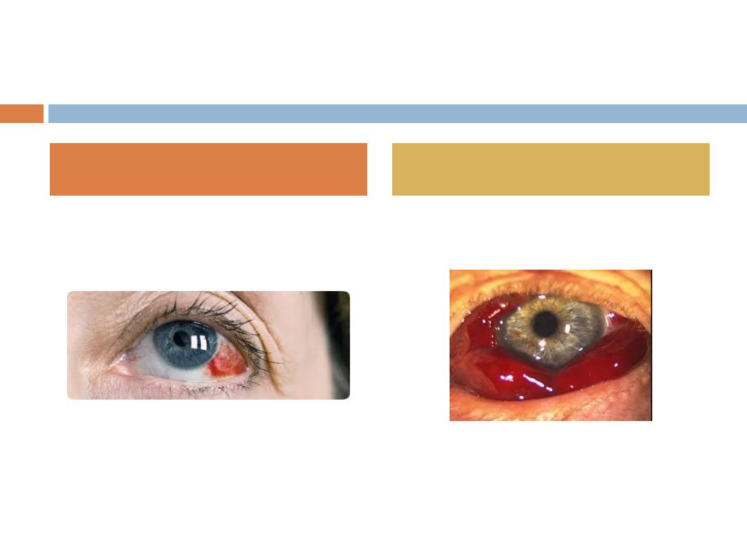
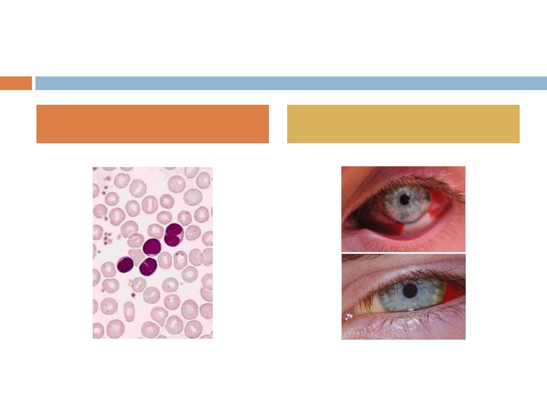
Other, less serious complications include
otitis
media, subconjunctival hemorrhage, and electrolyte
abnormalities.

LABORATORY DIAGNOSIS
Most patients who have Pertussis are diagnosed
clinically
because of the
paroxyms of cough,
especially when accompanied by whoop.
Diagnosis in the early stage of disease may be made
by culturing nasopharyngeal secretions.

TREATMENT
Infants younger than
6 months
of age who have
severe paroxysms
and
difficult
feeding
or
difficult
handling
secretions
may require hospitalization.
Antimicrobials given early may
moderate
symptoms,
but they are of little benefit in the paroxysmal
stage except to
limit
spread of organisms.
Erythromycin
40 to 50 mg/kg in four divided oral
doses is the drug of choice.
Corticosteroids
, and
albuterol
have been used in the
management of pertussis.

PREVENTION
Protective antibody to pertussis does not cross the
placenta. This means that the newborn is completely
vulnerable to infection.
Pertussis vaccine provides protection against whooping
cough. The immunity probably
is not as long-lasting
as
that following natural disease.
The immunization of the normal infant begins at the age
of 2 months , repeated at 8-week intervals for a total
of three doses with a combined vaccine (DPT).
Boosters are administered at ages 12 to 18 months and
4 to 6 years.

CONTROL OF OUTBREAKS
Close contacts younger than 7 years of age who have
not been immunized or who are immunized inadequately
should receive DPT vaccine.
All household and other close contacts, regardless of
vaccination status, should be given erythromycin 40 to
50 mg/kg per day, orally in four doses (for 14 days).
Children who have pertussis, if their medical condition
permits, may attend school after
5
days of antibiotic
treatment.
Patients not treated with antimicrobials are considered
contagious for 3 weeks after onset of paroxysms.

VARICELLA

Name !!!
The name chickenpox has been around for centuries,
it's from the blisters that are seen with the illness.
These red spots were once thought to look like
chickpeas (garbanzo beans). Another theory is that
the rash of chickenpox looks like the peck marks
caused by a chicken.

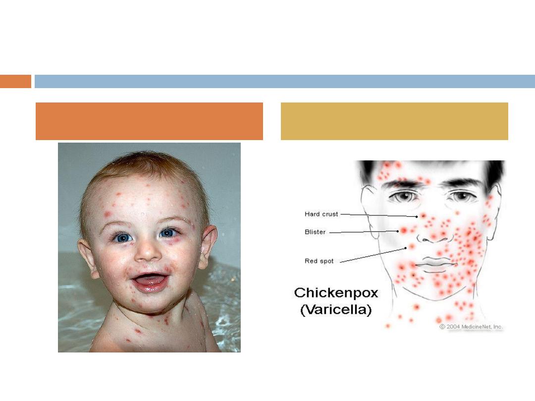
DEFINITIONS
Varicella
is a common exanthematous disease that
primarily affects children.
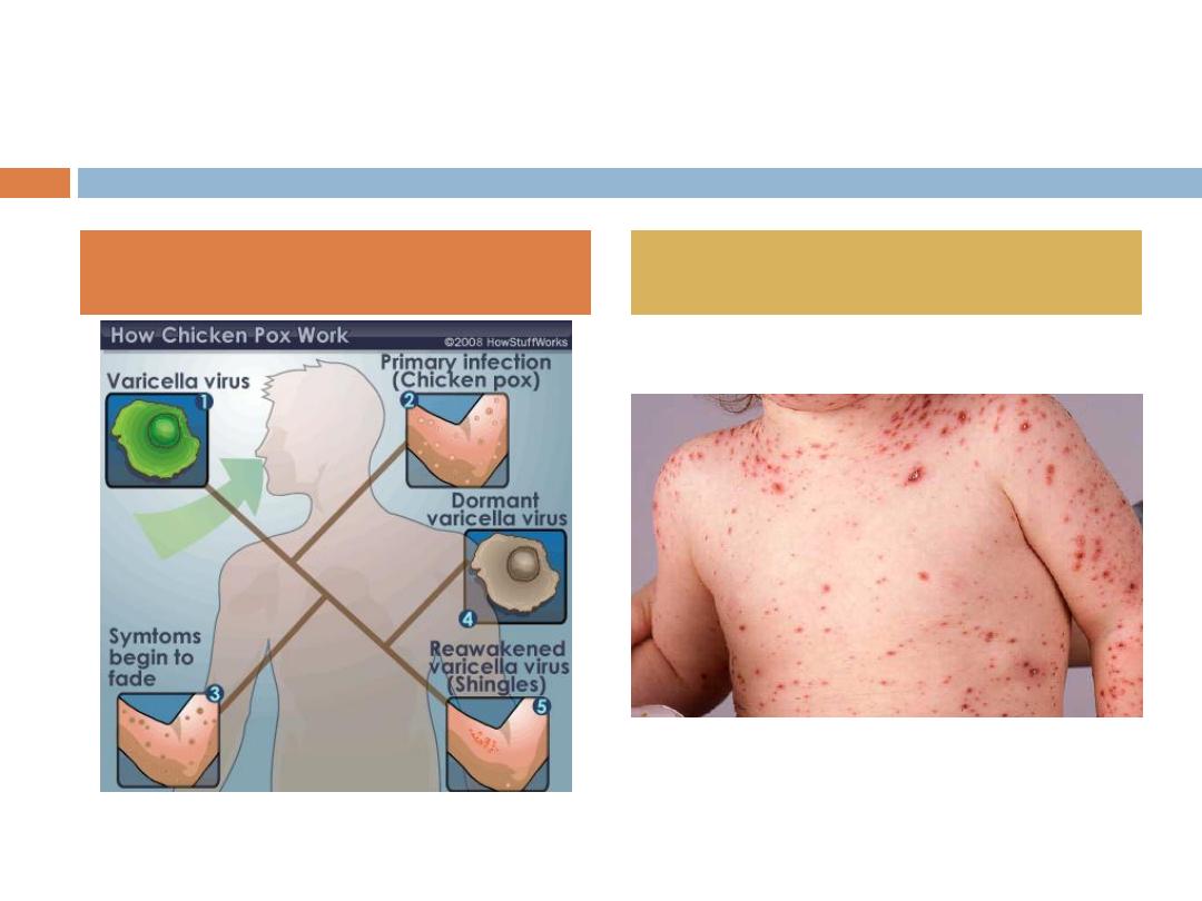
Chickenpox
is the clinical syndrome that results from
primary infection with varicella zoster virus.
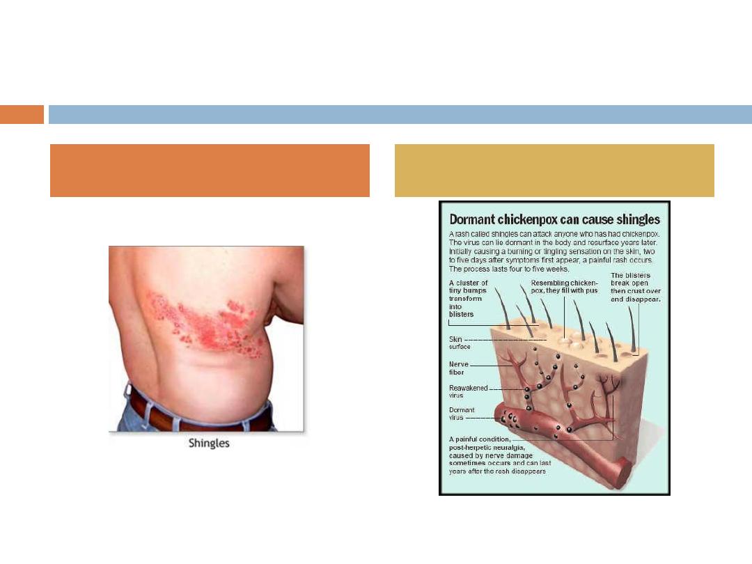
Herpes zoster
(“
shingles
” or “
zoster
”) after reactivation
of latent VZV, occur any time after a primary infection.
Disseminated zoster
in immunocompromised patients
consists of severe rash with systemic findings.
Congenital varicella syndrome
, or
varicella
embryopathy
,
is the result of varicella infection in a woman during the
first or second trimester of pregnancy.

EPIDEMIOLOGY
Varicella infection is more common during the late
winter and early spring.
The mode of transmission is
person to person
via
direct contact with infected mucosa or airborne
particles from respiratory secretions. Trans placental
passage of the virus also can occur.
Immunity after natural varicella infection is
considered lifelong.
The incubation period is
10
to
21
days.
Communicability is highest from
2 days prior
to rash to
2 days after
its onset.

Susceptible Healthy children may be considered
noncontagious after
all lesions have crusted
over,
although communicability may be prolonged in
immunocompromised patients who have severe
infection.
children may become infected with varicella after
direct contact with active zoster lesions because these
lesions contain infectious virus.
EPIDEMIOLOGY

PATHOGENESIS
After a person is exposed to VZV, the virus undergoes
two phases of replication during the incubation period.
Primary replication
, 3 to 4 days after exposure, occurs
in the oropharynx and regional LN, followed by a
primary viremia.
A secondary viremia
, with intracellular replication
within the reticuloendothelial system, occurs 10 to 21
days after exposure.
Late in the secondary viremic phase, the virus is
delivered to the skin, at which time the typical
cutaneous lesions become evident.

PATHOGENESIS
The virus also is carried to the respiratory mucosa
toward the end of the incubation period,
which is the
reason for communicability before the onset of rash
.
Following viremia, the virus becomes latent in
dorsal
root ganglia cells
. It remains there until reactivation,
at which time the virus travels back to the skin along
the sensory nerve.
Reactivation likely is due to
declining cell-mediated
immunity
, which explains the increased incidence in the
elderly and in immunocompromised patients.

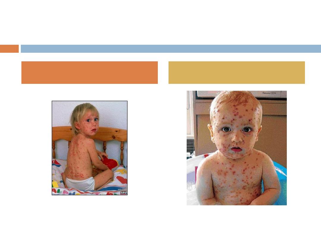
CLINICAL PRESENTATION
The rash of varicella often is preceded by a
24 – 48 hrs
period of fever, malaise, and other systemic symptoms.
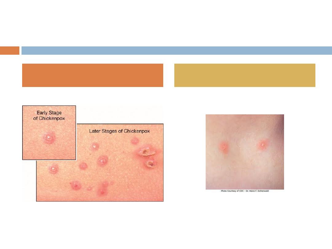
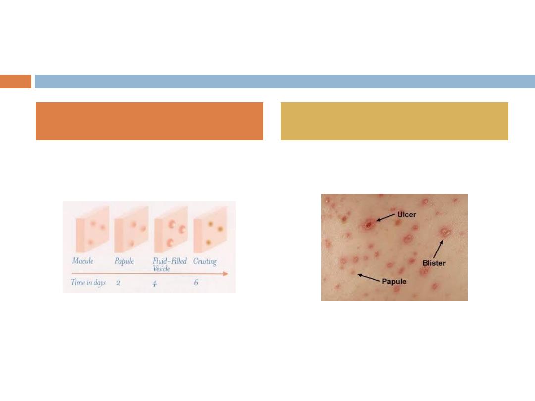
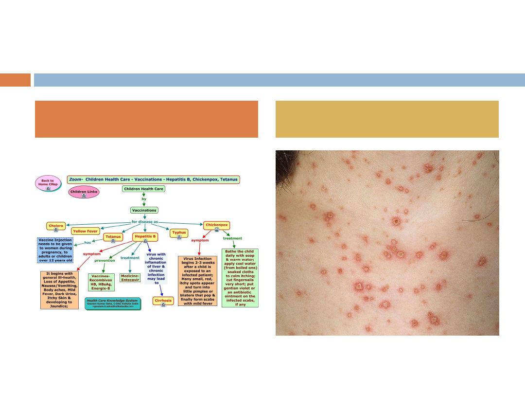
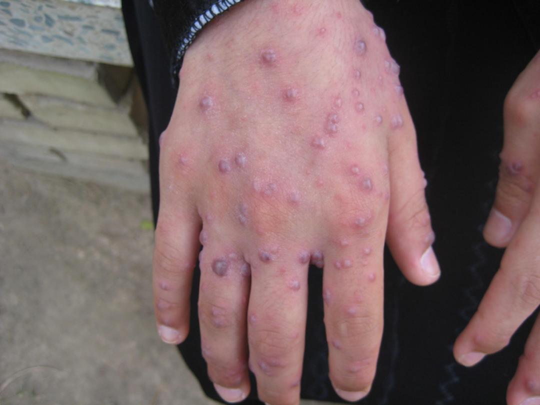
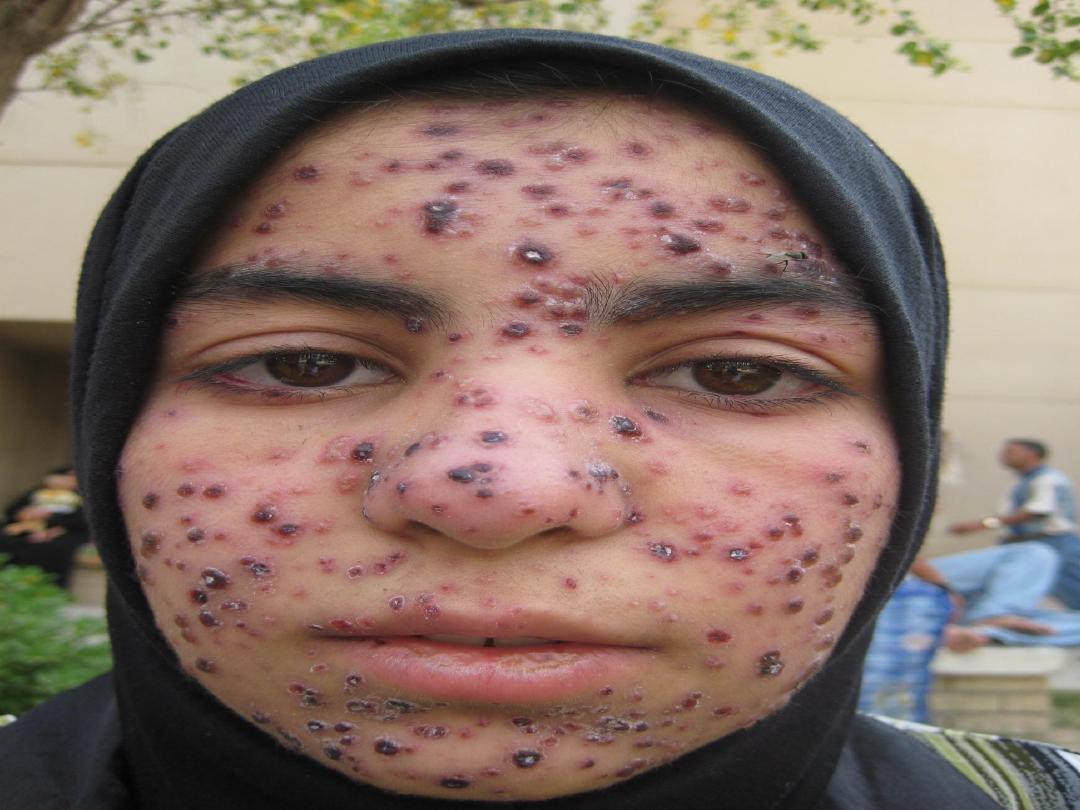
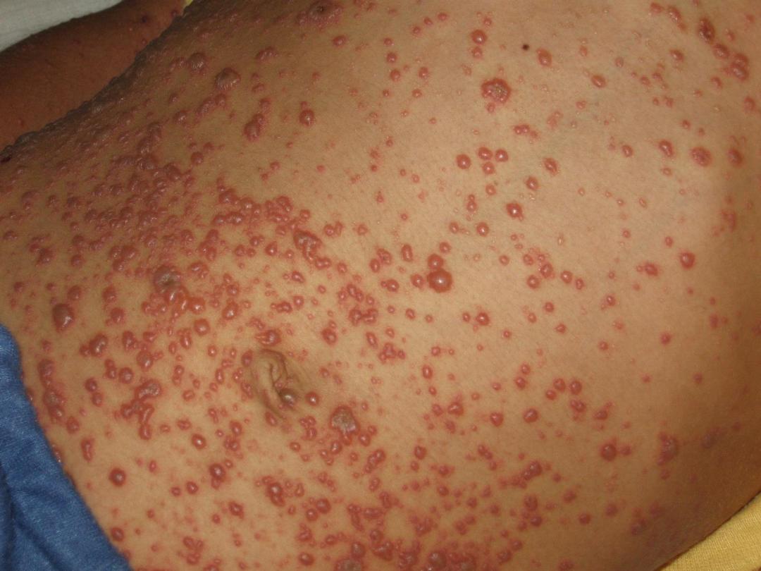
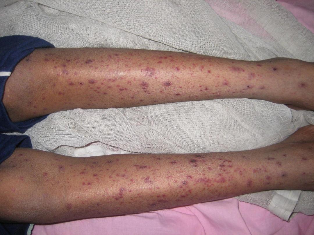
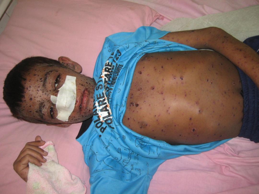
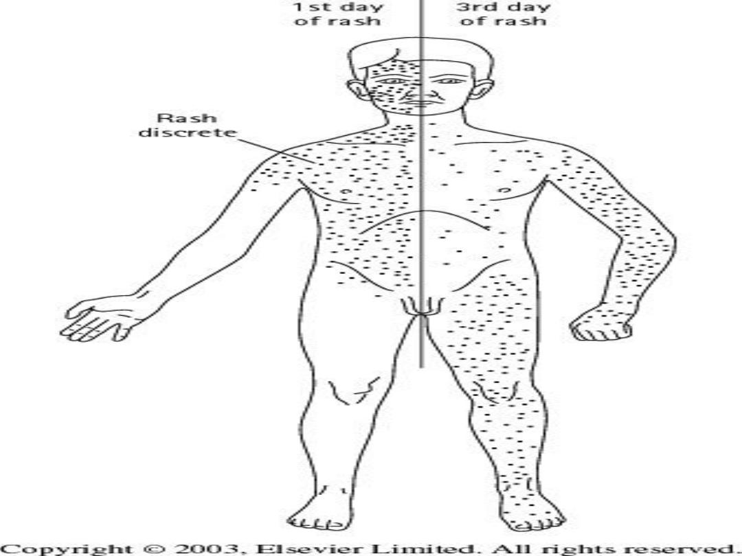
The typical exanthem begins as erythematous, pruritic
macules that develop into papules and fluid-filled
vesicles.
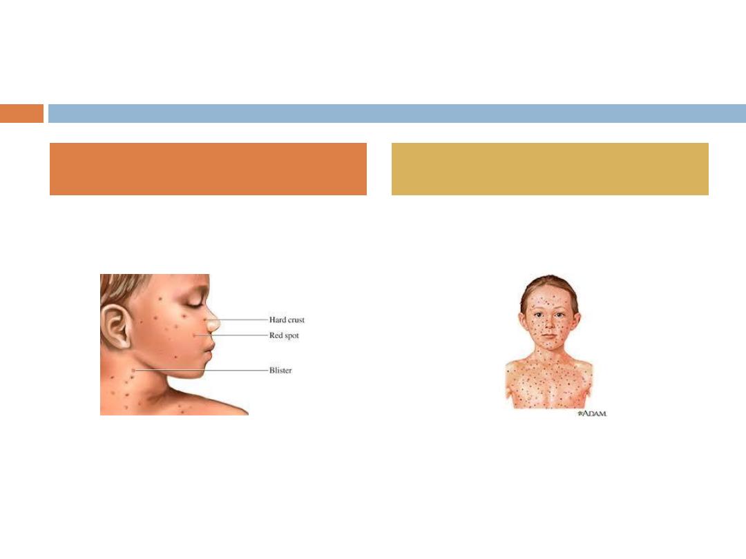
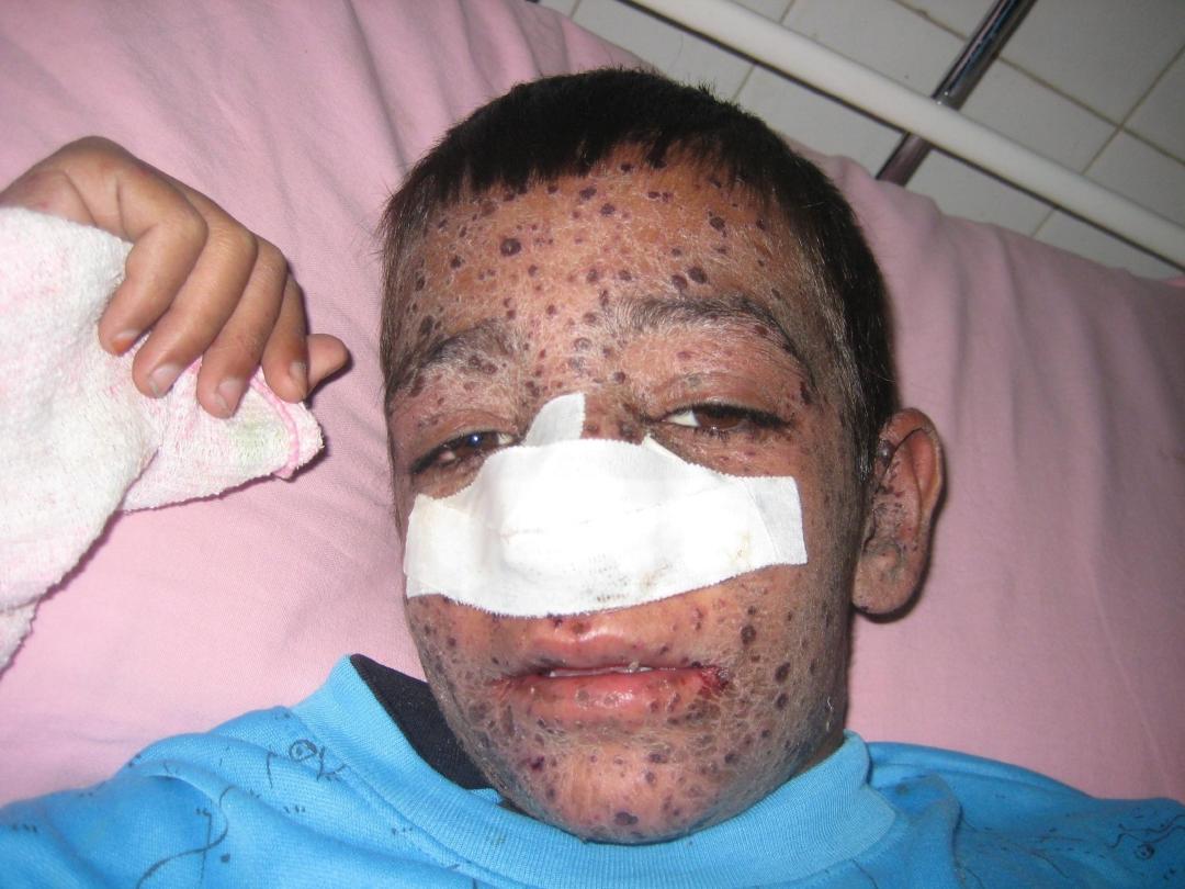
The crusting of the vesicles is the final stage of the
lesions before resolution, and scarring occurs
rarely
.
One of the most characteristic features of the
exanthem is the presence of all stages of lesions
simultaneously.

The average length of time from the development of
the initial lesion to the crusting of all lesions is
approximately 4 to 5 days.
Neonates born to women who develop primary
varicella infection 5 days before to 2 days after
delivery are at increased risk of severe varicella
infection, which may be fatal.
Clinical features of perinatally acquired varicella
include severe rash, pneumonia, hepatitis, and death in
30% of cases.
CLINICAL PRESENTATION

CLINICAL PRESENTATION
Congenital varicella syndrome results when a mother
is infected within the first 20 weeks of gestation.
“Zigzag” skin scarring and limb atrophy are
characteristic findings of this embryopathy.
Brain abnormalities, including hydrocephalus and
microcephaly, and eye abnormalities, such as
cataracts and chorioretinitis, also may occur.

COMPLICATIONS
The most common complication of varicella is
secondary bacterial infection, usually with
Strep
pyogenes
or
Staph aureus
.
Other invasive complications of varicella infection
include pneumonitis, meningoencephalitis, hepatitis,
arthritis, and glomerulonephritis.

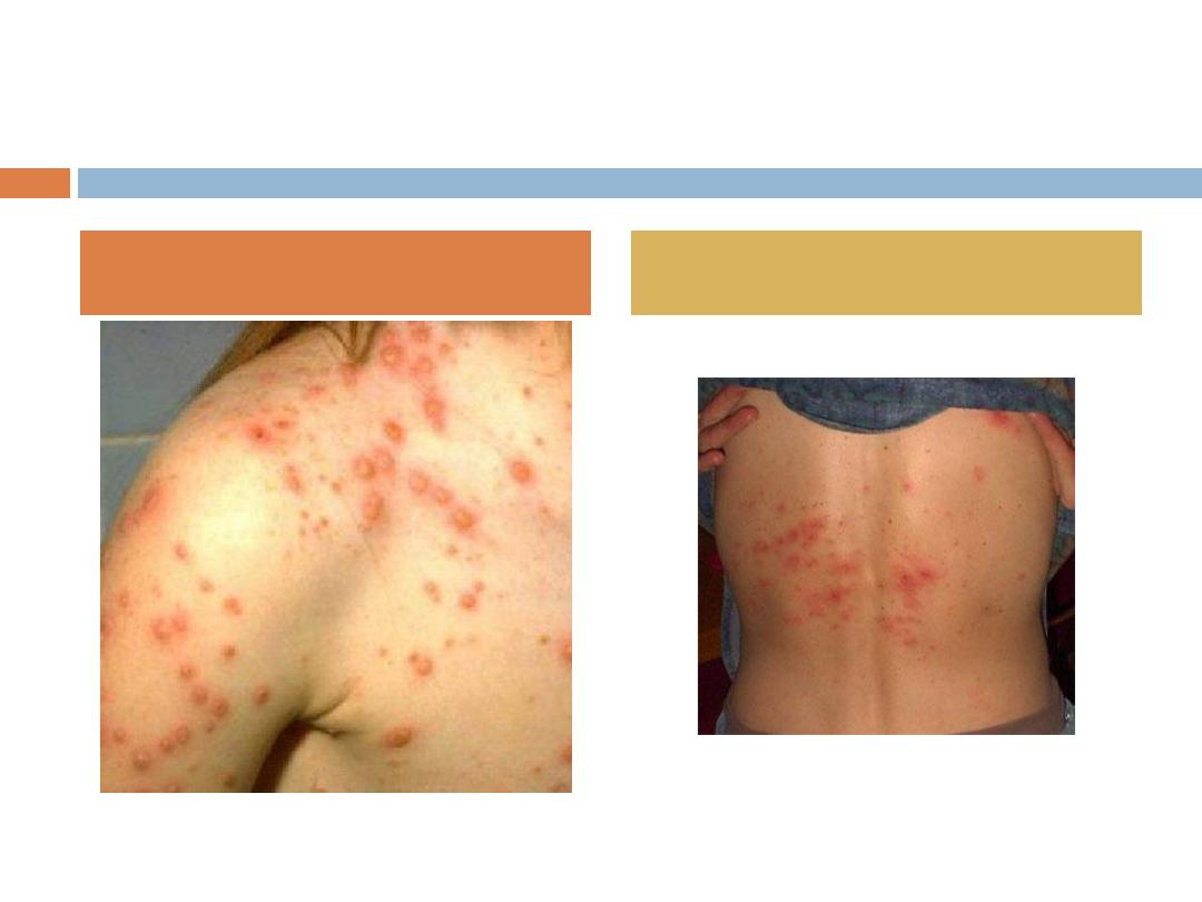
Herpes Zoster
Zoster, or “shingles,” is characterized by vesicles
clustered in a dermatomal distribution. The initial
presenting symptom frequently is pain at the future
site of the lesions, which usually arise within a few
days. Pruritus also may occur.
The most common sites are those supplied by the
trigeminal nerve and the thoracic ganglia.
New lesions occur over 2 to 3 days, then begin to crust
over the next week. Lesions resolve within 2 weeks.
The most common complication of herpes zoster is
postherpetic neuralgia, which is defined as pain that
lasts longer than 1 month. This is uncommon in
children.

DIAGNOSIS
The clinical appearances of primary varicella and
herpes zoster infections are so characteristic that
laboratory testing often is not necessary.
A Tzanck smear performed on a vesicle scraping shows
multinucleated giant cells, but it does not distinguish
between zoster and herpes simplex.
Viral culture, direct fluorescent antigen and PCR
performed on a vesicle scraping are of diagnostic help.

TREATMENT
The treatment of primary varicella-zoster infection is
supportive, including antipyretics and antihistamines or
oatmeal baths to control fever and itching.
Because of the risk of Reye syndrome, aspirin is
avoided.
The use of acyclovir is not routinely recommended for
healthy children younger than 12 years of age because
the disease generally has a benign, self-limited course.

TREATMENT
(cont.)
Oral acyclovir may be considered for those at risk
for more severe infection, including
1.
children older than age 12 years,
2.
persons who have chronic disease and
3.
persons who are taking chronic aspirin or
corticosteroid therapy.
Intravenous acyclovir should be used for
immunocompromised patients who have varicella-
zoster infection and should be initiated within 24
hours of the onset of the rash.

Varicella vaccine and varicella-zoster immune globulin
(VZIG) are both available for the prevention of
disease in susceptible persons exposed to VZV.
Significant exposure includes
1.
household contacts,
2.
close play or
3.
hospital contacts,
4.
newborns born to mothers who develop varicella 5
days prior to or 2 days after delivery, and
5.
those who have direct contact with active zoster
lesions.
POST EXPOSURE PROPHYLAXIS

ISOLATION
Airborne and contact precautions should be initiated
for hospitalized patients at least 5 days after the
onset of the rash and maintained until all vesicles are
crusted.

Thank you