Ophthalmology
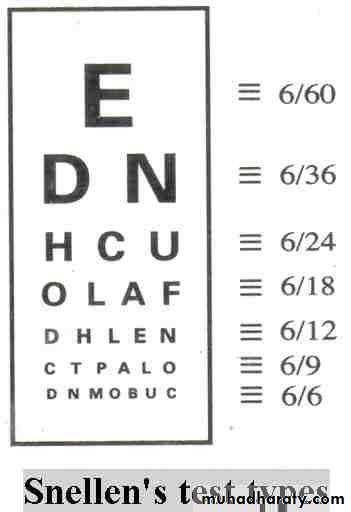
For assessment of visual acuity
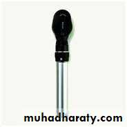

Direct ophthalmoscop:to examine fundus
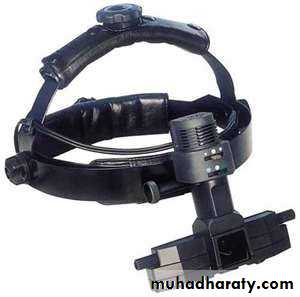
Indirect ophthalomscope: to examine fundusCondensed lens:it is used with the slit lamp to examine fundus
Goldman 3 mirrors contact lens: it is used with the slit lamp to examine fundus
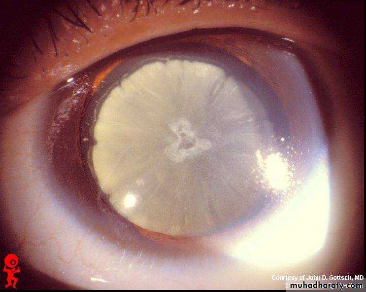
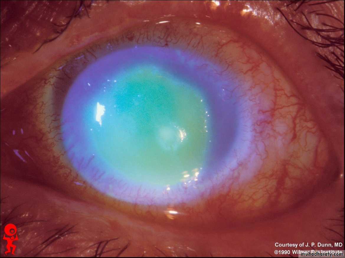
Dx :cataract+dilated pupil
Causes of cataract:1-age(commonest) 2-congenital 3-drugs like steriod 4-radiation 5-traumaCauses of dilated pupil:1-drugs 2-trauma 3- optic nerve damage 4- 3 nerve damage
Tx : surgery is the only tx for cataract
Assessment of patient:1-VA 2-light perception 3-pupillary reflex 4-color test through slit lamp 5- US to get idea about vitreous chamber and retian
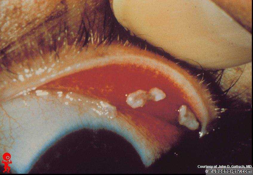
F.B. in the inner surface of upper lid
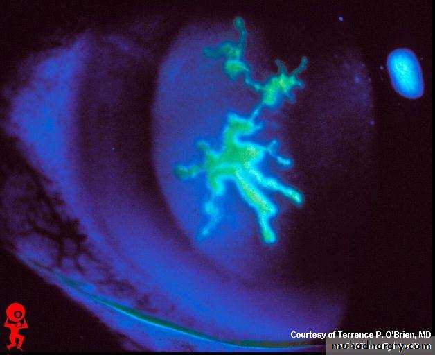
Flourescein stain showing ulcer in the retina(geographical ulcer)
Causes:1-steroids 2-chemical substances 3-bacterial infections 4-F.B. 5-trauma 6-contact lens usersTx : according to the cause
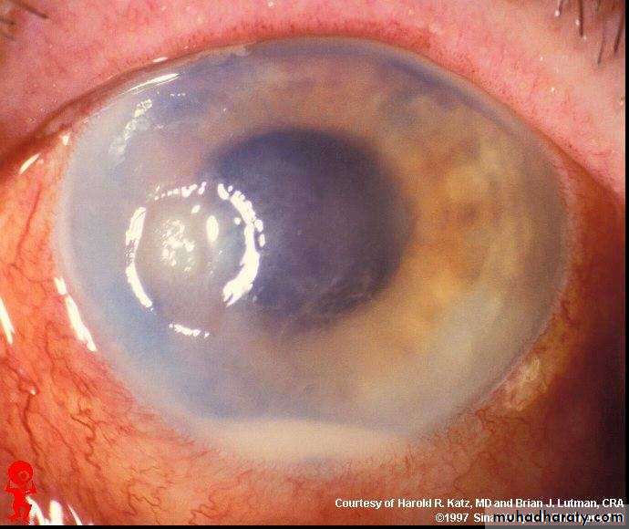
Hypopium (pus in the anterior chamber) with cloudy cornea +red eye
Causes of hypopium: 1-keratitis 2-iritis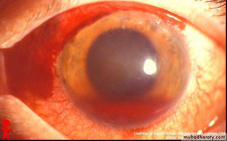
Dx : subconjunctival hemorrhage+hyphema(blood in the anterior chamber)+dilated pupil
Possible cause: traumaNext step in assessment: assess IOP
Tx :1-bed rest with head elevated 2-anti-glucoma drugs if increased IOP
3-drainage of bllod if high amount
Fluorescein stain showing corneal ulceration
Same causes of any ulcers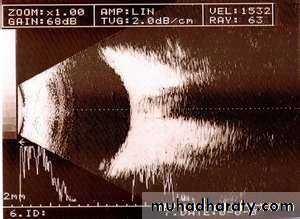
B scan sonography showing the vitreous cavity
Dx : peripheral laser iriditomy(YAG laser)
Ix : closure angle glucoma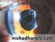
Dx :Posterior capsule opacification
Tx :YAG laser capsulotomy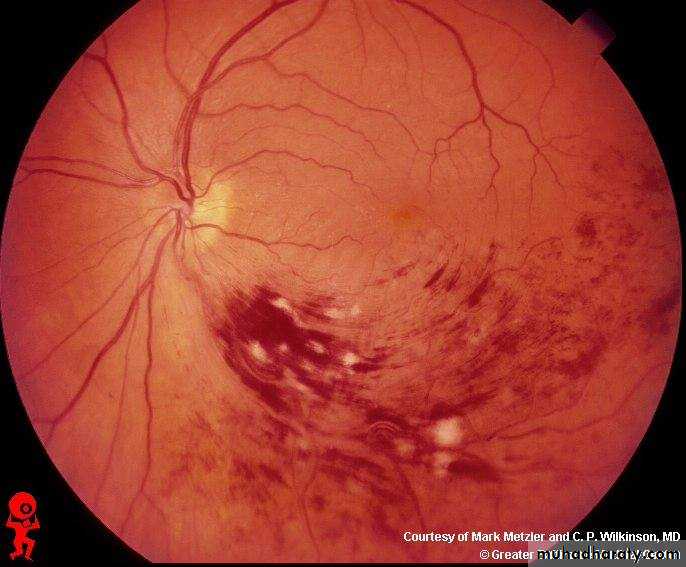
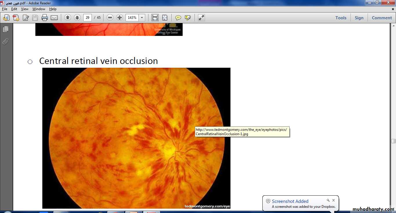
Occlusion of the lower temporal branch of CRV marked by hemorrhage and multiple dots of ischemia in the lower temporal zone of retina
CRVO
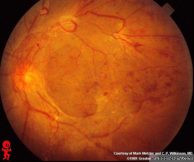
Proliferative diabetic retinopathy
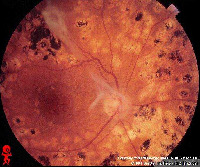
Proliferative diabetic retinopathy
هنا بعد استخدام الليز (المناطق السوداء ليزر قديم والمناطق البيضاء ليزر جديد)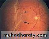
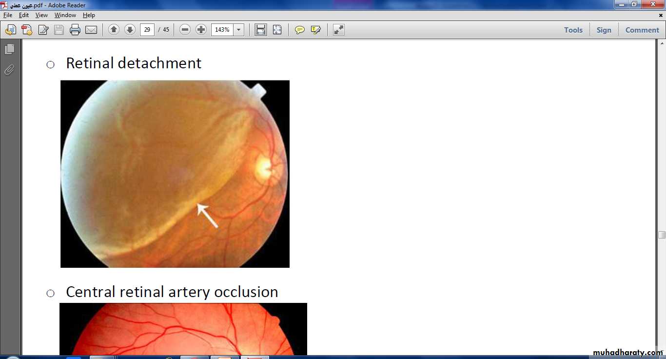
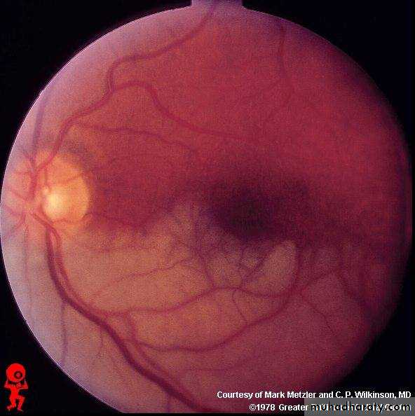
RETINAL DETACHMENT
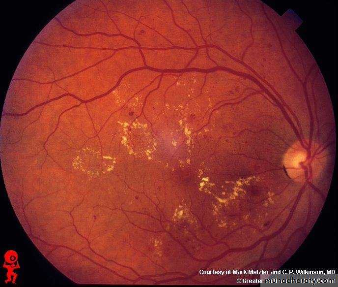
Non Proliferative diabetic retinopathy
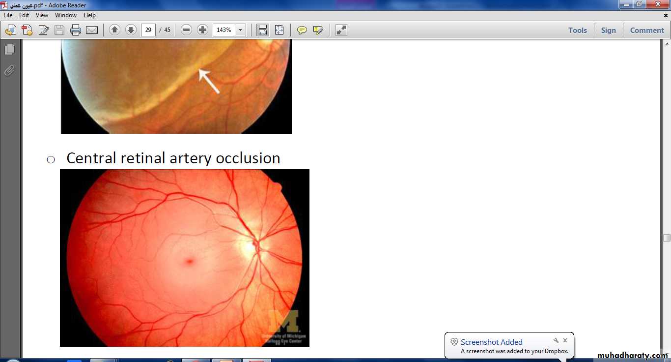
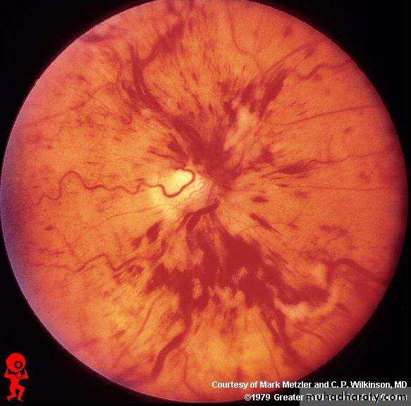
Occulosion of Lower temporal and lower nasal branches of CRA marked by complete ischemia