Practical session-2-
Knee exam
13-12-2015 Monday.
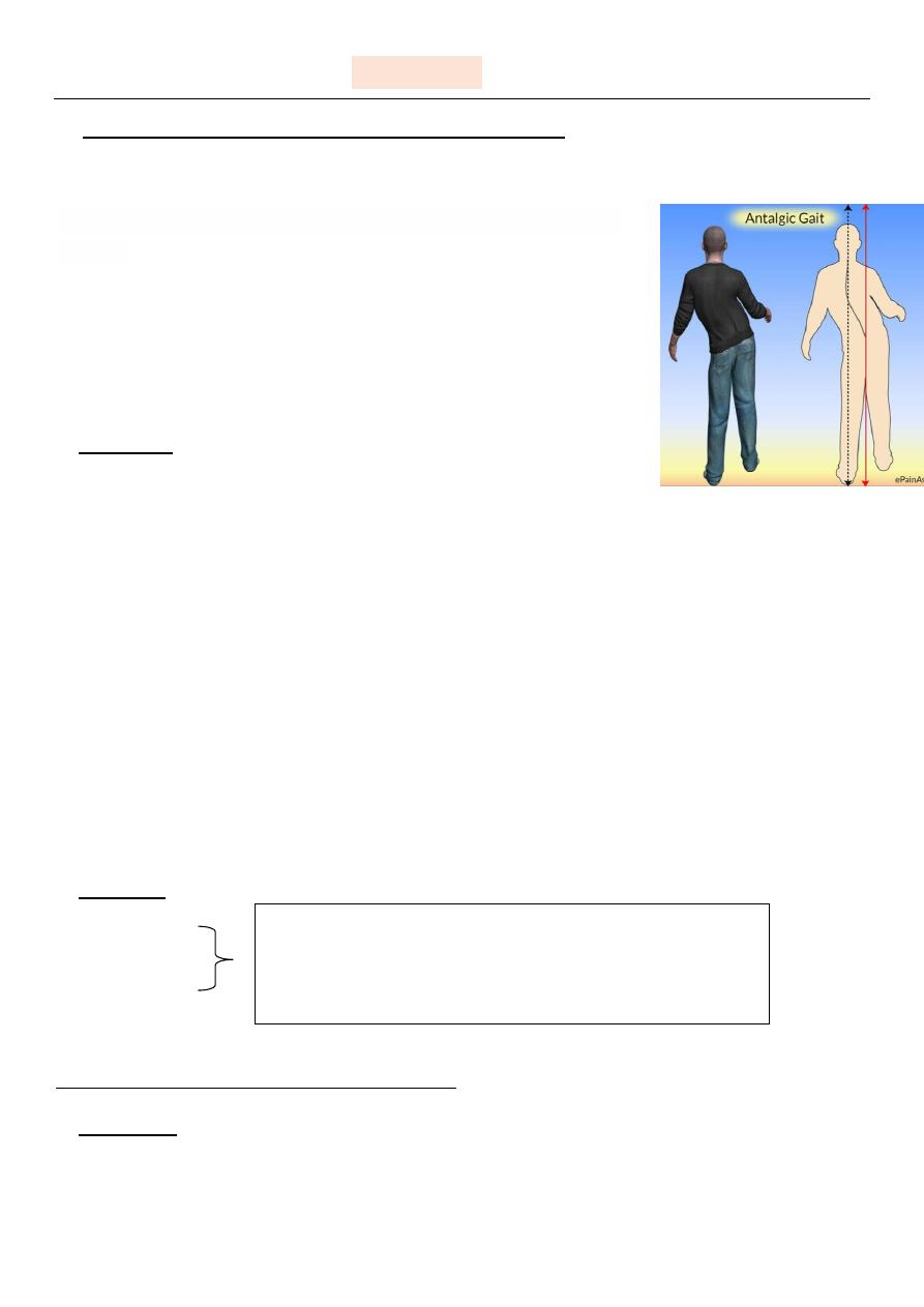
## Before all steps , ask the pt to walk & observe his gait .
(the patient we examined in the hospital was having "ANTALAGIC" gait .
The pt. doesn't want to spend time on the affected leg due
to pain.
1- inspection :-
a- swelling
localized:-trauma ,hemarthrosis ,pyarthrosis.
Generalized:-beyond joint margin:-bone disease ,lymphatic obstruction , systemic
inflammatory disease.
b- bruising
c- scar
d- wasting: can be confirmed by comparing two sides with tape measure.
((especially wasting of quadriceps:disuse ,prolonged bandaged,nerve injury))
e- deformity
genovarus :- deviation of distal ends of limb toward the midline .
Genovalgus :-deviation of distal ends of limb away "outward".
2- palpation :-
a-hotness
b-tenderness.
** always look to pt's face when you palpate.
3- movement :-
a- flexion
b- extension
Always compare the two sides with avoidance of
prolonged touching of the skin because this will mimic
pathological hotness of the area
important how to examine ligaments of
knee joints?
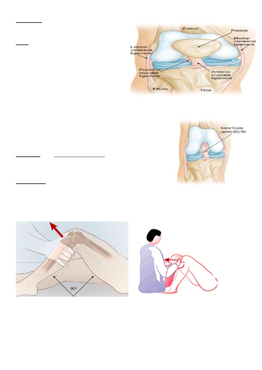
First ligaments of knee joints are :-
1) cruciate : anterior &posterior .
2) collateral :- medial & lateral .
1- cruciate :-
a- anterior cruciate ligament tear :-
Diagnosis :- by anterior drawer test .
Maneuver:-
With fixation of foot of the pt & flexing the knee joint , If the ACL is ruptured or injuried
the leg will slip"due to laxity" & can't resist the drawing action of the examiner with
accompanied severe pain .
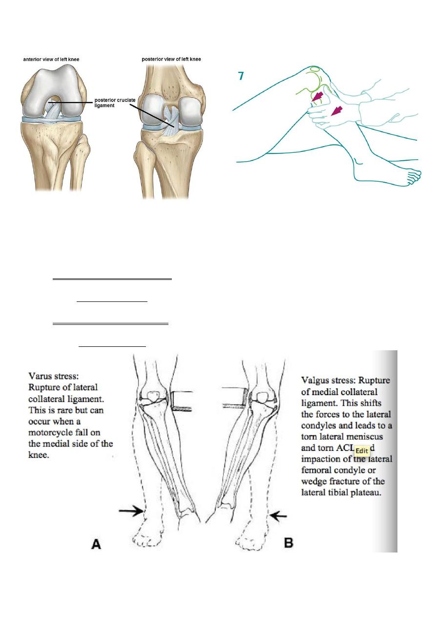
b- posterior cruciate ligament :-
The same steps are performed,but the slipping "laxity"of knee joint will be toward the force
of drawing "as in the picture above".
2- collateral:-
a- medial collateral ligament:-
test:- valgus stress test.
b- lateral collateral ligament:-
test:- varus stress test.
# As you perform the previously mentioned steps "in the picture above"pain will be
induced.
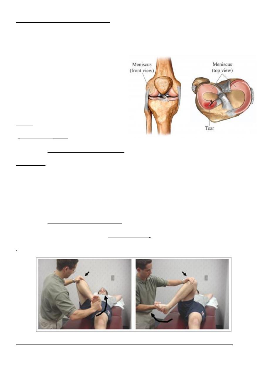
*menisci examination in knee joint :- very important in sport injuries.
What are mensci?
crescent-shaped fibrocartilaginous structure that, in contrast to articular disks, only partly
divides a joint cavity.
Exam:-
--
McMurray’s
test:-
a- medial menisci examination:-
maneuver:-
1. flex Knee joint.
2. external rotation of foot.
3. abduction.
* if pain is induced with click means injuried or tear mensci.
b- lateral menisci examination:-
same as the previous one
but with internal rotation.
'
Picture above illustrating the lateral menisci exam 'left' & medial menisci exam 'right”
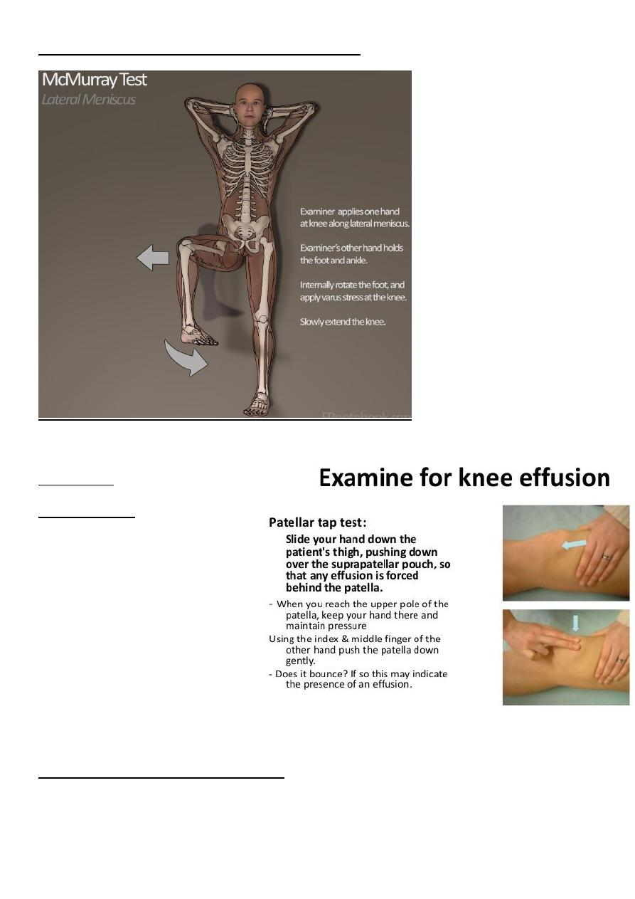
Picture illustrating the steps of lateral menisci exam
Important :-
Ballotment test "patellar tap test "
Aim :-to detect any knee effusion.
Maneuver :- →
Video for examination of knee joint >>>
https://drive.google.com/file/d/0B9zOkWPBzc66bkE5NjY3bkxXdVk/view?usp=sharing