Fractures of the Acetabulum
Fractures of the acetabulum occur when the head of the femur is driven into the pelvis.
This is caused either by a blow on the side (as in a fall from a height) or by a blow on the front of
the knee, usually in a dashboard injury when the femur also may be fractured.
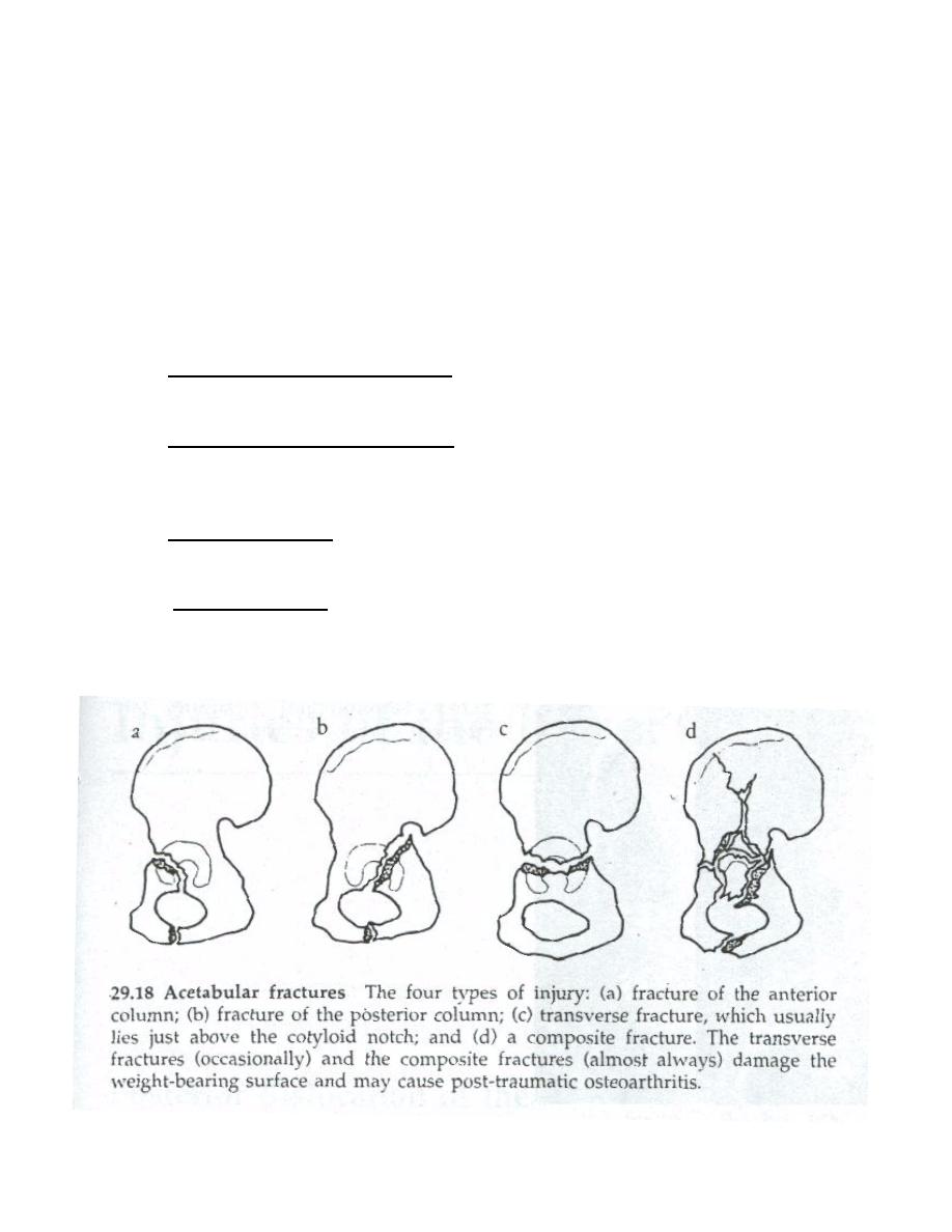
The types of fracture:
There are four main types of acetabular fracture: anterior, posterior, transverse and
combinations of these.
1. Anterior column and wall fracture; the fracture runs through the anterior part of the
acetabulum separating a segment. It is uncommon, does not involve the weight— bearing
area and has a good prognosis.
2. Posterior column and wall fracture; This fracture runs upwards separating the posterior
ischiopubic column of bone and breaking the weight-bearing part of the acetabulum. It is
usually associated with a posterior dislocation of the hip and may injure the sciatic nerve.
Treatment is more urgent and usually involves internal fixation to obtain a stable joint.
3. Transverse fracture; this is a fracture running transversely through the acetabulum and
separating the iliac portion above from the pubic and ischial portions below. It is usually
fairly easy to reduce and to hold reduced.
4. Complex fractures; some acetabular fractures are complex injuries which damage either
the anterior or the posterior segments (or both) as well as the roof of the acetabulurn. The
joint surface is disrupted; the fractures are difficult to reduce and the end result is likely
to he less than perfect.
!
1

Clinical features
There has usually been a severe injury —either a traffic accident or a fall from a height.
Associated fractures are not uncommon.
The patient may be severely shocked, and the complications associated with pelvic
fractures should be sought. There may be bruising around the hip and the limb may lie in internal
rotation (if the hip is dislocated). an attempt should be made to examine movements at the hip.
X-RAY
Several views of the hip, and sometimes a CT scan as well, may be needed before the
fracture can be accurately visualized.
Treatment
❖
Emergency treatment should consist only of counteracting shock and reducing a disloca-
tion.
❖
Traction is then applied to the limb (10 kg will suffice) and during the next 3 or 4 days
the patient’s general condition is brought under control.
❖
Definitive treatment of the fracture is delayed until he is fit and operating facilities are
optimal.
❖
With simple anterior or posterior fractures closed reduction under general anesthesia is
attempted. If this is successful, traction is maintained for a further 6 weeks.
❖
If closed reduction fails and adequate surgical expertise is available, operative reduction
and internal fixation with plates and screws is advisable.
❖
Complex fractures are difficult to reduce, and operative reduction and fixation is justified.
It should not be attempted in the absence of ideal operating facilities and adequate
surgical expertise.
❖
If comminution is so severe that the architecture cannot he restored, again operation
should not be attempted. Traction is continued and exercises started as soon as possible.
If necessary, arthroplasty of the hip can be carried out years later when union is complete.
Complications
1. Nerve injury: with posterior fractures the sciatic nerve may be injured. The nerve is usually
not severed, so the chances of recovery are good.
2. Avascular necrosis: As in all severe injuries of the hip, femoral head necrosis may occur. The
changes take months or even years to develop. If this progresses to fragmentation and collapse
of the head, arthroplasty may be indicated.
3. Osteoarthritis: Secondary osteoarthritis of the hip is a common (late) complication,
especially if the fracture involves the weight-bearing surface.
4. DVT and Pulmonary Embolism.
!
2

Injuries to the sacrum and coccyx
A blow from behind or a fall onto the ‘tail’ may fracture the sacrum or coccyx, or sprain
the joint between them.
If the fracture is displaced, reduction is worth attempting. The lower fragment may be
pushed backwards per rectum. The reduction is stable, which is fortunate. The patient is allowed
to resume normal activity, but is advised to use a U-shaped cushion when sitting.
Persistent pain, especially on sitting, is common after coccygeal injuries. If the pain is not
relieved by the use of a cushion or by the injection of local anaesthetic into the tender area,
excision of the coccyx may be considered.
Traumatic hip dislocation
Posterior dislocation of the hip
Four out of five traumatic hip dislocations are posterior (80%). Usually this occurs in a
road accident when someone seated in a truck or car is thrown forward, striking the knee against
the dashboard. The femur is thrust upwards and the femoral head is forced out of its socket; often
a piece of bone at the back of the acetabulum is sheared off as well (fracture-dislocation).
Special features
In a straightforward case the diagnosis is easy: the leg is short and lies adducted,
internally rotated and slightly flexed. However, if one of the long bones is fractured — usually
the femur — the hip injury can easily be missed. The lower limb should be examined for signs
of sciatic nerve injury.
X-RAY
In the anteroposterior film the femoral head is seen out of its socket and above the
acetabulum. Multiple views (and sometimes even a CTscan) may be needed to exclude a fracture
of the acetabular rim or the femoral head.
!
3

Treatment
❖
The dislocation must be reduced under general anesthesia. An assistant hold the pelvis;
the surgeon flexes the patient’s hip and knee to 90 degrees and pulls the thigh vertically
upwards.
❖
X-rays are essential to confirm reduction and to exclude a fracture.
❖
If it is suspected that bone fragments have been trapped in the joint CT is needed.
❖
Reduction is usually stable, but the hip has been severely injured and needs to be rested.
The simplest way is to apply traction and maintain it for 3 weeks.
❖
Movement and exercises are begun as soon as pain allows. At the end of 3 weeks the
patient is allowed to walk with crutches.
Complications
1. Fracture acetabulum: A fragment of the acetabulum may have been sheared off; if it is
large it should be fixed in place by screws. lf fragments of bone is small it should be
removed by operation.
2. Femoral head fracture: The segment of the femoral head if it does not fall back into place
when the dislocation is reduced, it must either be replaced and fixed in position or
removed.
3. Nerve injury: The sciatic nerve is sometimes damaged but it usually recovers sponta-
neously.
4. Avascular necrosis: The blood supply of the femoral head is seriously impaired in at least
10 per cent of traumatic hip dislocations. Avascular necrosis shows on x-ray as increased
density it may take weeks to show in x-ray, this may indicate osteotomy in young patients
and hip replacement in older patients.
5. Osteoarthiritis: Secondary osteoarthritis may occur due to any of the following: (1) carti-
lage damage, (2) retained fragments in the joint, (3) deformity of the acetabulum or
femoral head, (4) femoral head necrosis. Those may indicate hip replacement.
6. Vascular injury: superior gluteal artery.
!
4

Anterior dislocation of the hip
❖
This is rare compared with posterior dislocation. The leg lies externally rotated,
abducted and slightly flexed. Seen from the side, the anterior bulge of the dislocated
head is unmistakable.
❖
X-RAY In the anteroposterior view the dislocation is usually obvious, but occasionally
the head is almost directly in front of its normal position; a lateral film resolves any
doubt.
❖
Treatment: The way of reduction under anesthesia employed is almost identical with
that used to reduce a posterior dislocation, except that while the flexed thigh is being
pulled upwards, it should he adducted. The subsequent treatment is similar to that for
posterior dislocation. Avascular necrosis is the only complication.
Central dislocation of the hip
❖
This is not a true dislocation because the femoral head does not really come out of its
socket; it is driven through the medial wall of the fractured acetabulum into the pelvis.
❖
X-RAY Usually the floor of the acetabulum is shattered and comminuted.
❖
Treatment: An attempt should always be made to reduce the dislocation. Even if
secondary osteoarthritis is inevitable, a more or less normal anatomy will make recon-
structive surgery easier. Strong traction is applied and, if successful, maintained for 4—6
weeks. The hip is then mobilized and the patient is allowed up on crutches.
❖
If secondary osteoarthritis develops, this may need operative treatment; usually an
arthroplasty.
!
5