
1
4th stage
نسائية
Lec-5
د.اسماء
14/12/2015
Obstetric procedures
CAESAREAN SECTION
Is an operation used to deliver a potentially viable fetus through an
incision in the anterior abdominal wall and the uterus .
(operation for delivery of non viable fetus ~ hysterotomy ).
Types of C.S.
Classified according to the site of uterine incisions:
Lower segment C.S.
Transverse incision in the lower uterine segment .
Advantage : reduced chance of rupture .
reduced risk of bleeding ,peritonitis ,
paralytic ileus and bowel adhesion.
.
Upper segment (Classical C.S) :
Vertical incision in the upper uterine segment .
This incision may be made in the lower segment (low vertical incision)
but It’ll invariably extend into the upper segment .

2
Indication of C.S.
1.
Dystocia
(maternal/fetal )
CPD.
Failed induction of labour.
2. Maternal
-
Disease : PE , Eclampsia/DM/cardiac dis./ cervical CA .
-
Previous uterine surgery : Classical C.S. /Previous 2 C.S./
Previous myomectomy (Full thickness ) .
-
Obstruction to birth canal : fibroid / ovarian tumour.
3. Fetal :Fetal distress .
Cord prolapse.
Fetal malpresentation.
4. Placental : placenta previa .
abruptio placentae.
Indications for classical C.S.:
1. PTD of breech presentation with poorly formed
lower uterine segment ,when transverse incision may be
too narrow to allow a traumatic delivery .
2. Transverse lie (back inferior) with ruptured membrane.
3. Lower segment restricted because of fibroid ,
or dense adhesions .
4. Caesarean hysterectomy .
5. Post mortem C.S. to rescue the fetus from dead mother.
6. Invasive cervical cancer.

3
Technique of C.S. now favors :
Prophylactic antibiotics .
Cohen’s incision .
Delivery of the placenta by controlled cord traction .
Leaving the uterus in during repair .
Not reperitonealizing .
Preparation for C.S. :
Left lat. Position .
Empty the stomach and antacid .
Thrombo prophylaxis .
Prophylactic antibiotics .
Catheterization .
Skin preparation : shaving . iodine , chrorhexidine .
Skin incision :
Low transverse suprapubic incision .(more cosmotic , less
dehiscence and hernia ).
Cohen’s incision : less post operative febrile morbidity , shorter
operative time .
Midline or paramedian incision better exposure .
The skin wound is about 15 cm in length ,excise the scar of previous
C.S. operation .
Uterine incision :
Low transverse LSCS : less dissection of the bladder ,blood loss is
less ,lower incidence of uterine rupture .
Low vertical incision .
Classical or upper segment incision .

4
Risk of C. S. :
Maternal risk
Mortality after C.S. is 5-10X after normal vaginal delivery .
Risk is more after emergency than elective C.S..
Immediate complications :
1. Anasthesia , aspiation (Mendelson’s syndrome)
2. Haemorrhage (blood transfusion and shock)
3. Injury to adjacent organs .
4. Infection .
5. Post operative ileus .
6. Pulmonary embolism .
Remote : Rupture in pregnancy &labour , Placenta previa,
Intestinal obstruction and hernia, Risk of repeated C.S.
Fetal risk :
1. Risk of anasthesia .
2. Respiratory problems (transient tachypnea)
3. Intracranial haemorrhage (difficult delivery)
4. Prematurity ( inaccurate date ).

5
Delivery after previous C.S.
There is a tendency against repetition of C.S. in patient with
previous scar in order not to compromise the obstetric future of
the patient.
Vaginal delivery is allowed in patients with previous one lower
segment C.S., for non recurrent , indication ,with normal obstetric
situation.
Factors increase scar rupture are: type of C.S. (Risk of rupture of
scar is more in patient with classical C.S. (2.2%~ 0.5%) with
higher maternal and fetal mortality), sepsis after the previous
C.S., implantation of the placenta over the scar in the next
pregnancy.
Timing of elective C.S.
When C.S. indicated for maternal indication there is little choice in
timing delivery. If indication is for fetal interest :fetal maturity and
fetal condition determine the timing of C.S..
Maturity determined by proper pregnancy dating ,proved with
ultrasound ,and better in case of doubt is by amniocentesis and
L/S ratio.
Assisted Vaginal Delivery:
Vaccum (ventose)
Forceps
assisted vaginal delivery offers the option of operative procedure to
achieve delivery with the potential safety & quickly removing the infant,
mother & obstetrician from a difficult or hazardous situation when
spontaneous vaginal delivery does not occur within a reasonable time.
6
Indication for assisted delivery:
Maternal Indication :
1. Maternal distress during 2
nd
stage .
2. Prolonged 2
nd
stage .
3. Cardiopulmonary or vascular disease to
reduce the stress of the 2
nd
stage of labour .
4. Vaginal birth after previous lower segment
C.S. to reduce the stress on the scar .
5. Significant vaginal bleeding .
Fetal Indications :
1. Malposition of the fetal head (OP, OT)
2. Fetal distress ( bradycardia or deceleration )
and cord prolapse .
3. Preterm baby (1500 – 2500 Kg )
4. Vaginal delivery of breech : forceps for after coming head to avoid
traction on the trunk and the cervical spine and produce controlled
flexion of the head .
7
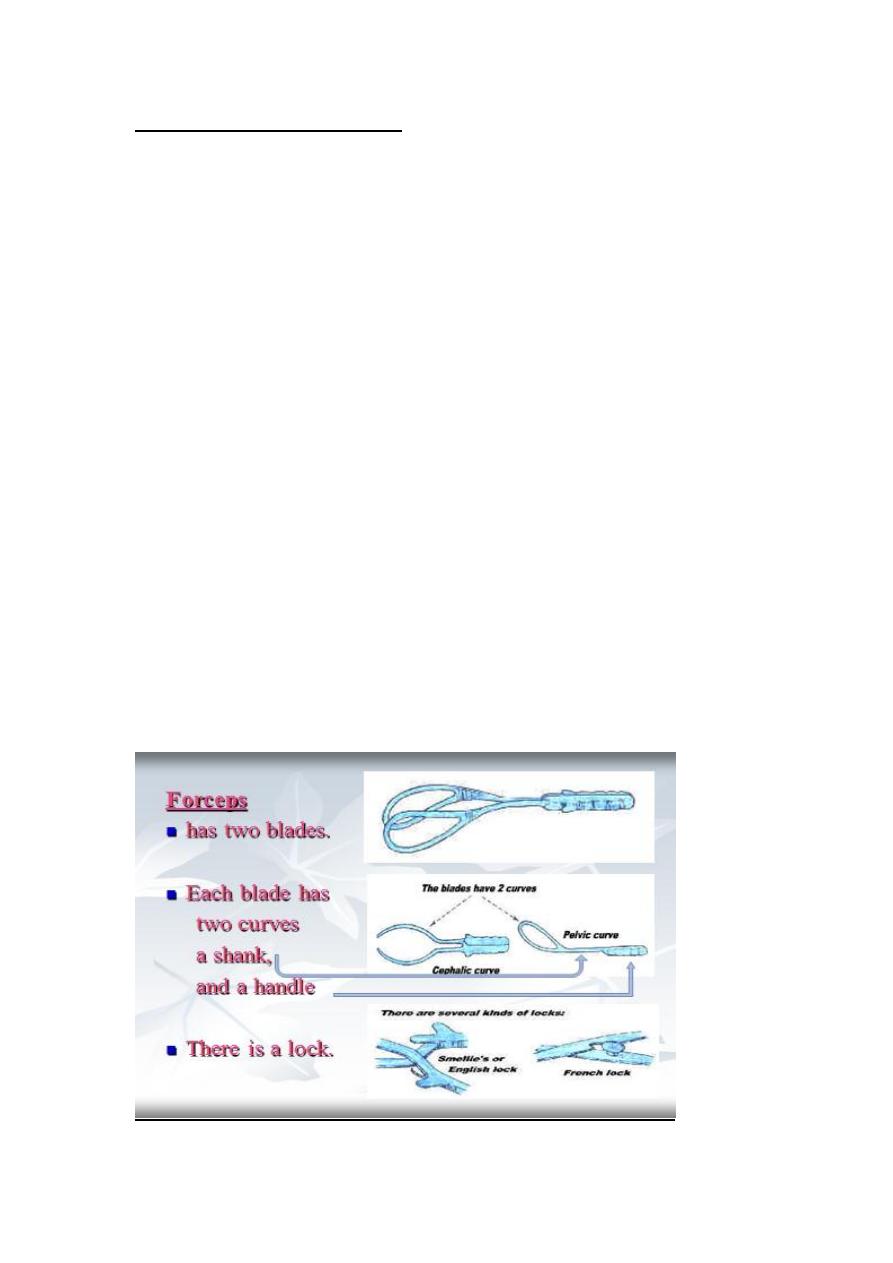
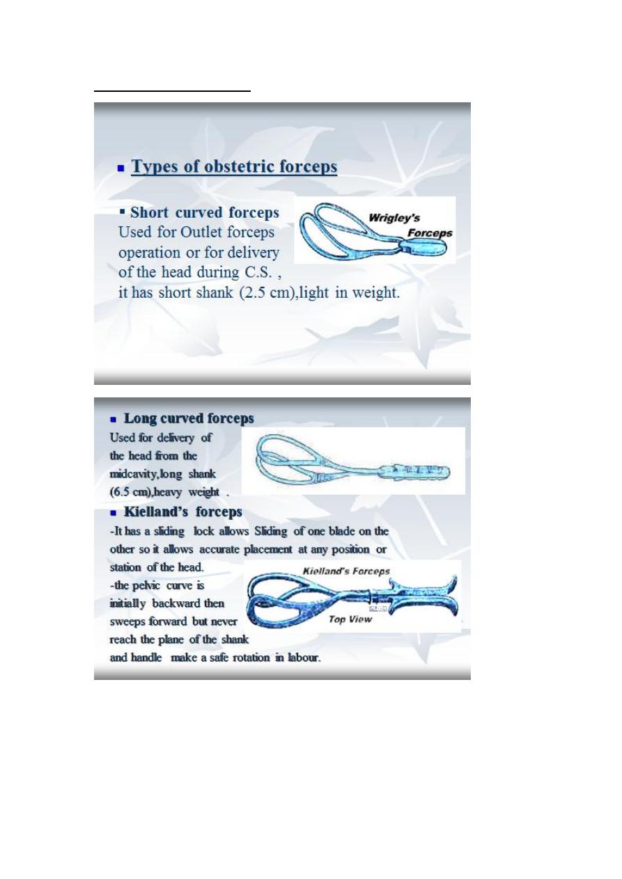
Types of obstetric forceps
.
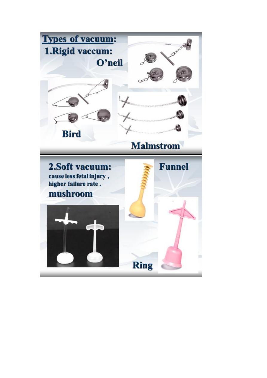
8
2.Soft

9
Contraindication :
Absolute :
1. lack of engagement .
2. Condition that contraindicate vaginal delivery.
(pelvic abnormality , fetopelvic disproportion)
3. Fetal malposition (face ,brow).
4. Dead fetus with postmortem changes .
5. Inability to diagnose the position of the fetal
head .
Relative :
1. Fetal macrosomia .
2. Lack of experience .
3. Repeated fetal scalp blood sampling or trauma .
4. Fetal bleeding or suspected coagulation defect .
5. Premature < 34 weeks or less than 1500 gm .
Prerequisites for delivery with forceps and vacuum:
Assisted delivery should be carried as a trial with cautious attempt
at delivery and is appropriately carried out in the operative
theater with immediate recourse to C.S. when unusual difficulty is
encountered at delivery.
11
The following steps should be certain before application of forceps :
1. Engaged head .
2. Position and attitude of the head .
3. Clinically adequate pelvis (mid ,outlet )
4. Empty bladder .
5. Ruptured membrane .
6. Cervix is fully dilated .
7. Appropriate anaesthesia (vacuum without )
8. Experience of the doctor .
9. Well informed patient .
10. working equipment .

11
Technique of vacuum
A betadine or antiseptic soap solution is applied to the rim of the
cup the cup slipped carefully in the vagina.
the cup is positioned over flexing point of fetal head
the negative pressure of this system is increased to 0.2kg/cm2
and perimeter of the cup is checked for intrapped cervical or
vaginal tissue
negative pressure is increased by 0.2kg/cm2 every 2 minute to a
maximum pressure of 0.8kg/cm2 in order to build a chignon.
then at this point the traction in the axis of birth canal can be
applied during uterine contraction
by placing hand in the vagina with the thumb in the cup an index
finger on the fetal scalp . traction force can be monitored in the
following manner
gradual increase in the traction force until the cup began to slip
away from the fetal scalp , diminished fetal traction are then hold
for the remainder of uterine contraction
Release traction between uterine contractions .
the expectation is that the presenting part should descend with
each push-pull event and the head will be delivered within
approximately 5 pulls.
the fetal heart is monitored through out the procedure with an
external fetal heart monitor once the head is delivered .the
suction is disconnected and the cup removed

12
Complication of assisted delivery :
Maternal complication : is more common with forceps than vacuum .
Soft tissue injuries includes :
Genital : uterine ,cervical ,vaginal ,perineal
lacerations .
Entrapment of the cervix is specific to the vacuum
Bladder and urethral injury : retention ,fistula .
Rectal injuries : laceration ,fistula ,defecation problems .
Fetal complication :
With forceps : 1. Transient facial marks.
2. Facial palsy .
3. Fracture of skull or facial bones .
4. Sever cervical cord damage .
With vacuum :
1. Scalp injury . 6. Fracture of skull .
2. Cephalhaematoma . 7. Neonatal jaundice
3. Subgleal haematoma . 8. Retinal haemorrhage
4. Intracranial haemorrhage .. 9. Brachial plexus injury .
5. Tentorial tears . 10. cerebral palsy .

13
Episiotomy :
Episiotomy :Is a surgical incision of the perinium to increase the
diameter of the vulval outlet during childbirth.
Indication :
Absolute :1. previous pelvic reconstruction.
2. pelvic floor surgery.
Relative : 1. short rigid perinium.
2. shoulder dystocia.
3. fetal distress.
4. instrumental or breech delivery.
Types of episiotomy:
Midline episiotomy :vertical incision towards the anus , less blood
loss ,easier repair ,quicker healing , less pain in the postpartum
period ,less dysparunia.
-risk of extension to the anus.
Medio lateral episiotomy : start at midline then laterally to avoid
the anal sphinctor .
Lateral episiotomy .
Technique of episiotomy
Consent.
Anaesthesia as local infiltration .
Performed at crowning of the head to reduce the bleeding.
Sharp scissors used to make single incision about 3-6 cm length
involving skin ,subcutaneous ,and superficial perineal muscules(2
nd
degree tear).
Immediate suturing after delivery to reduce blood loss ,using
interrupted or continuous suturing of the muscules and
subcutaneous tissues and subcuticular or interrupted suturing is
used for the skin.

14
Complication:
1.Difficult repair.
2.heavy bleeding.
3.extention to the anus.
4.infection.
5.pain and dyspareunia.
6.weak point in the perinium-tear.
7.Dryness from injury to bartholine gland.
After care:
1.analgesia,oral or suppositories.
2.prophylactic antibiotics.
3.washing with water and soap.
4.hot sitz bath.
5.removal of stitches at 5 days.